Abnormal Caudal Fixation of the Spinal Cord
- Tethered cord syndrome is the clinical syndrome produced by abnormal CAUDAL FIXATION (tethering) of the spinal cord, classically a LOW-LYING CONUS MEDULLARIS (ending below the normal L1-L2 level) with a THICKENED and often FATTY FILUM TERMINALE; the fixed, stretched distal cord suffers traction and impaired perfusion, which produces progressive symptoms, often precipitated or worsened by growth (in children) or by flexion/activity.
- The orthopaedic surgeon is often the first to suspect it, because the presenting clues are frequently MUSCULOSKELETAL: a unilateral or progressive CAVOVARUS foot, asymmetric calf/leg ATROPHY or a leg-length discrepancy, and SCOLIOSIS (especially atypical or left thoracic curves, or any curve with a neurological deficit) - so these findings should prompt a search for an underlying tethered cord.
- CUTANEOUS STIGMATA over the lumbosacral midline are an important external marker of underlying spinal dysraphism and tethering: a sacral DIMPLE (especially a large or high one), a HAIRY PATCH (hypertrichosis), a subcutaneous LIPOMA, a haemangioma/capillary malformation or a dermal sinus - their presence should raise suspicion and prompt imaging.
- Symptoms are PROGRESSIVE across three domains: NEUROLOGICAL (lower-limb weakness, sensory change, reflex changes), UROLOGICAL/bowel (neurogenic bladder, incontinence, recurrent urinary infection - a key reason for urological assessment), and PAIN (back and leg pain), and in children the deterioration is often linked to growth spurts.
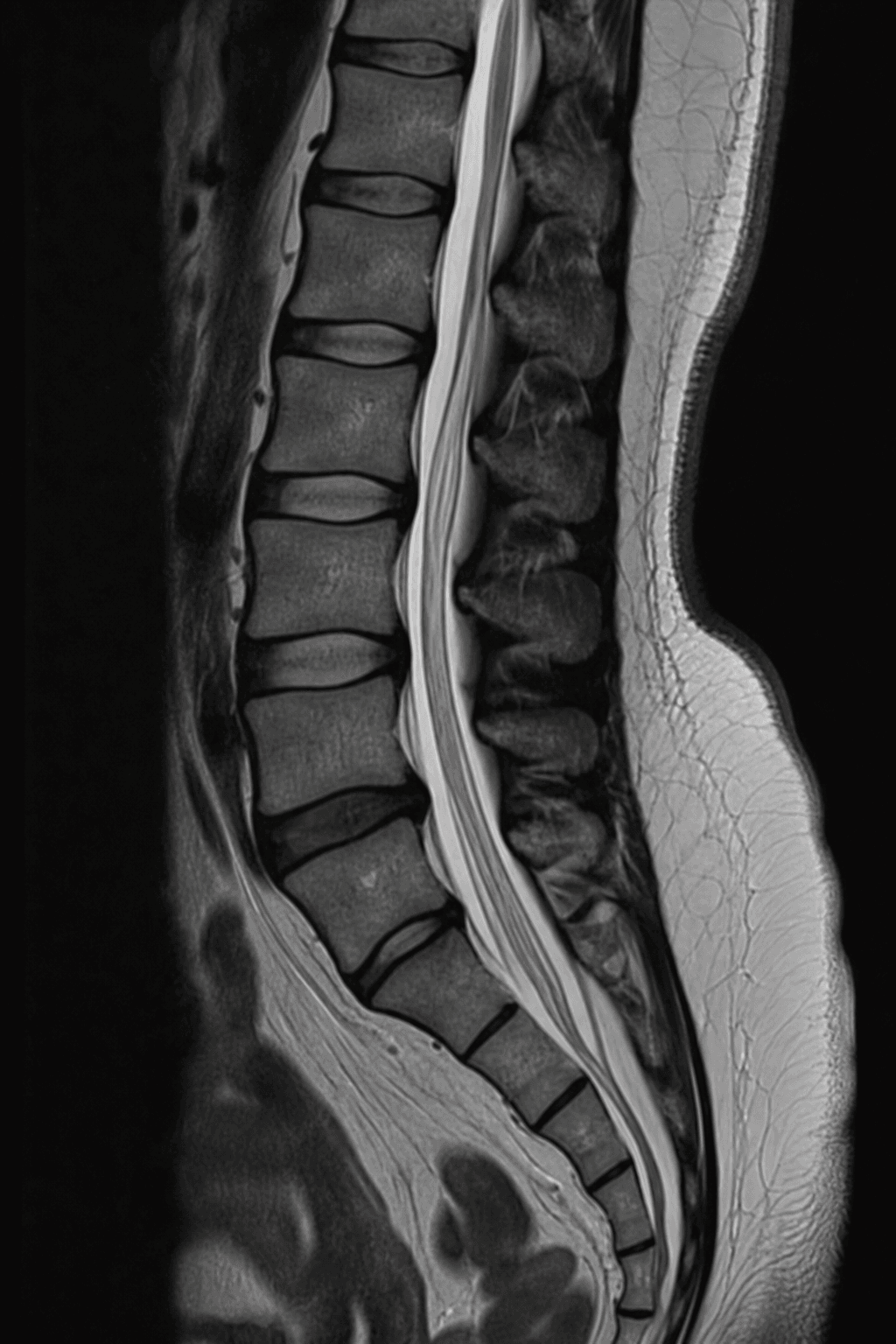
- MRI is the key investigation, demonstrating the LOW-LYING CONUS (below L2), a THICKENED filum terminale (a thickened/fatty filum, with a filum thickness threshold around 1.3 mm and over being associated with tethering/filum lipoma in studies), and any associated dysraphic lesion (lipomyelomeningocoele, dermal sinus, split-cord malformation, diastematomyelia); spinal ultrasound can screen infants before the posterior elements ossify.
- Treatment is surgical UNTETHERING - releasing the tethering element, most simply by sectioning a tight/thickened filum terminale (or addressing a more complex lesion) - performed to halt progression and, where possible, improve symptoms; the principle is that established deficits may not fully reverse, so timely surgery in a symptomatic or deteriorating patient is important, ideally within a multidisciplinary (neurosurgery, urology, orthopaedics) framework.
- “Tethered cord = abnormal caudal fixation: LOW conus (below L2) + THICKENED/fatty filum -> traction/ischaemia -> progressive neuro/uro/pain symptoms.
- “ORTHOPAEDIC clues: unilateral CAVOVARUS foot, asymmetric leg atrophy/leg-length difference, scoliosis (atypical/neurological). Plus cutaneous stigmata (dimple, hairy patch, lipoma).
- “MRI confirms (low conus, thick filum); treat by surgical UNTETHERING (filum section). Halts progression - established deficits may not fully reverse.
A unilateral/progressive cavovarus foot, asymmetric leg atrophy, scoliosis, and lumbosacral cutaneous stigmata (dimple, hairy patch, lipoma) - especially with bladder symptoms.
MRI: low conus (below L2), thickened/fatty filum. Treat by surgical untethering (filum section) - halts progression.
Definition, Clues & Imaging
Tethered cord syndrome is the syndrome of abnormal caudal fixation of the spinal cord - a low-lying conus (below L1-L2) with a thickened, often fatty filum terminale - so the distal cord is under traction with impaired perfusion, producing progressive symptoms, often worsened by growth. The orthopaedic surgeon frequently suspects it from the periphery: a unilateral or progressive cavovarus foot, asymmetric calf atrophy or a leg-length difference, and scoliosis (especially atypical or neurological curves). Lumbosacral cutaneous stigmata - a sacral dimple, hairy patch, lipoma, haemangioma or dermal sinus - are an important external marker. Symptoms span neurological, urological and pain domains. MRI confirms the low conus and thickened filum and defines any dysraphic lesion; spinal ultrasound screens infants.
Management
- Confirm and characterise on MRI - the low conus, thickened/fatty filum and any associated lesion (lipomyelomeningocoele, dermal sinus, split-cord/diastematomyelia); ultrasound screens infants before the posterior elements ossify.
- Assess all three domains - neurological examination, urological assessment (urodynamics/bladder), and the orthopaedic deformities (foot, leg, spine).
- Surgical untethering - release the tethering element, most simply by sectioning a tight/thickened filum terminale, or addressing a more complex lesion; the aim is to halt progression and, where possible, improve symptoms.
- Act before deficits become fixed - established neurological/urological deficits may not fully reverse, so timely surgery in a symptomatic or deteriorating patient matters; manage within a multidisciplinary team (neurosurgery, urology, orthopaedics).
The recurring orthopaedic lesson of tethered cord syndrome is that a unilateral cavovarus foot, asymmetric leg atrophy, or an atypical/neurological scoliosis is frequently the FIRST sign of an underlying tethered cord, so these findings - especially with lumbosacral cutaneous stigmata or any bladder symptoms - should prompt a neuro-axis MRI rather than treating the foot or curve in isolation. Operating on the foot or spine without recognising and addressing the tethered cord risks ongoing progression and poorer outcomes. Because the syndrome is progressive and established deficits may not fully reverse, timely untethering in a symptomatic or deteriorating patient, coordinated with neurosurgery and urology, is the priority.
Evidence & Key Studies
Cutaneous markers and filum terminale thickness in identifying tethered cord (children with sacral dimples)
- In children with sacral dimples, tethered cord syndrome requiring surgery was identified in 4.6%, supporting the link between lumbosacral cutaneous markers and underlying tethering.
- A dimple long diameter of 5 mm or more was significantly associated with tethered cord syndrome.
- A filum terminale thickness cutoff of 1.3 mm or more predicted filum terminale lipoma with about 93% sensitivity and 80% specificity, and spinal ultrasound can screen for it.
Surgical management of tethered spinal cord through a minimally invasive approach (filum release)
- Tethered cord release can be performed by hemilaminectomy, durotomy and careful separation/sectioning of the filum terminale, here via a minimally invasive approach.
- The case (a young adult with chronic back pain and thigh numbness) had a midline fusion defect and abnormal conus medullaris termination, illustrating the typical pathology.
- Postoperative recovery was rapid without complications, illustrating the principle of untethering to relieve traction on the cord.
According to PubMed, the association of lumbosacral cutaneous markers (sacral dimple size) with tethered cord and the filum terminale thickness threshold (around 1.3 mm) with spinal ultrasound screening come from the cited Watanabe study; the surgical principle of untethering by sectioning/separating the filum terminale, and the typical pathology (abnormal conus termination with a dysraphic defect), from the cited Ozer report. The low-conus/thickened-filum definition, the orthopaedic clues (cavovarus foot, asymmetric leg, scoliosis), the neuro/urological/pain symptom triad and the multidisciplinary, progression-halting principle are standard, well-established teaching. (See also our Spinal Dysraphism and Cavovarus Foot topics.)
Clinical Decision Scenarios
Practise clinical reasoning and management decisions out loud
“A child has a unilateral cavovarus foot and a sacral dimple. What underlying diagnosis must you consider and how do you investigate?”
“How is tethered cord syndrome managed, and what do you tell the family about outcome?”
Mnemonics & Memory Aids
TETHER
Hook:TETHER: Thick filum/low conus, External skin stigmata, Toes (cavovarus), Hold of bladder lost, Examine/MRI, Release (untether).
Definition
- Abnormal caudal fixation of the cord (traction/ischaemia)
- Low-lying conus (below L2) + thickened/fatty filum terminale
- Progressive; often worsened by growth
Clues
- Orthopaedic: unilateral cavovarus foot, asymmetric leg atrophy/leg-length, scoliosis
- Cutaneous: sacral dimple, hairy patch, lipoma, haemangioma, dermal sinus
- Urological: neurogenic bladder, incontinence, recurrent UTI
Diagnosis
- MRI: low conus, thickened/fatty filum, associated dysraphic lesion
- Spinal ultrasound screens infants (before ossification)
- Urodynamics for bladder assessment
Management
- Surgical untethering (section the filum / release the lesion)
- Halts progression; established deficits may not fully reverse - act timely
- Multidisciplinary; monitor for re-tethering and manage foot/spine/bladder