Central Ray Deficiency
- Typical Cleft: Isolated, sporadic, U-shaped, good first web, better function.
- Atypical Cleft: Syndromic, familial, V-shaped, poor first web, worse function.
- Manske-Halikis I-V: Based on thumb web (normal→suppressed) and cleft severity.
- EEC Syndrome: Ectrodactyly-Ectodermal dysplasia-Clefting - autosomal dominant.
- Surgical Goals: Close cleft, deepen first web, release syndactyly, maintain function.
- “Typical = U-shaped, good function, sporadic
- “Atypical = V-shaped, syndromic, familial
- “Snow-Littler procedure = cleft closure technique
- “EEC syndrome triad: limbs, skin/hair, facial cleft
- “First web space depth determines function
Overview and Epidemiology
What it is. Cleft hand, also called ectrodactyly, split hand or lobster claw deformity, is a congenital longitudinal deficiency in which one or more of the central rays, metacarpals and phalanges, have failed to form. The result is a V-shaped or U-shaped cleft in the central portion of the hand.
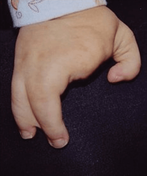
How common. Split-hand/split-foot malformation (SHFM), the umbrella term that encompasses cleft hand, occurs in approximately 1 in 8,500 to 1 in 25,000 newborns and accounts for approximately 15% of all limb reduction defects (Holder-Espinasse et al, Eur J Hum Genet 2019). Both hands are frequently involved, many patients have concurrent foot involvement, and the sexes are broadly equally affected.
Typical and atypical. The historical split into typical and atypical cleft hand is descriptive rather than diagnostic. The "atypical cleft hand" has now largely been reclassified as symbrachydactyly, a transverse or dysplastic process rather than a true central longitudinal deficiency, and the Differential Diagnosis section below sets out how the two are told apart.
Where its siblings are. Cleft hand sits inside the congenital hand overview and is classified alongside the other failures of formation and differentiation: syndactyly, which is almost always present within the cleft and is often the thing you actually operate on; polydactyly; thumb hypoplasia, because first-web suppression is what determines function here; and clinodactyly and macrodactyly as the other digit-shape anomalies a parent will ask you to distinguish it from.
Genetics and Associated Syndromes
Inheritance. Most commonly autosomal dominant with incomplete penetrance; X-linked and autosomal recessive forms also occur (Holder-Espinasse et al, 2019). Incomplete penetrance is why an apparently isolated case can still be familial, and why relatives are examined.
The loci. The recognised SHFM loci, SHFM1-6:
- SHFM1 at 7q21.2 (DLX5)
- SHFM2 at Xq26
- SHFM3 at 10q24 duplication, described in that paper as "one of the most frequent" causes
- SHFM4 at 3q27 (TP63)
- SHFM5 at 2q31
- SHFM6 (WNT10B, 12q13)
EEC syndrome. Ectrodactyly-ectodermal dysplasia-clefting is the most common syndromic association. It is autosomal dominant and caused by TP63 mutation at the 3q27 (SHFM4) locus.
EECEEC Syndrome Triad
Hook:EEC: Ectrodactyly (limbs), Ectodermal dysplasia (skin/hair/teeth), Clefting (facial)
The other associations. SHFM (split hand-split foot malformation) denotes isolated limb defects. Cornelia de Lange syndrome and Adams-Oliver syndrome are the other syndromes to know.
Why you look beyond the limb. In the 32-case 10q24 series, 5 had findings outside the limb: renal dysplasia, cutis aplasia, hypogonadism, and agenesis of the corpus callosum with hydrocephalus (PMID 30622331). The screening advice later on this page is not a formality.
Anatomy and Pathophysiology
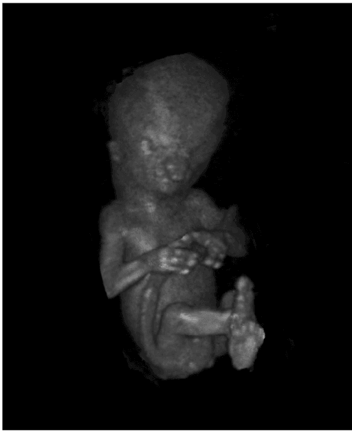
Embryology. The deficiency arises during limb bud formation at 4-8 weeks of gestation. Five digital rays form from radial (thumb) to ulnar (small finger), the apical ectodermal ridge (AER) and the zone of polarising activity (ZPA) controlling proximal-distal and radial-ulnar patterning, while programmed cell death clears the interdigital tissue. In cleft hand the AER and ZPA are disrupted, and the central rays, index, middle and ring (rays 2, 3 and 4), are suppressed or absent during differentiation; in atypical forms the development of the first web space is affected as well.
What is missing. One or more of rays 2, 3 and 4 are completely or partially absent, and the loss may involve metacarpals, phalanges or both. The soft-tissue cleft, V-shaped or U-shaped, extends proximally.
- Typical cleft: a wider, shallower U-shaped cleft, often with a single ray absent, most often the middle finger (ray 3); the deficiency is isolated to the central rays and the first web is preserved, so opposition and grasp are relatively preserved
- Atypical cleft: a narrow, deep V-shaped cleft extending to the carpus, often with 2 or 3 central rays absent, abnormal transverse metacarpal elements, and a tight, narrow, adducted first web, so opposition and grasp are poor
The soft tissues. The border digits are often joined by syndactyly (index-middle or ring-small) and are often stiff, with joint contractures. Neurovascular bundles may run an abnormal course around the cleft borders, extrinsic tendons may be absent or misdirected, and the thumb may be radially deviated or adducted.
Why the first web decides everything. The depth of the thumb-index web is the critical determinant of hand function: a preserved web goes with good opposition and grasp, a suppressed one with poor. Its suppression is variable, and grading it is the basis of the classification that follows.
Classification Systems
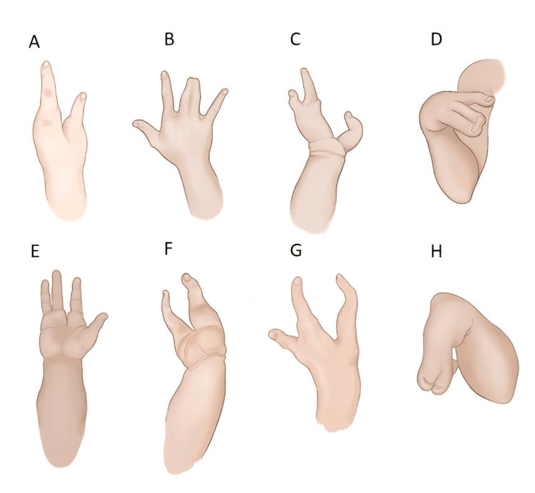
The system that decides treatment. Manske and Halikis graded the cleft hand by the state of the first web rather than the width of the central defect, because the web is what determines function. The grade therefore reads straight across into a decision: Types I and II function well and may not require surgery; Types III and IV benefit from surgery for function and appearance; Type V needs complex reconstruction or prosthetic consideration.
- First Web Space
- Normal thumb web
- Cleft Description
- Central cleft only
- Function
- Good - normal pinch/grasp
- First Web Space
- Mildly narrowed
- Cleft Description
- Central cleft + mild web suppression
- Function
- Good - slightly reduced
- First Web Space
- Moderately narrowed
- Cleft Description
- Central cleft + moderate suppression
- Function
- Moderate - reduced pinch
- First Web Space
- Severely suppressed
- Cleft Description
- Deep cleft + severe web contracture
- Function
- Poor - minimal pinch
- First Web Space
- Absent (monodactyly)
- Cleft Description
- Single digit only (thumb or finger)
- Function
- Very poor - no opposition
Clinical Presentation
History. The cleft is an obvious deformity visible at birth, so the history is about what surrounds it. Ask for a family history, because atypical forms are familial; ask about skin, hair and teeth (ectodermal dysplasia) and about cleft palate, because these point to a syndrome. Motor milestones are usually normal.
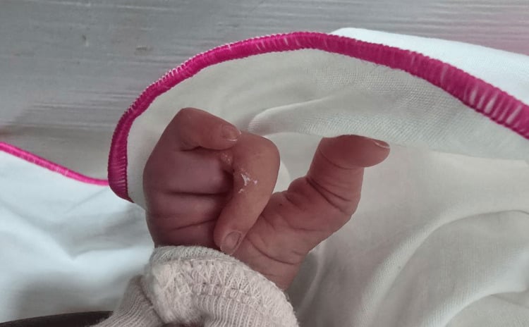
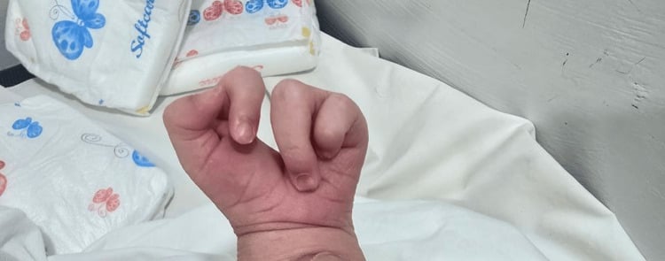
Examination. Examine both hands and both feet. On inspection and palpation, document:
- The configuration of the cleft, V-shaped or U-shaped
- The number of digits present and which rays are missing
- The depth and width of the first web space
- Syndactyly of the border digits
- Thumb position, looking for radial deviation or an adduction contracture
- Which metacarpals are present, and whether abnormal transverse metacarpal elements can be felt (atypical forms)
- The tightness of the first web skin
- The digital arteries and nerves, which may be anomalous
Function is the examination that matters most. Can the thumb oppose the remaining fingers? Assess tip, key and three-point pinch, power grip with the remaining digits, and the first web span measured between thumb and finger at maximal abduction. Then watch the child: can they manipulate objects, feed themselves and grasp toys?
Associated findings. 60% have a split foot deformity. Syndactyly of the border digits is common, polydactyly may coexist with central ray absence, the thumb may be underdeveloped, and the border digits often have joint contractures.
Red flags for a syndrome. Any of the following should prompt a syndrome work-up:
- Sparse hair or teeth (ectodermal dysplasia, EEC syndrome)
- Cleft lip or palate (EEC syndrome)
- Cleft feet (split hand-split foot malformation)
- Multiple limb defects (a broader genetic syndrome)
- Developmental delay (a syndromic aetiology)
Flatt called the typical cleft hand "a functional triumph and a social disaster": it usually works remarkably well, because the preserved first web gives good opposition and grasp, but it looks abnormal and attracts attention.
That paradox governs the operation. In a well-functioning (Manske I-II, typical) hand, surgery is largely for appearance, so you are operating on a working hand and risking stiffness, web contracture and neurovascular injury to functioning border digits; the decision is a careful shared one that weighs cosmesis and social factors against making a good hand worse. In the atypical or severe (Manske III-V) hand the first web is suppressed, surgery genuinely improves function, and the indication is functional.
When counselling: most children adapt and develop excellent compensatory function; address the family's distress and social concerns explicitly; avoid over-promising a "normal-looking" hand; and, if cosmetic surgery is chosen, time it before school-age social awareness.
Investigations
Radiographs. AP and lateral views document the bony anatomy. The external cleft must be matched to the film: read it ray by ray from radial to ulnar, identify which metacarpals and phalanges are present, absent or hypoplastic, and define the first web before planning anything. Look also for carpal anomalies, which may occur in atypical forms, for abnormal transverse metacarpal elements, for a short or radially deviated first metacarpal, and at any syndactyly to decide whether the fusion is bony or soft tissue. The typical cleft usually shows absence of the middle finger ray (ray 3) alone; the atypical cleft shows multiple absent rays, transverse bones and carpal anomalies.
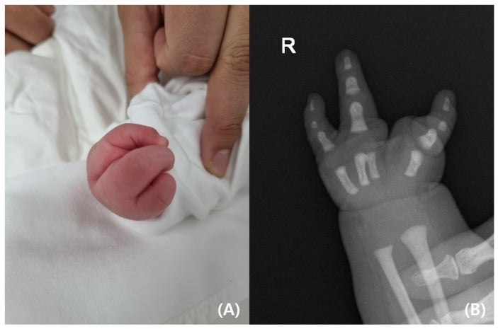
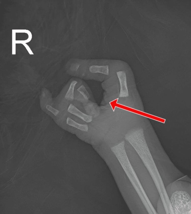
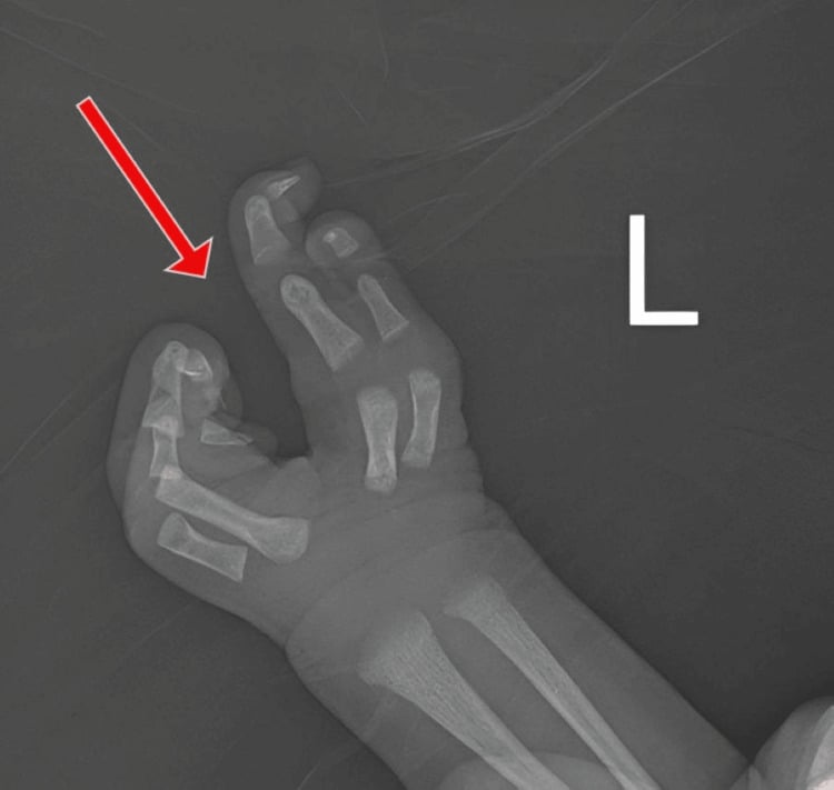
Genetic testing. Indicated for a familial pattern, syndromic features or bilateral involvement. TP63 testing looks for the EEC mutation and chromosomal microarray for deletions and duplications; genetic counselling is essential for familial cases.
Syndromic work-up. When indicated, it covers:
- Skin, hair and teeth, for ectodermal dysplasia
- Craniofacial evaluation for cleft lip or palate
- Both feet, for cleft feet
- Ophthalmology, for the eye anomalies of some syndromes
- Audiology, if syndromic
Functional baseline. Age-appropriate hand function tests, grip and pinch strength where the child will cooperate, and a video recording of function before surgery, so that the result can be judged against something.
Differential Diagnosis
Cleft hand must be distinguished from other central and transverse failures of formation, because management and prognosis differ markedly. The single most important modern distinction is between true central longitudinal deficiency (cleft hand) and symbrachydactyly of the cleft/atypical type, which is a transverse/dysplastic process and follows symbrachydactyly principles.
- Pattern of deficiency
- Central rays (2-4) absent, V- or U-shaped cleft
- Distinguishing features
- Well-formed border digits, transverse metacarpal bones may be present, foot often involved
- Laterality / inheritance
- Often bilateral; frequently familial / autosomal dominant
- Pattern of deficiency
- Transverse arrest with short or absent central digits
- Distinguishing features
- Nubbins / ectodermal remnants, short fingers, no familial pattern, foot usually spared
- Laterality / inheritance
- Almost always unilateral; sporadic
- Pattern of deficiency
- Amputation-like loss distal to a transverse level
- Distinguishing features
- No proximal digital elements beyond the arrest, may have nubbins
- Laterality / inheritance
- Unilateral; sporadic
- Pattern of deficiency
- Acrosyndactyly, distal ring constrictions, irregular digit loss
- Distinguishing features
- Constriction grooves, distal fusion, lymphoedema; no metacarpal cleft
- Laterality / inheritance
- Unilateral or asymmetric; sporadic, non-genetic
- Pattern of deficiency
- Extra (not absent) central rays, often hidden within syndactyly
- Distinguishing features
- Excess rather than deficient tissue, HOXD13 in synpolydactyly
- Laterality / inheritance
- Often bilateral; autosomal dominant
The classic exam trap is calling a unilateral, sporadic, nubbin-bearing hand with a central gap a "cleft hand" — if it is unilateral, non-familial and the foot is spared, think symbrachydactyly (cleft type), not true central longitudinal deficiency.
Management Algorithm
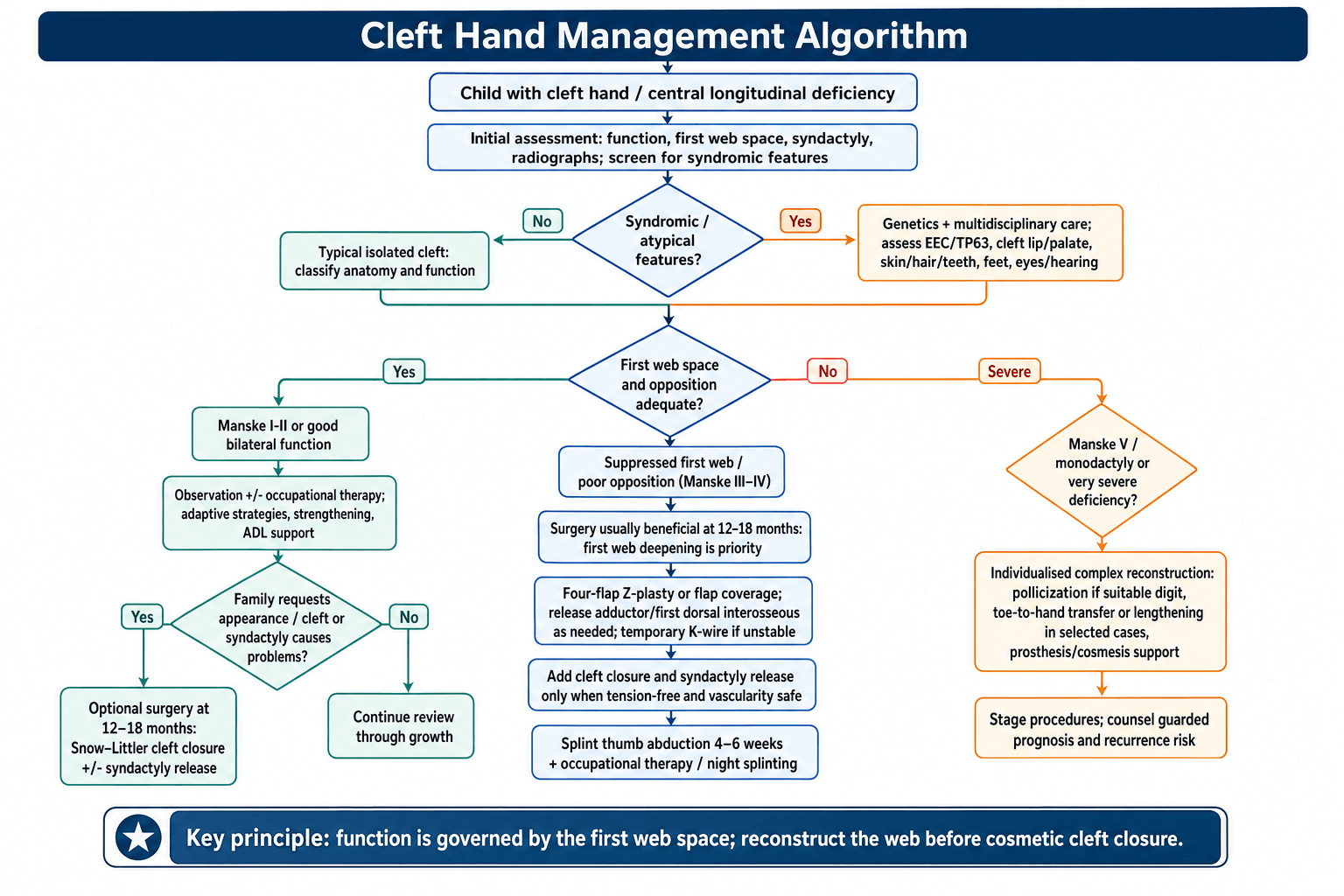
The goals, in order. Function comes first, appearance second, and growth is preserved throughout:
- Function: maximise opposition (thumb-finger pinch), improve grasp and release, and deepen the first web if it is suppressed
- Appearance: close the cleft, restore a more normal hand contour and improve social acceptance
- Growth: protect the growth plates and avoid damage to viable digits
Who is observed. A Manske Type I hand has good function and a normal first web, so surgery is optional; a Type II hand has mild impairment and may not require surgery; a unilateral cleft in a child with good bilateral hand function may not need surgery on the non-dominant hand. Occupational therapy teaches adaptive strategies, strengthens the existing digits, stretches a mildly contracted first web and trains feeding, dressing and writing.
Timing. Surgery is best done at 12-18 months: early enough to integrate the hand before fine motor development, late enough for safe anaesthesia and tissue handling, and before social awareness at 2-3 years. Type V reconstruction is considered at 18-24 months.
- First Web Status
- Normal thumb web
- Treatment Priority
- Cleft closure only (if desired)
- Timing
- 12-18 months
- First Web Status
- Mild narrowing
- Treatment Priority
- Cleft closure + web release
- Timing
- 12-18 months
- First Web Status
- Moderate suppression
- Treatment Priority
- First web deepening priority
- Timing
- 12-18 months
- First Web Status
- Severe suppression
- Treatment Priority
- Complex reconstruction required
- Timing
- Staged procedures
- First Web Status
- Absent (monodactyly)
- Treatment Priority
- Consider pollicization vs prosthesis
- Timing
- 18-24 months
What the operation sets out to do. Four things, and the second outranks the first. Close the cleft, which improves appearance and prevents trauma to the cleft margins. Deepen the first web space, which improves opposition and grasp and is the most important step for function. Release syndactyly of the border digits if present. And maintain viability by preserving the neurovascular supply to the digits.
Cleft hand is frequently part of split-hand/split-foot malformation, so the feet are commonly involved too, and the management principle is the opposite of the hand's. A cleft foot is generally well tolerated: children walk well, and a foot bears weight rather than performing precision grip, so there is no equivalent of the first web to reconstruct. The default is non-operative, with accommodative or extra-depth footwear, custom shoes or simple insoles, and many cleft feet need no surgery at all.
Surgery is reserved for shoe-fitting problems and progressive deformity: a very wide forefoot that will not fit footwear (forefoot narrowing or cleft closure), a protruding medial ray or hallux varus, or a deep cleft with skin breakdown. The goal is a plantigrade, shoeable foot, not fine function.
Always examine both feet and the contralateral hand, and use foot involvement as a prompt for genetic assessment (SHFM/EEC).
Surgical Techniques
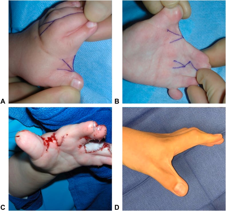
The classic cleft closure. The Snow-Littler procedure closes the cleft and narrows the hand, and is the gold standard for cleft closure in a functional hand. It suits the Manske Type I-III hand with a wide cleft and a good or only mildly narrowed first web. It is contraindicated where the first web is severely suppressed (Manske IV-V), because deepening the web is the priority, and in monodactyly (Type V), where there is insufficient tissue.
The steps.
- Incisions: longitudinal along the ulnar border of the radial digit and the radial border of the ulnar digit, designed as opposing flaps that close the cleft in a zigzag
- Dissection: identify and preserve the neurovascular bundles to the border digits, excise any rudimentary central ray tissue, and preserve the periosteum of the metacarpals
- Transverse bone resection, the key step: if an abnormal transverse metacarpal bone is present, resect it, which closes the gap between the metacarpal heads and lets the border digits come together
- First web deepening, if needed: release a suppressed web, by Z-plasty or four-flap Z-plasty, to at least the level of the metacarpal necks
- Cleft closure: approximate the border digits with the opposing zigzag flaps, and skin graft if the closure is under excessive tension
- Syndactyly release, if present: separate fused border digits with zigzag incisions and skin grafts as needed
The pearls. The first web is always addressed before the cleft is closed. Tight closure is avoided, with skin grafts used if needed. The neurovascular bundles may run an abnormal course, so they are found before anything is divided. And the incisions zigzag, because a linear scar across a web contracts.
Afterwards. Splint in the position of function for 3-4 weeks, then gentle range of motion, with occupational therapy for strengthening and daily-living training.
Complications and Outcomes
In theatre. The anomalous course of the neurovascular bundles increases the risk of injury (1-2%), and vascular anomalies make haemostasis difficult. The other intraoperative failures are an incomplete first web release and a skin flap that will not survive because of tension or poor design.
Early, within 6 weeks.
- Flap necrosis in 2-5%, from poor flap design or tension
- Infection in 1-3%, treated with antibiotics and debridement if needed
- Haematoma, rare, evacuated if it compresses neurovascular structures
- Wound dehiscence, more common after a closure under excessive tension
- Skin graft loss in 5-10% of grafts after syndactyly release
Late, after 6 weeks. Recurrent web space contracture is the most common complication, in 10-20%, especially in atypical clefts. Linear scars contract, which is why Z-plasties are used; border digits may become stiff (5-10%); a physis injured during surgery can disturb growth, rare with careful technique; vascular insufficiency in the border digits causes cold intolerance; and a nerve transected or trapped in scar forms a neuroma.
What to expect. In the typical cleft, near-normal hand function is achieved in 70-80%, with a good cosmetic result and improved opposition and grasp. In the atypical cleft, function is improved but not normal in 50-60%, the first web may re-contract and need revision, and multiple operations are often needed.
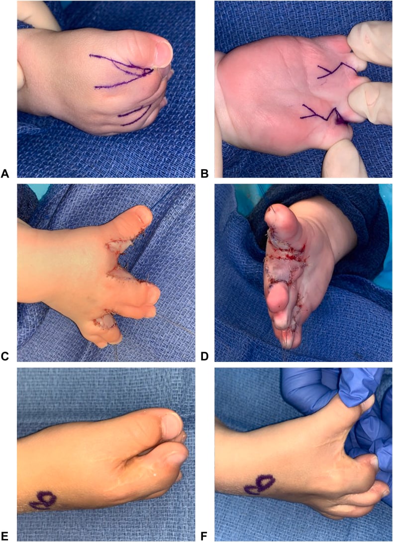
Revision. Indicated for web space re-contracture, inadequate initial correction or scar contracture, after waiting at least 6-12 months for the scar to mature. The techniques are a repeat Z-plasty, skin grafting or free flap coverage, and the success rate is lower than for primary surgery.
Postoperative Care and Rehabilitation
The principle. The tissues are allowed to heal before they are stretched, and then the first web is stretched aggressively to prevent re-contracture.
- Well-padded splint in the position of function, thumb abducted if the first web was deepened
- Elevation to reduce swelling
- Paracetamol for pain; avoid NSAIDs, which may increase bleeding
- Dressing kept clean and dry; first change at 7-10 days, or earlier if there are concerns
- Splint removed at 3-4 weeks after cleft closure; the thumb stays abducted for 4-6 weeks after web deepening
- Gentle passive range of motion and oedema control; no forceful stretching
- Scar massage once the incisions have healed
- Occupational therapy begins
- Active range of motion and strengthening with therapy putty
- Fine motor skill development, with use encouraged in age-appropriate play and daily activities
- Aggressive first web stretching to prevent re-contracture
- Advanced strengthening and bilateral hand coordination
- Age-appropriate writing and drawing; adaptive equipment trialled if needed
- Night abduction splint for 3-6 months
- Scar management with silicone gel sheets, massage and moisturisation
- Annual review through skeletal maturity, assessing function at each visit
- Further surgery planned as the child grows
- Psychosocial support for appearance concerns and school integration
Guidelines, Registries & Global Practice
Global epidemiology
- Split-hand/split-foot malformation (SHFM), the umbrella entity for cleft hand, affects approximately 1 in 8,500 to 1 in 25,000 newborns and represents about 15% of all limb reduction defects (Holder-Espinasse et al, Eur J Hum Genet 2019).
- Inheritance is most often autosomal dominant with incomplete penetrance; this drives the need for genetic counselling and examination of relatives in apparently isolated cases.
- Cleft hand is a central longitudinal deficiency in the IFSSH-endorsed OMT classification (Tonkin et al, J Hand Surg Am 2013).
Guidance and standards, side by side
There are no disease-specific randomised guidelines for cleft hand; practice is built on classification systems and specialist consensus. Standards that genuinely shape practice are summarised below.
- Position relevant to cleft hand
- Cleft hand classified as central longitudinal deficiency; standard global nomenclature
- Evidence basis
- Consensus + reliability study, 101 patients (Level IV)
- Position relevant to cleft hand
- Treatment driven by thumb-web status (types I-V); web reconstruction prioritised over cleft closure
- Evidence basis
- Original cohort of 46 hands (Level IV)
- Position relevant to cleft hand
- Surgery individualised to function; first-web deepening, cleft closure and syndactyly release typically 1-2 years of age; non-operative care acceptable when function is good
- Evidence basis
- Expert consensus / case series (Level IV-V)
- Position relevant to cleft hand
- Offer TP63 / chromosomal microarray testing and counselling for familial, bilateral or syndromic (EEC, SHFM) presentations
- Evidence basis
- Guideline-level genetic practice
Registry evidence
There is no dedicated international cleft-hand registry, and joint-replacement registries (NJR, AJRR, AOANJRR, SHAR) do not apply. Population-level data derive instead from congenital anomaly surveillance networks (for example EUROCAT in Europe and the ICBDSR international clearinghouse), which capture SHFM among limb reduction defects and underpin the 1 in 8,500-25,000 incidence figure.
Global practice variation
- High-resource settings: early multidisciplinary assessment (hand surgery, plastics, genetics, occupational therapy), functional surgery individualised to thumb-web status, and access to four-flap Z-plasty, microsurgical toe-to-hand transfer and pollicization for severe cases.
- Limited-resource settings: emphasis on the highest-yield functional interventions (first-web deepening, syndactyly release) with less reliance on microsurgical reconstruction; genetic testing and prosthetics may be unavailable.
- Across all settings the shared principle is constant: function is governed by the thumb web, so web reconstruction takes priority over cosmetic cleft closure.
Manske-Halikis Classification: Surgical Classification of Central Deficiency by Thumb Web
OMT (Oberg-Manske-Tonkin) Classification of Congenital Upper Limb Anomalies
EEC Syndrome: Clinical Spectrum in 24 Cases
Duplication of 10q24 locus: broadening the clinical and radiological spectrum
Snow-Littler Procedure: Clinical Experience and Outcomes
Exam Viva Scenarios
Practise clinical reasoning and management decisions out loud
“A 15-month-old child is referred with unilateral cleft hand. On examination, there is a U-shaped cleft with absence of the middle finger. The first web space is normal depth. The thumb and index finger are separate, and the ring and small fingers show simple syndactyly. X-ray confirms isolated absence of ray 3. Parents are asking about treatment options.”
“An 18-month-old presents with bilateral cleft hands. Both hands show V-shaped clefts extending to the carpus with absence of rays 2, 3, and 4. The first web spaces are severely narrowed with the thumbs held in adduction. The child cannot oppose the thumbs to the small fingers. The child also has sparse hair and bilateral cleft lip. X-rays show multiple central ray absence and abnormal transverse metacarpal bones. What is your assessment and management?”
“A 2-year-old child has a cleft hand with only a single central digit present (small finger). The thumb and all other rays are absent. X-ray confirms single 5th ray only. The parents are distressed and asking if the child will be able to use the hand. How would you manage this case?”
Classification
- Typical (70%): U-shaped, sporadic, normal first web, good function
- Atypical (30%): V-shaped, familial/syndromic, poor first web, worse function
- Manske I: Normal first web - surgery optional
- Manske II: Mild narrowing - good prognosis
- Manske III: Moderate suppression - surgery beneficial
- Manske IV: Severe suppression - complex reconstruction
- Manske V: Monodactyly - pollicization vs observation
Key Anatomy
- Central ray absence (rays 2, 3, 4) - index, middle, ring fingers
- First web depth = MOST important functional determinant
- Transverse metacarpal bones in atypical forms
- Neurovascular bundles may have anomalous course
- Border digits often have syndactyly
EEC Syndrome
- Ectrodactyly (cleft hand/foot) - 90% have limb defects
- Ectodermal dysplasia (sparse hair, absent teeth, dry skin)
- Clefting (cleft lip/palate)
- TP63 gene mutation - autosomal dominant
- Multidisciplinary care essential
Surgical Principles
- Timing: 12-18 months optimal (before fine motor development)
- FIRST WEB DEEPENING is more important than cleft closure for function
- Snow-Littler: classic cleft closure technique
- Four-flap Z-plasty: gold standard for first web deepening
- Syndactyly release: separate fused border digits
Key Numbers
- Incidence: approximately 1 in 8,500-25,000 (SHFM, Holder-Espinasse 2019)
- Bilateral: hands frequently both involved
- Cleft feet: 60% also have split foot
- Surgery timing: 12-18 months
- Good outcomes: 70-80% typical, 50-60% atypical
Complications
- Web contracture recurrence: 10-20% (most common, especially atypical)
- Flap necrosis: 2-5% (tension, poor design)
- Neurovascular injury: 1-2% (anomalous anatomy)
- Stiffness: 5-10% of border digits
- Revision surgery: needed in 15-20% of atypical clefts
VIVA ESSENTIALS
- Typical vs Atypical FIRST - determines prognosis and approach
- First web depth determines function - prioritize this over cleft closure
- Screen for EEC syndrome: sparse hair, cleft palate, dry skin, absent teeth
- Manske Type I-II may not need surgery - counsel about optional nature
- Atypical clefts: first web deepening BEFORE cleft closure
- Bilateral cases: stage procedures 3-6 months apart
References
-
Tonkin MA, Tolerton SK, Quick TJ, et al. Classification of congenital anomalies of the hand and upper limb: development and assessment of a new system. J Hand Surg Am. 2013;38(9):1845-1853.
-
Manske PR, Halikis MN. Surgical classification of central deficiency according to the thumb web. J Hand Surg Am. 1995;20(4):687-697.
-
Buss PW, Hughes HE, Clarke A. Twenty-four cases of the EEC syndrome: clinical presentation and management. J Med Genet. 1995;32(9):716-723.
-
Barsky AJ. Cleft hand: classification, incidence, and treatment. Review of 149 cases. J Bone Joint Surg Am. 1964;46:1707-1720.
-
Miura T, Nakamura R, Tamura Y. Long-term follow-up of cleft hands: a review of 40 clefts in 25 patients. J Hand Surg Am. 1990;15(5):739-743.
-
Ogino T, Minami A, Fukuda K, et al. Congenital anomalies of the upper limb among the Japanese in Sapporo. J Hand Surg Br. 1986;11(3):364-371.
-
Nutt JN, Flatt AE. Congenital central hand deficiency: a classification based on anatomical patterns and treatment. J Hand Surg Br. 1981;6(1):48-58.
-
Blauth W, Borisch N. Cleft hands: classification and incidence. Handchir Mikrochir Plast Chir. 1990;22(1):2-7.
-
Bouvet JP, Lemerle P, Lortat-Jacob A. Le traitement des mains botes centrales (central deficiency). Ann Chir Plast. 1980;25(3):240-247.
-
Al-Qattan MM, Al-Thunayan A, De Cordier M, et al. Classification of the typical form of symbrachydactyly. J Hand Surg Br. 1998;23(6):801-804.
-
Snow JW, Littler JW. Surgical treatment of cleft hand. Plast Reconstr Surg. 1967;40(5):413-418.
-
Buck-Gramcko D. Pollicization of the index finger: method and results in aplasia and hypoplasia of the thumb. J Bone Joint Surg Am. 1971;53(8):1605-1617.
-
Flatt AE. The Care of Congenital Hand Anomalies. 2nd ed. St Louis: Quality Medical Publishing; 1994.
-
Oberg KC, Feenstra JM, Manske PR, et al. Developmental biology and classification of congenital anomalies of the hand and upper extremity. J Hand Surg Am. 2010;35(12):2066-2076.
-
Kay SPJ, McCombe D, Kozin SH, et al. Deformities of the hand and fingers. In: Wolfe SW, Hotchkiss RN, Pederson WC, Kozin SH, eds. Green's Operative Hand Surgery. 7th ed. Philadelphia: Elsevier; 2017:1267-1316.
-
Holder-Espinasse M, Jamsheer A, Escande F, et al. Duplication of 10q24 locus: broadening the clinical and radiological spectrum. Eur J Hum Genet. 2019;27(4):525-534. PMID: 30622331.
-
Rider MA, Grindel SI, Tonkin MA, Wood VE. An experience of the Snow-Littler procedure. J Hand Surg Br. 2000;25(4):376-381. PMID: 11058008.
-
Manske PR, Goldfarb CA. Congenital failure of formation of the upper limb. Hand Clin. 2009;25(2):157-170. PMID: 19380058.
Further Reading
- Tonkin MA. Failure of differentiation part II: Cleft hand. Hand Clin. 2009;25(2):195-213.
- Goldfarb CA, Manske PR, Busa R, et al. Upper-extremity phocomelia reexamined: a longitudinal dysplasia. J Bone Joint Surg Am. 2005;87(12):2639-2648.
- Elliott AM, Evans JA. Genotype-phenotype correlations in mapped split hand foot malformation (SHFM) patients. Am J Med Genet A. 2006;140(13):1419-1427.