The workhorse controlled femoral osteotomy for implant and cement removal in revision hip arthroplasty
- Core principle: the lateral third of the proximal femur is elevated as a vascularised osteomuscular sleeve with the vastus lateralis and abductors left attached — this vascularity drives the high union rate
- Indications: removal of well-fixed cementless or cemented stems, distal cement extraction, varus proximal femoral remodelling, exposure in periprosthetic infection, and improved acetabular exposure
- Length is templated preoperatively: distal enough to clear the stem tip or cement column, but leaving at least 4-6 cm of intact diaphysis for fixation of the revision stem
- A prophylactic distal cerclage cable placed 1-2 cm below the transverse limb BEFORE reaming and stem insertion prevents distal crack propagation — the single most examinable technical step
- Revision stems must bypass the osteotomy and achieve diaphyseal fixation: modular fluted tapered titanium or extensively porous-coated cobalt-chrome stems; proximally coated stems are contraindicated
- Complications, in the order Abdel's 612 hips actually found them: post-operative greater trochanteric fracture 7 per cent, fragment migration beyond 1 cm 7 per cent, INTRA-OPERATIVE fracture of the diaphyseal fragment 4 per cent, nonunion only 2 per cent. Fracture of the fragment - not escape, not nonunion - is the commonest complication, and most of it happens on the table
- “The anterior hinge is created by controlled anterior perforations and gentle osteotome levering — an uncontrolled hinge fracture converts a planned osteotomy into an unplanned periprosthetic fracture
- “In infection, ETO is safe and does not compromise two-stage exchange; it dramatically speeds removal of well-fixed implants and cement, reducing cortical perforation and operative time
- “Wagner described a straight lateral transfemoral approach; the Paprosky/Younger posterior-based ETO is the technique most commonly described in modern revision practice
The vastus lateralis and gluteus medius/minimus must remain attached to the fragment throughout. Stripping the fragment devascularises it and converts a greater than 95 percent union rate into a non-union and escape problem.
Place a cerclage cable or wire 1-2 cm distal to the transverse limb before reaming, trialling or impacting the revision stem. Hoop stresses during insertion otherwise propagate a longitudinal crack distally and destroy the fixation zone.
The proximal femur cannot support a stem after ETO. Use a stem gaining 4-6 cm of scratch-fit diaphyseal fixation distal to the osteotomy — modular fluted tapered or extensively porous-coated. Cemented or proximally coated primary stems will subside and fail.
The anterior cortex is perforated with a drill or pencil-tip burr and levered open with wide osteotomes in unison along the length. Cracking it open at a single point causes an uncontrolled fracture of the fragment or anterior cortex.
Rationale and Indications
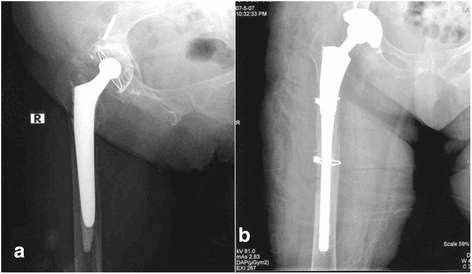
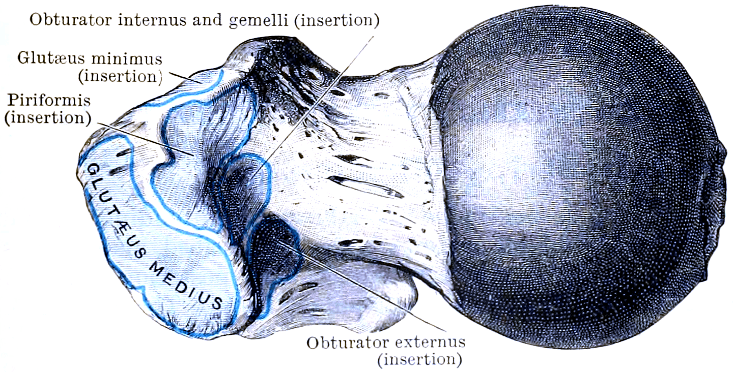
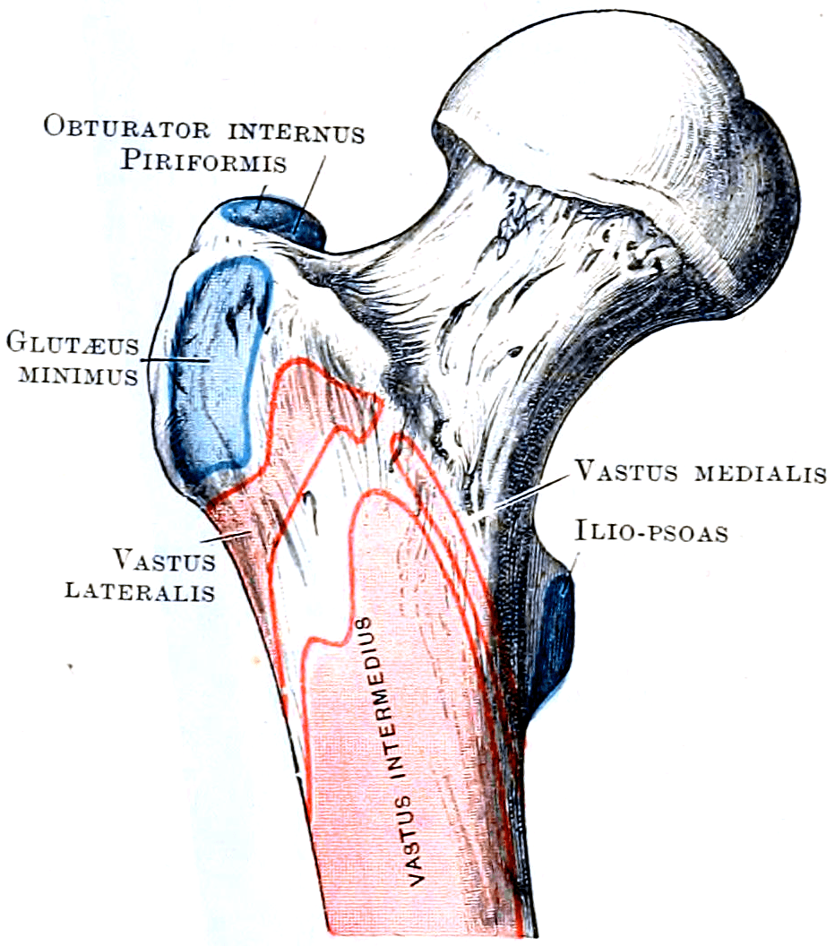
- 1Step 1 — Define the problem
Failed femoral component requiring stem removal: extensively porous-coated cementless stem, well-fixed cemented stem with a distal cement column, or infected implant at first-stage exchange
If it cannot, trephines and flexible osteotomes from above risk cortical perforation, eccentric reaming and greater trochanteric fracture — plan an ETO
- 2Step 2 — Assess proximal femoral geometry
A loose stem remodels the proximal femur into varus, so straight-line reaming from above aims at the lateral cortex
Neutral alignment of the new stem rather than lateral cortical perforation
- 3Step 3 — Other pro-ETO drivers
Stiff or protrusio hip needing acetabular access, a stiff arthroplasty where forced dislocation risks femoral fracture, or a selected Vancouver B2 or B3 periprosthetic fracture
Complete implant and cement removal under direct vision — critical in infection, where retained cement is a common cause of persistent sepsis
- 4Step 4 — Check the distal diaphysis (the go or no-go question)
Is there an intact supportive isthmus distal to the planned osteotomy exit? Less than 4 cm of intact diaphysis is inadequate
4 cm or more of supportive isthmus — proceed with ETO and a distally fixing stem. Less than 4 cm — go to step 6
- 5Step 5 — Check bone quality of the fragment
Severely osteoporotic or ectatic bone will fragment further when the lid is raised
If the fragment cannot be trusted, endoprosthetic replacement is the safer plan
- 6Step 6 — Inadequate distal bone stock (Paprosky IIIB or IV)
No diaphyseal segment will hold a scratch-fit, so an ETO alone leaves nothing to fix into
The osteotomy may still be used purely for exposure, but fixation must come from an alternative construct
- 7Step 7 — Protect the soft-tissue sleeve
The fragment survives on the abductor insertion above (gluteus medius and minimus) and the vastus lateralis origin below
A vascularised fragment that unites when closed and cabled back over the new stem
The ETO converts a hostile femoral revision into a controlled, reproducible exposure. Rather than fighting a well-fixed stem or distal cement mantle from above — risking cortical perforation, eccentric reaming and greater trochanteric fracture — the surgeon opens the lateral femur like the lid of a box, works on the implant-bone or cement-bone interfaces under direct vision, then closes and cables the lid.
Indications
- Removal of a well-fixed cementless stem — especially extensively porous-coated stems, where the distal interface cannot be reached from the shoulder of the implant
- Removal of a well-fixed cemented stem and distal cement column — direct access to the cement mantle avoids perforation and cortical windows
- Varus proximal femoral remodelling — a loose stem remodels the proximal femur into varus; straight-line reaming from above would perforate the lateral cortex, whereas ETO allows neutral alignment of the new stem
- Periprosthetic joint infection — rapid, complete removal of implants and cement at first-stage exchange; retained cement is a common cause of persistent infection
- Exposure — improved acetabular access in stiff or protrusio hips, and safe dislocation of a stiff arthroplasty where forced dislocation risks femoral fracture
- Selected Vancouver B2/B3 periprosthetic fractures — the fracture may be incorporated into the osteotomy
Relative contraindications
- Severely osteoporotic or ectatic bone where the fragment will fragment further (consider cortical struts or endoprosthetic replacement)
- Inadequate intact diaphysis distal to the planned osteotomy (less than 4 cm of supportive isthmus — Paprosky IIIB/IV femora may need impaction grafting, a cemented long stem or a proximal femoral replacement)
Preoperative Planning
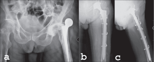
- Full-length AP and lateral femoral radiographs including the entire stem and cement column
- Mark the tip of the greater trochanter as the proximal reference
- The osteotomy must extend distal to the stem tip and any distal cement plug so the interfaces can be accessed under vision; typical length 12-15 cm from the tip of the greater trochanter
- Then confirm that at least 4-6 cm of intact, supportive diaphysis remains between the transverse limb and the point where the canal flares or bone quality deteriorates — this is the fixation zone for the revision stem
- Template the revision stem: it should engage the isthmus with a scratch fit over 4-6 cm; if the templated stem cannot achieve this, reconsider the reconstruction (longer stem, struts, or megaprosthesis)
- In varus remodelling, plan the transverse limb distal to the apex of the varus deformity so reaming re-establishes a neutral axis
Wagner (Anteriorly Based) versus Paprosky (Laterally Based) ETO
- Wagner transfemoral osteotomy
- Direct lateral / transgluteal
- Paprosky/Younger posterior-based ETO
- Posterior (posterolateral)
- Wagner transfemoral osteotomy
- Anterolateral femur
- Paprosky/Younger posterior-based ETO
- Posterolateral femur, just anterior to the linea aspera
- Wagner transfemoral osteotomy
- Posterior soft tissue hinge
- Paprosky/Younger posterior-based ETO
- Controlled anterior bony hinge; anterior cortex perforated with drill holes and levered open
- Wagner transfemoral osteotomy
- Anterolateral fragment with vastus lateralis attached
- Paprosky/Younger posterior-based ETO
- Lateral one third of femur with greater trochanter, abductors and vastus lateralis in continuity
- Wagner transfemoral osteotomy
- Relies on posterior soft tissue attachments
- Paprosky/Younger posterior-based ETO
- Abductor-trochanter-vastus sleeve stays intact and hinges forward on anterior periosteum, maximising fragment blood supply
- Wagner transfemoral osteotomy
- Historic description; the basis of the transfemoral concept
- Paprosky/Younger posterior-based ETO
- Standard technique in contemporary revision practice; flows directly from the posterior revision exposure
- Wagner transfemoral osteotomy
- Abductor dysfunction from the lateral / transgluteal approach
- Paprosky/Younger posterior-based ETO
- Uncontrolled anterior hinge fracture if the perforating drill holes are omitted
- Wagner transfemoral osteotomy
- Direct lateral / transgluteal
- Paprosky/Younger posterior-based ETO
- Posterior (posterolateral)
- Wagner transfemoral osteotomy
- Anterolateral femur
- Paprosky/Younger posterior-based ETO
- Posterolateral, just anterior to linea aspera
- Wagner transfemoral osteotomy
- Posterior soft tissue hinge
- Paprosky/Younger posterior-based ETO
- Controlled anterior bony hinge, perforated and levered open
- Wagner transfemoral osteotomy
- Anterolateral fragment with vastus lateralis attached
- Paprosky/Younger posterior-based ETO
- Lateral one third with greater trochanter, abductors and vastus lateralis attached
- Wagner transfemoral osteotomy
- Historic description; basis of the concept
- Paprosky/Younger posterior-based ETO
- Standard technique in contemporary revision practice; compatible with posterior revision exposure
- Wagner transfemoral osteotomy
- Abductor dysfunction from lateral approach
- Paprosky/Younger posterior-based ETO
- Uncontrolled anterior hinge fracture if perforations omitted
Use the published names in an exam. Abdel's 612-hip series names the two osteotomies Paprosky - laterally based and Wagner - anteriorly based. "Posterior-based ETO", used loosely on this page and in many units, describes the approach the osteotomy flows from rather than where the fragment is based, and it is not the term you will find in the literature.
The cut has also inverted since the original description. Younger and Paprosky in 1995 cut the anterolateral proximal femur for one third of its circumference and levered it open on an anterolateral hinge. Contemporary practice, working through a posterior approach, places the longitudinal limb posterolaterally just anterior to the linea aspera and hinges the fragment forward on the anterior cortex and periosteum. The fragment, the one-third circumference and the abductor-trochanter-vastus sleeve are unchanged; the side the surgeon cuts from is not.
Why the modern version: most femoral revisions use a posterior approach, and hinging forward keeps the entire abductor-trochanter-vastus sleeve in continuity on intact anterior periosteum and perforated anterior cortex, maximising fragment vascularity.
Operative Technique (Posterior-Based ETO)
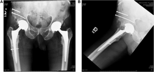
Posterior-based ETO: sequential operative stages
Lateral decubitus on a radiolucent table with the pelvis rigidly secured and checked as truly vertical (acetabular orientation errors follow pelvic malposition). Fluoroscopy available, full revision instrumentation checked and opened. Extended posterior approach: fascia lata incised in line with the femur, short external rotators and posterior capsule taken as a flap for later repair, and the fascial and skin incision extended distally along the shaft to the planned osteotomy level. Dislocate posteriorly if the hip is mobile; if stiff, cut the osteotomy first and dislocate with the fragment elevated to protect the femur from torsional fracture.
Measure 12 to 15 cm (as templated) from the tip of the greater trochanter and mark the transverse limb with diathermy. Elevate only the posterior edge of vastus lateralis off the linea aspera, then cut the posterior cortex with an oscillating saw from trochanter to transverse level, angling the blade to capture one third of the circumference. Perforating branches of profunda femoris pierce the posterior intermuscular septum — ligate or coagulate under vision; protect the sciatic nerve during posterior dissection.
Make the distal transverse cut rounded or bevelled with saw or pencil-tip burr — never a square corner, which is a stress riser. Perforate the anterior cortex with multiple drill holes or a burr along the planned anterior line, working through vastus lateralis or by sliding a saw or drill deep to it.
Insert several wide osteotomes along the posterior limb and lever in unison, hinging the fragment anteriorly on its perforated cortex and soft tissues. The fragment rotates forward with vastus lateralis and abductors intact. Levering at a single point risks fracturing the anterior fragment.
With the fragment hinged forward the stem is exposed along its lateral aspect. Cementless stems: divide the bone-implant interface with flexible osteotomes, Gigli saw proximally and a pencil-tip burr around ingrowth surfaces; extensively coated stems may need transection with a metal-cutting burr then trephining of the distal cylinder over guide wires. Cemented stems: extract the stem, remove the mantle under direct vision with splitters and reverse hooks, and drill, tap and back-slap the distal plug or clear it with ultrasonic instruments, with fluoroscopy confirming complete clearance. Debride the canal; in suspected infection take five or more deep tissue samples before antibiotics and debride membrane and cement aggressively.
Pass a cable or heavy wire around the femur 1 to 2 cm distal to the transverse limb and tension it before any reaming or stem insertion — this is the single step that prevents longitudinal fracture propagation from hoop stress during broaching and impaction. Pass cable passers hugging bone to avoid injuring the perforators.
Straight reamers by hand into the diaphysis to a scratch fit over 4 to 6 cm, correcting varus remodelling by lateralising the entry through the osteotomy bed; avoid power reaming in varus bone (cortical perforation). Implant either a modular fluted tapered titanium stem (axial stability from the taper, rotational stability from the flutes, proximal body sized independently for leg length and version) or an extensively porous-coated cobalt-chrome stem requiring 4 to 6 cm of intimate cortical contact. The stem must bypass the transverse limb by at least two cortical diameters. Trial reduction confirms stability, leg length, version and abductor tension; under-sizing causes subsidence.
Reduce the fragment over the stem — mild lateral gapping over a wide revision body is acceptable — and secure with 2 to 3 cerclage cables or wires, avoiding cables directly over the vastus ridge where they cause bursitis; use luque wires or a cable-grip or claw device for a deficient trochanter. Repair the posterior capsule and short external rotators to the trochanter through bone tunnels, then layered closure over drains as per unit practice. Salvage: an intraoperative distal crack is treated by extending cerclage fixation and lengthening the stem to bypass by two cortical diameters; inadequate distal fit needs a longer stem, strut augmentation, or conversion to a distally locked or megaprosthetic reconstruction.
- Position: lateral decubitus on a radiolucent table, pelvis rigidly secured; check the pelvis is truly vertical (acetabular orientation errors follow pelvic malposition)
- Imaging/equipment: fluoroscopy available; full revision instrumentation checked and opened
- Approach: extended posterior approach; incise fascia lata in line with the femur; release short external rotators and posterior capsule as a flap for later repair; extend the fascial and skin incision distally along the femoral shaft to the planned osteotomy level
- Dislocate the hip posteriorly if mobile; if stiff, perform the osteotomy first and dislocate with the fragment elevated — this protects the femur from torsional fracture
The commonest catastrophic intraoperative error is longitudinal fracture propagation from hoop stress during broaching or stem impaction. The prophylactic cable distal to the transverse limb is cheap insurance and is expected in any viva answer on ETO technique.
ETO in the Infected Hip
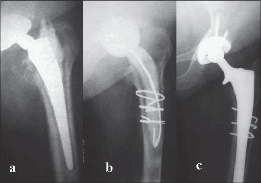
- ETO is safe in periprosthetic joint infection and is often essential for complete removal of well-fixed implants and cement — retained infected cement is a recognised cause of failed two-stage exchange
- Reported union rates after ETO in staged revision for infection remain high, provided the vascular sleeve is preserved
- At first stage: cables can be retained if needed for femoral integrity; an articulating or static antibiotic spacer is placed; the osteotomy is cabled closed around the spacer
- At second stage: the healed or healing osteotomy usually does not need to be re-opened unless further debridement or cement removal is required; the definitive diaphyseal-fitting stem is inserted
- Single-stage exchange with ETO is practised in some European centres with comparable infection control in selected patients
Complications: Prevention and Management
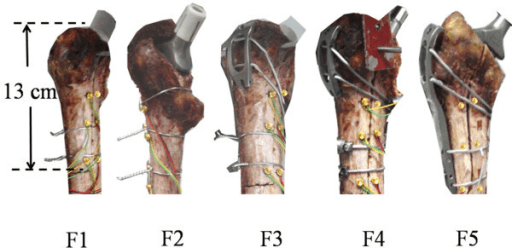
- Mechanism
- Stripping of the vastus lateralis and gluteus medius sleeve strips the blood supply to the osteotomised fragment
- Prevention
- Keep the abductor-vastus sleeve in continuity with the fragment; bevel or round the transverse limb; stable cable fixation across a vascularised bed
- Management if it occurs
- Asymptomatic fibrous union — observe; painful non-union — revise fixation with claw or cable-grip plate, bone graft and exclude infection
- Mechanism
- Hoop stress from reaming, broaching or stem impaction runs a crack down from the sharp transverse limb
- Prevention
- Prophylactic distal cerclage placed BEFORE reaming and impaction; hand reaming; round off the transverse limb corner
- Management if it occurs
- Additional cerclage wires; longer stem bypassing the fracture by at least two cortical diameters; strut allograft if comminuted
- Mechanism
- Unopposed abductor pull displaces the fragment proximally when fixation is inadequate or loading is premature
- Prevention
- Preserve the soft tissue sleeve; two to three cables; anatomic reduction; no early active abduction
- Management if it occurs
- Minimally symptomatic — observe; symptomatic escape — trochanteric claw or cable-grip revision fixation plus or minus bone graft
- Mechanism
- Over-levering the fragment forward on the anterior cortical hinge, or a thin fragment cut too anteriorly
- Prevention
- Take one third of the femoral circumference; osteotomise with multiple drill holes rather than forced levering; hinge gently on the anterior cortex
- Management if it occurs
- Cable the fragment in pieces to the intact femur; add claw fixation or strut graft; protect weight bearing and abduction
- Mechanism
- Posterior to the osteotomy; at risk from retractor placement, limb lengthening and posterior fragment displacement
- Prevention
- Keep the hip extended and knee flexed; place posterior retractors on bone under direct vision; monitor limb length restoration
- Management if it occurs
- Immediate exploration if a retractor or cable is implicated; flex the knee and relax length; most traction palsies recover over months
- Mechanism
- Cross the linea aspera at the level of the distal transverse limb and are cut or avulsed during exposure and cerclage passage
- Prevention
- Subperiosteal dissection at the distal limb; pass cerclage passers directly on bone hugging the cortex
- Management if it occurs
- Direct ligation or clipping; do not blindly diathermy into muscle; if the vessel retracts, extend the exposure to control it
- Mechanism
- Loss of the abductor lever arm from proximal fragment position, or under-restored offset and length
- Prevention
- Keep abductors attached to the fragment; anatomic reduction; restore offset, length and version
- Management if it occurs
- Abductor strengthening programme; persistent weakness with escape — reattachment or claw fixation; dual mobility or constrained bearing if unstable
- Mechanism
- Prominent cable crimps over the lateral cortex and vastus ridge irritate the overlying soft tissues
- Prevention
- Sink the crimps away from the lateral prominence; avoid cables sitting over the vastus ridge
- Management if it occurs
- Analgesia, physiotherapy, image-guided injection; remove cables once the osteotomy has united, usually after 6 months
- Mechanism
- Inadequate diaphyseal scratch fit distal to the osteotomy in an unsupportive proximal femur
- Prevention
- Achieve 4 to 6 cm of scratch fit; seat the tapered stem to templated depth; check with an intraoperative radiograph
- Management if it occurs
- Minor early subsidence of tapered stems may self-stabilise; progressive subsidence — revise to a larger or longer stem
- Mechanism
- Loss of offset, length or version combined with a compromised posterior repair and abductor deficiency
- Prevention
- Restore offset, length and version; meticulous posterior soft tissue repair; abduction precautions
- Management if it occurs
- Closed reduction and bracing; recurrent — assess component position and consider dual mobility or a constrained liner
- Prevention
- Preserve soft tissue sleeve; 2-3 cables; avoid early active abduction
- Management
- If minimally symptomatic, observe; symptomatic escape — trochanteric claw/cable-grip revision fixation plus or minus bone graft
- Prevention
- Vascularised fragment, stable cable fixation, bevelled transverse limb
- Management
- Asymptomatic fibrous union — observe; painful non-union — revise fixation, graft, exclude infection
- Prevention
- Prophylactic distal cerclage BEFORE reaming/impaction; hand reaming; rounded transverse limb
- Management
- Additional cerclage; longer stem bypassing the fracture by two cortical diameters; strut allograft if comminuted
- Prevention
- Keep abductors attached to fragment; anatomic reduction; correct offset and length
- Management
- Abductor strengthening programme; persistent weakness with escape — reattachment/claw fixation; consider constrained or dual mobility bearing if instability
- Prevention
- Sink cable crimps away from the lateral prominence; avoid cables over vastus ridge
- Management
- Analgesia, physiotherapy, image-guided injection; removal of cables once osteotomy united (usually after 6 months)
- Prevention
- Adequate scratch fit 4-6 cm; tapered stem seated to templated depth; intraoperative radiograph
- Management
- Minor early subsidence of tapered stems may self-stabilise; progressive subsidence — revise to larger/longer stem
- Prevention
- Restore offset, length, version; meticulous posterior repair; abduction precautions
- Management
- Closed reduction and bracing; recurrent — assess component position; dual mobility or constrained liner
Postoperative Protocol
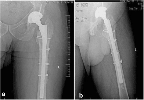
Protected weightbearing (touch to partial, unit-dependent) with crutches or frame. Posterior hip precautions if posterior approach. No active abduction and no abduction against resistance — the abductors pull directly on the fragment. Check radiographs before discharge and at 6 weeks.
If radiographs show maintained fragment position and no stem subsidence, progress to weightbearing as tolerated. Begin gentle active abduction from around 6-8 weeks once early consolidation is visible.
Osteotomy union typically evident radiographically by 3-6 months. Abductor strengthening progresses; gait re-education for residual Trendelenburg pattern.
Confirm union, stem osseointegration (spot welds, absence of subsidence or lucency). Consider cable removal only for persistent symptomatic bursitis after confirmed union.
SLICEETO Planning Essentials
Hook:You SLICE the lateral femur open like a lid — but only after planning the cut, the cable and the stem.
CIVICIndications for ETO
Hook:ETO serves the whole revision CIVIC duty — implant out, deformity corrected, infection cleared, exposure gained.
Guidelines, Registries & Global Practice
- Global epidemiology: revision hip arthroplasty volumes are rising worldwide with the ageing primary arthroplasty population; femoral revision for aseptic loosening, infection and periprosthetic fracture are the dominant indications in which ETO is used
- Society guidance: no society issues a stand-alone ETO guideline; the technique is embedded in revision arthroplasty teaching from AAOS, EFORT and BOA-affiliated revision networks. AO principles govern the associated periprosthetic fracture scenarios (Vancouver-based management, where B2/B3 fractures may incorporate an ETO with a diaphyseal-fitting stem). International Consensus Meeting (ICM) statements on periprosthetic joint infection support complete removal of implants and cement — the practical justification for ETO in the infected femur
- Registry evidence: national registries (NJR, AOANJRR, AJRR, SHAR, Norwegian, NZJR) capture revision stem type and survivorship rather than the osteotomy itself; they consistently show good mid-term survivorship of fluted tapered titanium revision stems, the constructs most often paired with ETO
- Practice variation by resource setting: where modular tapered systems, cable systems or ultrasonic cement removal are unavailable, surgeons rely on Wagner-type monoblock tapered stems, stainless wire cerclage and mechanical cement removal; the underlying principles — vascularised fragment, distal cerclage, diaphyseal bypass fixation — are unchanged and should be stated as universal
- Centralisation: several health systems concentrate complex femoral revision (including ETO with Paprosky IIIB/IV femora) in revision networks or specialist centres, reflecting the volume-outcome relationship in revision arthroplasty
Controversies & Areas of Uncertainty
- Cables versus monofilament wires: cables give stronger, more fatigue-resistant fixation but generate metal debris if they fray and are implicated in trochanteric bursitis; wires are cheaper and less irritant but weaker — no high-quality comparative trial exists
- Modular versus monoblock fluted tapered stems: modularity allows independent adjustment of length, offset and version but introduces taper junction corrosion and fracture risk; modern monoblock tapered stems show comparable survivorship, and practice is shifting in some centres
- Length of stem bypass: two cortical diameters is the widely taught rule but derives from biomechanical extrapolation rather than robust clinical evidence
- ETO versus episiotomy/cortical window for cemented stem removal: shorter controlled splits (femoral episiotomy) may suffice for loose cemented stems with limited cement, avoiding a full ETO — the threshold between the two is judgement-based
- Weightbearing after modern tapered stems: several units now allow early weightbearing as tolerated with tapered stems even after ETO, arguing the taper is axially stable; traditional protocols remain protective for 6 weeks, and evidence is limited to series data
- ETO in single-stage septic exchange: increasingly reported with acceptable infection control, but patient selection criteria are not standardised
MCQ Practice Points
Q: What is the typical planned length of an ETO? A: 12 to 15 cm from the tip of the greater trochanter, individualised to extend beyond the stem tip or cement column while preserving at least 4 to 6 cm of supportive diaphysis for revision stem fixation.
Q: What preserves vascularity of the ETO fragment? A: Maintaining the continuous soft tissue sleeve of vastus lateralis and gluteus medius/minimus on the fragment, with an anterior soft tissue and periosteal hinge — this underlies union rates above 90-95 percent.
Q: What single step prevents distal crack propagation during revision stem insertion? A: A prophylactic cerclage cable placed and tensioned 1 to 2 cm distal to the transverse limb before reaming, trialling or impaction.
Q: Which stems are appropriate after ETO? A: Stems achieving 4 to 6 cm of diaphyseal fixation distal to the osteotomy — modular fluted tapered titanium or extensively porous-coated cobalt-chrome — bypassing the osteotomy by at least two cortical diameters. Proximally coated or standard cemented primary stems are inappropriate.
Q: How much of the femoral circumference should the fragment include, and why is the distal limb bevelled? A: Approximately one third of the circumference; the transverse limb is rounded or bevelled to avoid a stress riser and distal fracture propagation.
Q: Which movement is specifically restricted after ETO and why? A: Active and resisted abduction for approximately 6 weeks, because the abductors insert on the osteotomised fragment and can displace it proximally before union.
Exam Viva Scenarios
Practise clinical reasoning and management decisions out loud
“A 68-year-old requires revision of a well-fixed extensively porous-coated femoral stem for recurrent instability with malversion. How will you remove the stem and reconstruct the femur?”
“During impaction of a fluted tapered revision stem after an ETO, you feel a change in pitch and see a longitudinal crack extending 3 cm distal to the transverse limb. How do you manage this?”
“A 74-year-old has a chronically infected cemented total hip with a well-fixed stem and a long distal cement column. Justify your use of an ETO at first-stage exchange and describe the sequence.”
“Six months after ETO revision, a patient has lateral hip pain, a Trendelenburg gait, and radiographs show the trochanteric fragment migrated 2 cm proximally with broken cables. How do you assess and manage this?”
Indications (CIVIC)
- Well-fixed cementless stem removal (especially extensively coated)
- Cemented stem and distal cement column extraction
- Varus proximal femoral remodelling — realign the reaming axis
- Infection — complete implant and cement clearance at staged exchange
- Exposure of the stiff hip and acetabulum
Planning Numbers
- Length 12-15 cm from tip of greater trochanter, beyond stem tip/cement
- Fragment = lateral one third of femoral circumference
- Preserve 4-6 cm intact diaphysis distal to osteotomy for stem fixation
- Revision stem bypasses transverse limb by at least two cortical diameters
Technique Keys
- Posterior-based (Paprosky/Younger) ETO through extended posterior approach
- Bevelled/rounded transverse limb — no square stress riser
- Perforate anterior cortex; lever osteotomes in unison for controlled anterior hinge
- Keep vastus lateralis-abductor sleeve attached — vascularised fragment
- Prophylactic distal cerclage BEFORE reaming and stem impaction
- Fix fragment with 2-3 cables; modular fluted tapered or extensively porous-coated stem
Outcomes and Complications
- Union greater than 90-95 percent with vascularised fragment and cable fixation
- Fragment migration/escape — claw or cable-grip revision if symptomatic
- Non-union usually fibrous and often asymptomatic — exclude infection
- Fracture propagation — extra cerclage plus longer bypassing stem plus or minus struts
- Cable bursitis — inject; remove cables only after union
- Abductor weakness — restrict active abduction 6 weeks, then strengthen
Aftercare
- Protected weightbearing approximately 6 weeks, then progress with radiographic union
- No active or resisted abduction for 6 weeks
- Posterior precautions if posterior approach
- Radiographs at 6 weeks, 3 months, then until united; watch for stem subsidence
Outcomes and Evidence
Extended Proximal Femoral Osteotomy - a New Technique for Femoral Revision Arthroplasty
- The founding description. The ANTEROLATERAL proximal femur is cut for ONE THIRD of its circumference, extended distally, and levered open on an ANTEROLATERAL hinge of periosteum and muscle.
- That creates the intact muscle-osseous sleeve the technique depends on: gluteus medius, greater trochanter, anterolateral femoral diaphysis and vastus lateralis, in continuity.
- It exposes the whole fixation surface and the distal cement, and allows canal machining under direct vision.
- Named advantages beyond exposure: varus placement of the new stem becomes impossible, a weakened trochanter is protected from iatrogenic injury, the proximal femur can be closed down onto the prosthesis, and soft-tissue tension can be adjusted.
- Repaired with cerclage wires or cables. The first 20 patients showed excellent cement and component removal with reliable healing and no change to the post-operative regimen.
Extended Slide Trochanteric Osteotomy for Revision Total Hip Arthroplasty
- 46 hips in 45 patients, mean age 66.3 years, followed a mean of 44 months.
- Union of the distal osteotomy site in 98 per cent (44 of 45 hips), with NO change in femoral component position.
- Time to union was NOT significantly correlated with the number of cables used, the pre-operative cortical bone thickness, or the pre-operative cancellous quality of the greater trochanter.
- Time to bridging-callus union was significantly LONGER in hips with a strut allograft than without (p = 0.04).
- Two fractures of the osteotomy fragment occurred; neither required further revision.
Extended Trochanteric Osteotomy in Revision THA - Contemporary Outcomes of 612 Hips
- 612 ETOs at one institution from 2003 to 2013 by two techniques: PAPROSKY (laterally based) in 367 and WAGNER (anteriorly based) in 245 - this is the authoritative modern naming for the two osteotomies.
- Indications: aseptic loosening 65 per cent, periprosthetic joint infection 18 per cent, periprosthetic fracture 6 per cent, femoral implant fracture 5 per cent, other 6 per cent.
- Union in 98 per cent at a mean of 6 months (range 1 to 24 months). Nonunion in only 9 hips (2 per cent).
- Mean migration of the proximal fragment before union was 3 mm; migration greater than 1 cm occurred in 7 per cent (37 of 501 analysed).
- THE COMMONEST COMPLICATIONS ARE FRACTURES OF THE FRAGMENT, NOT NONUNION OR ESCAPE: intra-operative fracture of the ETO diaphyseal fragment in 22 hips (4 per cent), post-operative diaphyseal fragment fracture in 3 (0.5 per cent), and post-operative greater trochanteric fracture in 41 (7 per cent).
- Ten-year survivorship: 97 per cent free of revision for aseptic femoral loosening, 91 per cent free of component removal or revision for any reason, and 82 per cent free of REOPERATION for any reason.
- Harris hip score improved from 57 to 77.