The Fibrous Arch That Traps the Motor Nerve
- Origin from the hook of the hamate and the palmar surface of the flexor retinaculum; insertion into the ulnar side of the base of the proximal phalanx of the little finger.
- The muscle shares its origin and its insertion with abductor digiti minimi, and the two are frequently inseparable.
- Its fibrous arch of origin from the hook of hamate is one of the two named structures that compress the deep motor branch of the ulnar nerve in Guyon canal zone 2.
- Innervated by the deep motor branch of the ulnar nerve, C8-T1, supplied early in that nerve's course alongside abductor digiti minimi and opponens digiti minimi.
- It is absent or fused with abductor digiti minimi in a significant minority of hands — a normal variant, not a pathological finding.
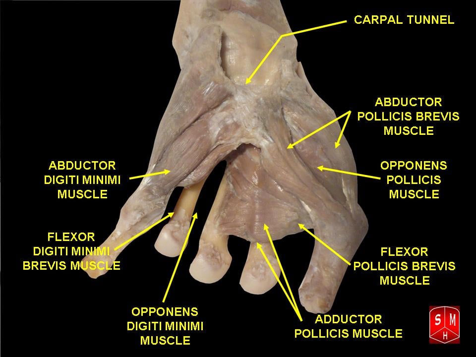
- “Flexor digiti minimi brevis and abductor digiti minimi together form the middle layer of the hypothenar eminence; opponens digiti minimi lies deep to both, directly on the fifth metacarpal.
- “The deep motor branch passes BETWEEN abductor digiti minimi and flexor digiti minimi brevis before piercing opponens digiti minimi.
- “Releasing the fibrous arch of flexor digiti minimi brevis is the operative step that actually decompresses a zone 2 lesion — dividing the volar carpal ligament alone is not enough.
- “The hypothenar eminence is the donor field for the Huber opponensplasty; the abductor is harvested, but the surgeon works through the plane between it and flexor digiti minimi brevis, where the nerve lies.
Overview
Flexor digiti minimi brevis is a small, slender muscle of the hypothenar eminence lying radial to abductor digiti minimi and superficial to opponens digiti minimi. Together with the abductor it forms the middle layer of the eminence, and the two share both an origin and an insertion; a fascial plane, sometimes barely present, separates them.
Its own function is straightforward — flexion of the little finger metacarpophalangeal (MCP) joint. Its surgical significance lies in the corridor it creates: the deep motor branch of the ulnar nerve passes between flexor digiti minimi brevis and abductor digiti minimi as it dives from Guyon canal into the palm, and the fibrous arch of origin of flexor digiti minimi brevis from the hook of hamate is one of the two named bands that can strangle that nerve.
Every finger has extrinsic flexors (flexor digitorum superficialis and profundus) and an intrinsic MCP flexor from the interossei and lumbricals. The little finger has, in addition, a dedicated intrinsic short flexor. Why?
- The little finger is a border digit with a mobile carpometacarpal joint (20-30 degrees of flexion-extension, far more than the index or middle). It needs an intrinsic that flexes the ray and the MCP joint against that mobility, otherwise the whole ulnar column collapses in power grip.
- The ulnar border of the hand is the pressure-bearing side in hammer grip and in the golf or racquet grip. Flexor digiti minimi brevis and abductor digiti minimi together buttress that border, converting the mobile fifth ray into a stable post at the moment of grip.
- Only the third palmar interosseous acts on the little finger's MCP joint in adduction; there is no fourth dorsal-type abductor of the little finger other than abductor digiti minimi itself. The hypothenar three therefore carry a disproportionate share of the little finger's intrinsic control, which is why Wartenberg sign — persistent little finger abduction — is such an early and visible sign of ulnar motor palsy.
Please Feel All OverHypothenar Eminence — Superficial to Deep
Hook:The deep motor branch runs between F and A, then through O.
Attachments, Innervation and Relations
Origin
- The hook (hamulus) of the hamate, on its palmar and radial surface.
- The adjacent palmar surface of the flexor retinaculum (transverse carpal ligament).
- The origin is partly fibrous and arch-shaped, spanning from the hook to the retinaculum. The deep motor branch of the ulnar nerve and the deep branch of the ulnar artery pass beneath this arch as they turn radially and dorsally. This arch is the single most important surgical fact about the muscle.
Insertion
- The ulnar side of the base of the proximal phalanx of the little finger, blending with the insertion of abductor digiti minimi.
- Slips extend into the extensor expansion (dorsal apparatus) of the little finger, so the muscle contributes to interphalangeal extension in the same way an interosseous does.
Relationship to abductor digiti minimi
- The two muscles share an insertion, are frequently continuous at their origins, and act as a functional unit.
- Abductor digiti minimi originates from the pisiform, the pisohamate ligament and the flexor carpi ulnaris tendon; flexor digiti minimi brevis originates from the hook of hamate and the retinaculum. The origins are the way to tell them apart on dissection: pisiform is abductor, hook is flexor.
Variations
- Absent or vestigial in a significant minority of hands; reported absence rates vary widely between series.
- Fused with abductor digiti minimi so that no separate belly is identifiable — the commonest variant.
- Occasional accessory slips from the pisohamate ligament or from a persistent median artery sheath.
- Clinical implication of absence: there is no functional deficit, and the finding at operation should not be reported as pathological.
The distal boundary of Guyon canal.
- A fibrous arch spanning from the hook of the hamate to the flexor retinaculum, the tendinous origin of the muscle.
- The deep motor branch and the deep ulnar artery pass beneath it as they turn radially.
- Dividing this arch is the operative step that decompresses a zone 2 lesion. Releasing the volar carpal ligament alone leaves it intact and the release fails.
The second gate.
- A fibrous band within or at the proximal edge of opponens digiti minimi, sometimes described as the pisohamate hiatus.
- The nerve pierces the muscle or passes beneath this band immediately after clearing the first arch.
- Follow the nerve until it lies free on the interossei — anything less is an incomplete release.
Action and Biomechanics
Actions
- Action
- Flexion
- Co-actors
- Third palmar interosseous, fourth lumbrical, extrinsic flexors
- Consequence of loss
- Contributes to clawing of the little finger in ulnar palsy
- Action
- Weak abduction and rotation toward the thumb
- Co-actors
- Abductor digiti minimi, opponens digiti minimi
- Consequence of loss
- Loss of ulnar border cupping
- Action
- Extension via slips into the dorsal apparatus
- Co-actors
- Interossei and lumbricals
- Consequence of loss
- Loss of IP extension with the MCP flexed — the intrinsic-minus posture
- Action
- Assists flexion of the ray
- Co-actors
- Opponens digiti minimi
- Consequence of loss
- Reduced palmar cup
The intrinsic mechanism at the little finger
Understanding what flexor digiti minimi brevis does at the interphalangeal joints requires understanding the extensor mechanism:
- The intrinsic muscles (interossei, lumbricals and, at the little finger, the hypothenar muscles) insert palmar to the axis of the MCP joint and dorsal to the axes of the interphalangeal joints via the lateral bands.
- The result is the defining intrinsic action: MCP flexion with simultaneous PIP and DIP extension.
- Lose the intrinsics and the extrinsics act unopposed: the extensor digitorum communis hyperextends the MCP, and the flexor digitorum profundus and superficialis flex the interphalangeal joints. That posture — MCP hyperextension with IP flexion — is the claw (intrinsic-minus) hand.
The ulnar claw and the ulnar paradox
- In a low ulnar lesion (wrist, Guyon canal) the flexor digitorum profundus to the ring and little fingers is intact, so it pulls the interphalangeal joints into flexion against paralysed intrinsics. Clawing is marked.
- In a high ulnar lesion (elbow, cubital tunnel) that same profundus is also paralysed, so the interphalangeal joints cannot be pulled into flexion and clawing is less marked despite the more proximal, more extensive injury.
- This is the ulnar paradox: the more proximal the lesion, the less the deformity. As a high lesion recovers and the profundus reinnervates before the intrinsics, the clawing worsens — an alarming but expected sign of recovery.
Wartenberg sign
- The little finger drifts into persistent abduction and cannot be actively adducted to the ring finger.
- Mechanism: the third palmar interosseous (the little finger's only adductor) is paralysed, while the extensor digiti minimi, which has an abduction moment at the little finger MCP joint, is radial-nerve innervated and unopposed.
- It is one of the earliest and most visible signs of ulnar motor palsy and is functionally troublesome — the abducted little finger catches in pockets.
Synergists and antagonists
- Synergists: abductor digiti minimi, opponens digiti minimi, the fourth lumbrical, the third palmar interosseous and the extrinsic flexors.
- Antagonists: extensor digiti minimi and the extensor digitorum communis slip to the little finger.
Surface Anatomy and Examination
Landmarks and palpation
- The hypothenar eminence is the fleshy ridge along the ulnar border of the palm.
- Pisiform: palpable at the distal wrist crease on the ulnar side, within the flexor carpi ulnaris tendon.
- Hook of hamate: approximately 1-1.5 cm distal and radial to the pisiform, palpated by placing the interphalangeal joint of your own thumb over the patient's pisiform, pointing your thumb toward the patient's index web, and pressing with the pulp.
- Flexor digiti minimi brevis is not separately palpable. Ask the patient to flex the little finger MCP joint while keeping the interphalangeal joints straight and palpate the radial side of the hypothenar mass; the whole eminence firms up.
- Hypothenar wasting appears as flattening and hollowing along the ulnar palmar border with prominence of the fifth metacarpal, best appreciated by comparing both palms side by side in oblique light.
Named tests around the ulnar hand
- How to perform
- Hand flat, fingers adducted; ask the patient to hold them together
- Positive finding
- Little finger abducts and cannot be adducted
- What it means
- Third palmar interosseous weakness with unopposed extensor digiti minimi — ulnar motor palsy
- False positives
- Congenital little finger abduction; extensor digiti minimi subluxation; little finger MCP arthritis
- How to perform
- Key pinch on a card against resistance; watch the thumb IP joint
- Positive finding
- IP flexion during pinch
- What it means
- Adductor pollicis weakness — deep ulnar motor branch involved
- False positives
- Painful thumb MCP joint; Riche-Cannieu anastomosis gives false negatives
- How to perform
- Resist little finger abduction with the hand flat
- Positive finding
- Weakness
- What it means
- Deep branch lesion at or proximal to the hypothenar branches
- False positives
- Little finger MCP arthritis limiting the movement
- How to perform
- Resist index abduction with the hand flat
- Positive finding
- Weakness with a hollow first web dorsally
- What it means
- Earliest and most sensitive sign of ulnar motor palsy
- False positives
- Thumb MCP or first web pain
- How to perform
- Passively hold the MCP joints in flexion and ask the patient to extend the PIP joints
- Positive finding
- PIP joints extend fully — a positive (correctable) test
- What it means
- The extensor mechanism is intact; an MCP anti-claw procedure alone will work
- False positives
- Fixed PIP contracture makes it uninterpretable
- How to perform
- Percuss between pisiform and hook of hamate
- Positive finding
- Paraesthesia into the little and ulnar ring finger
- What it means
- Ulnar nerve irritation at the wrist
- False positives
- Present in a proportion of normal subjects
- How to perform
- Occlude both arteries, exsanguinate, release each in turn
- Positive finding
- Delayed refill on releasing the ulnar artery
- What it means
- Ulnar artery occlusion — hypothenar hammer syndrome
- False positives
- Incomplete arch as a normal variant; wrist over-extension
Grading hypothenar function
- Test little finger MCP flexion with the interphalangeal joints held straight, which removes the extrinsic flexors from the movement.
- Test abduction and adduction separately: abduction is abductor digiti minimi (deep branch, early), adduction is the third palmar interosseous (deep branch, later). A dissociation between them helps localise within the deep branch's course.
- Record grip strength and specifically ulnar-sided grip; hypothenar loss reduces the power of a hammer or racquet grip disproportionately to the loss recorded on a standard dynamometer.
Complications
- Mechanism
- Blind dissection between abductor digiti minimi and flexor digiti minimi brevis, or blind excision of a hook fragment
- Prevention
- Identify the nerve proximal to the canal and follow it distally under vision
- Salvage
- Direct repair if recognised; late intrinsic reconstruction, anti-claw procedure and adductorplasty
- Mechanism
- Volar carpal ligament divided but the fibrous arch of flexor digiti minimi brevis left intact
- Prevention
- Follow the deep branch until it lies free on the interossei
- Salvage
- Revision release with wider distal exposure
- Mechanism
- Operating without pre-operative cross-sectional imaging
- Prevention
- Image every pure motor ulnar palsy
- Salvage
- Re-exploration and excision
- Mechanism
- The artery lies radial and superficial to the nerve
- Prevention
- Identify the bundle proximally; do not dissect blindly at the hook
- Salvage
- Direct repair or vein graft; ligate only with a confirmed patent radial supply
- Mechanism
- Skeletonising or kinking the proximal ulnar pedicle, or a tight subcutaneous tunnel
- Prevention
- Preserve a cuff of tissue around the pedicle; make a generous tunnel; check perfusion before closure
- Salvage
- Return to theatre, release the tunnel; if the muscle is lost, an alternative opponensplasty
- Mechanism
- Transverse incision across the flexion crease
- Prevention
- Cross the crease obliquely or with a step
- Salvage
- Z-plasty scar revision
- Mechanism
- Crush injury without compartment assessment
- Prevention
- Assess all ten hand compartments; release through a longitudinal ulnar incision along the fifth metacarpal
- Salvage
- Late release plus contracture management
- Mechanism
- Ulnar-sided wrist incision carried too dorsally
- Prevention
- Stay palmar to the flexor carpi ulnaris axis
- Salvage
- Neuroma excision and burial in muscle or bone
Clinical Relevance
Ulnar tunnel syndrome by zone
- Anatomy
- Proximal to the bifurcation, at the pisiform level
- Deficit
- Mixed: all ulnar intrinsics weak PLUS palmar little and ulnar ring sensory loss
- Typical causes
- Ganglion, hook of hamate fracture, distal radius fracture, lipoma
- Anatomy
- Deep motor branch, around the hook, beneath the FDMB arch, through ODM
- Deficit
- Pure motor: clawing, weak pinch, positive Froment, first dorsal interosseous wasting. Sensation NORMAL
- Typical causes
- Ganglion (commonest), hook of hamate non-union, fibrous arches, anomalous muscle
- Anatomy
- Superficial branch, distal and ulnar with the ulnar artery
- Deficit
- Pure sensory over the palmar little and ulnar ring finger, plus palmaris brevis weakness
- Typical causes
- Ulnar artery thrombosis or aneurysm, ganglion
The critical negative
Dorsal ulnar hand sensation is normal in every Guyon canal zone. The dorsal cutaneous branch arises 5-8 cm proximal to the ulnar styloid and passes dorsally beneath flexor carpi ulnaris, entirely outside the canal. A patient with numbness on the dorsum of the ulnar hand has a lesion at or proximal to the distal forearm — most often the cubital tunnel.
Causes, in the order you should think of them
- Ganglion from the pisotriquetral or hamate-metacarpal joint — the commonest identified cause.
- Hook of hamate fracture or non-union — sporting injury, missed on plain films, diagnosed on CT.
- Repetitive external compression — cyclist's palsy, vibrating tools, wheelchair use.
- Anomalous muscle — accessory abductor digiti minimi (roughly 20-25% of limbs), hypertrophic palmaris brevis, an accessory flexor digiti minimi slip.
- Ulnar artery pathology — thrombosis or aneurysm from hypothenar hammer syndrome.
- Fracture — distal radius, pisiform, fifth carpometacarpal fracture-dislocation.
- Idiopathic, with fibrous arch compression.
Imaging rule
Image every pure motor ulnar palsy before operating. Radiographs including a carpal tunnel view, then ultrasound or MRI of the wrist and palm, and CT if a hook fracture is suspected.
Surgical Relevance
Guyon canal decompression
- 1Step 1 — Image first
Nerve conduction studies to confirm the level; radiographs with a carpal tunnel view; ultrasound or MRI for any pure motor deficit; CT if a hook of hamate fracture is suspected.
Never operate on an unimaged pure motor palsy
- 2Step 2 — Incision
Curvilinear along the ulnar palm, starting proximal to the wrist crease over flexor carpi ulnaris, crossing the crease obliquely, curving radially along the hypothenar crease.
Avoids a contracting scar across the crease
- 3Step 3 — Identify the bundle proximally
Find the ulnar nerve and artery proximal to the canal, radial to flexor carpi ulnaris; the nerve is ulnar and deep to the artery.
Known to unknown
- 4Step 4 — Release the roof
Divide the volar carpal ligament and split palmaris brevis in the line of its fibres.
Opens zone 1
- 5Step 5 — Find the bifurcation
At the level of the distal pole of the pisiform, roughly 1 cm distal to it.
The zone 1 and 2 boundary
- 6Step 6 — Divide the fibrous arch of flexor digiti minimi brevis
Follow the deep branch around the hook and divide the fibrous arch of origin of flexor digiti minimi brevis, and the fibres of abductor digiti minimi if needed.
This is the step that decompresses zone 2
- 7Step 7 — Release the opponens arch
Continue distally, dividing the fibrous arch within opponens digiti minimi until the nerve lies free on the interossei.
Anything less is incomplete
- 8Step 8 — Deal with the cause
Excise a ganglion at its stalk; excise a hook of hamate non-union subperiosteally; resect and vein-graft a thrombosed ulnar artery segment.
Release alone fails if the mass stays
- Hook of hamate: approximately 1-1.5 cm distal and radial to the pisiform.
- Ulnar nerve bifurcation: at the distal pole of the pisiform, roughly 1 cm distal to the pisiform, at the proximal edge of the hook.
- Fibrous arch of flexor digiti minimi brevis: immediately distal to the bifurcation, spanning from the hook to the retinaculum; this is the distal boundary of Guyon canal, roughly 4-4.5 cm from the proximal edge of the palmar carpal ligament.
- Deep motor branch crossing the palm: roughly 1-2 cm distal to the distal edge of the transverse carpal ligament, running on the interossei, deep to the flexor tendons.
- Deep palmar arch: approximately 1 cm proximal to the superficial arch, which itself lies about 1 cm distal to the transverse carpal ligament at Kaplan's cardinal line.
- Dorsal cutaneous branch of the ulnar nerve: arises 5-8 cm proximal to the ulnar styloid; outside this field but at risk in ulnar-sided wrist approaches.
The Huber transfer and the hypothenar donor field
The Huber (Nicolaysen) opponensplasty transfers abductor digiti minimi on its neurovascular pedicle across the palm to the thumb, principally for congenital thumb hypoplasia (Blauth II and IIIA). Flexor digiti minimi brevis is not the donor, but it defines the dissection:
- Detail
- Abductor digiti minimi, not flexor digiti minimi brevis
- Why it matters
- The abductor has the bulk, the length and a reliable single dominant pedicle; FDMB is small and often absent
- Detail
- The ulnar artery branch and the motor branch enter proximally near the pisiform, within about 1 cm of the muscle origin
- Why it matters
- The muscle is rotated about this proximal pedicle like turning a page; the pedicle limits the arc and must not be skeletonised
- Detail
- Between abductor digiti minimi and flexor digiti minimi brevis
- Why it matters
- The deep motor branch lies in exactly this plane as it dives — identify it before separating the muscles
- Detail
- Released from the pisiform and the flexor carpi ulnaris tendon
- Why it matters
- Adequate release is needed for the arc; over-release proximally endangers the pedicle
- Detail
- Subcutaneous tunnel across the palm, superficial to the palmar fascia
- Why it matters
- A tight tunnel strangles the muscle; make it generous and check for kinking of the pedicle at the corner
- Detail
- Abductor pollicis brevis tendon and the dorsoulnar extensor hood at the thumb MCP
- Why it matters
- The dorsoulnar limb supplies pronation; insertion into abductor pollicis brevis alone gives abduction only
- Detail
- Loss of little finger abduction
- Why it matters
- Usually well tolerated and rarely a complaint; counsel the family beforehand
- Detail
- An unstable trapeziometacarpal joint (Blauth IIIB and beyond)
- Why it matters
- Pollicisation is required instead; a transfer into an unstable basal joint fails
Other operations in the hypothenar field
- Hypothenar fat pad flap for revision carpal tunnel surgery: raised superficial to the hypothenar muscle fascia and rotated radially over the median nerve. Keeping the plane superficial to the fascia protects the deep motor branch and the deep ulnar artery.
- Abductor digiti minimi muscle flap for coverage of the ulnar wrist and proximal palm.
- Pisiformectomy for pisotriquetral arthritis: the pisiform is shelled out from within the flexor carpi ulnaris tendon, preserving the tendon; the ulnar nerve lies immediately radial and must be identified.
- Hook of hamate excision: subperiosteal, staying on bone, after identifying the deep motor branch which lies immediately deep and radial to the hook.
Guidelines, Registries & Global Practice
Anatomical variation across populations
- Absence or fusion of flexor digiti minimi brevis with abductor digiti minimi is a well-recognised variant reported across cadaveric series worldwide. Reported rates vary widely because series differ in how strictly a separate belly is defined. The fibrous arch of origin persists even when the muscle belly does not, which is the point of surgical relevance.
- Accessory abductor digiti minimi is reported in roughly 20-25% of limbs across populations and is a reversible cause of ulnar tunnel syndrome identified only at surgery.
- The Martin-Gruber anastomosis is present in approximately 15-20% of limbs in most populations and confounds electrodiagnostic localisation of hypothenar and interosseous weakness everywhere.
Differences in described technique
- There is no dedicated AAOS or BOA/BSSH guideline on ulnar tunnel syndrome; practice is anatomically driven and based on case series. The consistent teaching across AO, AAOS instructional material and European sources is that release must extend distally through the hypothenar fibrous arch, and that a pure motor deficit demands imaging before surgery.
- Adjunctive carpal tunnel release at the time of Guyon decompression is commonly performed for idiopathic cases on the basis that the transverse carpal ligament forms the canal floor. It is not universal and is not supported by randomised data.
- Anti-claw surgery shows the widest international variation. High-volume leprosy reconstruction programmes in South Asia and Brazil have the largest experience and favour the Zancolli lasso and Brand transfers for their simplicity and low equipment requirement; European and North American practice more often uses modified Stiles-Bunnell transfers in post-traumatic palsy.
Global context
- Leprosy remains the largest global cause of ulnar intrinsic palsy and of clawing. Reconstruction in these programmes is entirely implant-free, which makes the anatomical rules on this page directly transferable to any setting.
- Cyclist's palsy and vibration-induced ulnar neuropathy are recognised occupational conditions in many jurisdictions; prevention through equipment modification is more effective than surgery and should be the first advice given.
- Resource considerations: Guyon canal decompression requires no implants, but the recommendation to image a pure motor palsy holds universally, because the consequence of missing a ganglion is a failed operation. Where MRI is unavailable, high-resolution ultrasound is an adequate substitute and is widely available.
MCQ Practice Points
Q: What distinguishes the origin of flexor digiti minimi brevis from abductor digiti minimi? A: Flexor digiti minimi brevis arises from the hook of hamate and the retinaculum; abductor digiti minimi arises from the pisiform.
Q: Which fibrous structure forms the distal boundary of Guyon canal? A: The fibrous arch of origin of the hypothenar muscles, principally flexor digiti minimi brevis, spanning from the hook of hamate to the flexor retinaculum.
Q: Between which two muscles does the deep motor branch of the ulnar nerve pass? A: Abductor digiti minimi and flexor digiti minimi brevis, before piercing opponens digiti minimi.
Q: How long is Guyon canal? A: Approximately 4-4.5 cm, from the proximal edge of the palmar carpal ligament to the fibrous arch of the hypothenar muscles.
Q: What causes Wartenberg sign? A: Paralysis of the third palmar interosseous with unopposed extensor digiti minimi, producing persistent little finger abduction.
Q: What does a positive Bouvier test tell you? A: The claw is correctable — the extensor mechanism is intact and a metacarpophalangeal flexion block or simple dynamic transfer will suffice.
Q: Where must an anti-claw tendon transfer be routed? A: Volar to the deep transverse metacarpal ligament. Routed dorsal to it, it extends the MCP joint and worsens the claw.
Q: Is absence of flexor digiti minimi brevis pathological? A: No. Absence or fusion with abductor digiti minimi is a recognised normal variant with no functional deficit.
Q: Why does a proximal ulnar lesion claw less than a distal one? A: The flexor digitorum profundus to ring and little is also paralysed in a high lesion, so it cannot flex the interphalangeal joints against the paralysed intrinsics.
Q: Which hypothenar muscle is used for the Huber opponensplasty? A: Abductor digiti minimi, rotated on its proximal neurovascular pedicle.
Exam Viva Scenarios
Practise clinical reasoning and management decisions out loud
“A 44-year-old had a Guyon canal release nine months ago for clawing and weak pinch. His symptoms are unchanged. Sensation was and remains normal. What went wrong and what will you do?”
“A 35-year-old has a low ulnar nerve palsy following a laceration at the wrist repaired 18 months ago. The ring and little fingers claw. When you hold the MCP joints flexed, he can fully extend the PIP joints. How will you reconstruct?”
“You are performing a Huber opponensplasty in a 2-year-old with Blauth type II thumb hypoplasia. Take me through the hypothenar dissection and the structures you must protect.”
Anatomy
- Origin: hook of hamate and flexor retinaculum
- Insertion: ulnar base of the little finger proximal phalanx
- Middle hypothenar layer, radial to abductor digiti minimi
- Absent or fused with the abductor in a minority of hands
Nerve
- Deep motor branch of the ulnar nerve, C8-T1
- Supplied early, within 1 cm of the hook of hamate
- The nerve passes BETWEEN this muscle and abductor digiti minimi
- Its fibrous arch of origin compresses the nerve in zone 2
Guyon Canal
- Length approximately 4-4.5 cm
- Roof: volar carpal ligament and palmaris brevis
- Floor: transverse carpal ligament, pisohamate and pisometacarpal ligaments
- Distal boundary: the hypothenar fibrous arch — divide it
Clinical
- Zone 2 lesion: pure motor, normal sensation
- Wartenberg sign: little finger abduction, third palmar interosseous
- Bouvier test decides the anti-claw operation
- Huber transfer uses abductor digiti minimi, not this muscle
Evidence Base
The Anatomy of the Distal Ulnar Tunnel
- Defined the distal ulnar tunnel as a region of the wrist 4 to 4.5 cm in length in which the nerve is vulnerable to external compression
- Described three zones based on the relationship to the bifurcation of the ulnar nerve
- All 36 zone 2 lesions produced intrinsic paralysis; hypothenar involvement depended on the position of the lesion within zone 2
- All 39 cases of combined motor and sensory deficit were zone 1 lesions, and zone 3 lesions were purely sensory
- Correlated the internal topography of the nerve with the structures forming the tunnel and the predicted clinical deficit
Architectural Design of the Human Intrinsic Hand Muscles
- Twenty muscles studied (18 intrinsics and 2 thumb extrinsics, 180 muscles in total): length, mass, pennation angle, fibre length and sarcomere length measured, with physiological cross-sectional area calculated
- The first dorsal interosseous and adductor pollicis had physiological cross-sectional areas comparable to extrinsic muscles and much greater than the other intrinsic muscles
- The interossei had relatively high physiological cross-sectional areas with low fibre length to muscle length ratios, indicating adaptation for high force and low excursion
- The lumbricals had an extremely high fibre length to muscle length ratio, implying a design toward high excursion
- Intrinsic muscle lengths were relatively similar to one another, interpreted as a space constraint within the hand
A Modification in Technique of Abductor Digiti Minimi (Huber) Opponensplasty
- The Huber opponensplasty detaches abductor digiti minimi distally and reroutes it through a subcutaneous tunnel to the thumb metacarpophalangeal joint, restoring opposition after median nerve trauma or in congenital thenar deficiency.
- THE PIVOT POINT FOR THE ROTATION IS THE PISIFORM, AND IT IS THERE THAT THE ULNAR NERVE IS VULNERABLE TO COMPRESSION BY THE TRANSFERRED MUSCLE.
- The described modification is COMPLETE ISLANDING of the muscle without re-inserting its proximal end, which removes the pivot and therefore the compression risk.
- The authors report this eliminates the complication without reducing the opposition achieved.
Anomalous Flexor Digiti Minimi Brevis in Guyon's Canal
- In an adult male cadaver, the flexor digiti minimi brevis arose abnormally high - from the superficial transverse septum in the DISTAL FOURTH OF THE FOREARM, not from the hook of hamate and retinaculum.
- The muscle had two strata at its origin: a thin superficial layer of transversely running fibres confined to the forearm, NOT PREVIOUSLY REPORTED, and a thick deep longitudinal layer forming the bulk of the muscle.
- It traversed GUYON'S CANAL SUPERFICIAL TO THE ULNAR NERVE AND VESSELS to reach the hypothenar eminence - a potential cause of ulnar tunnel syndrome.
- The ulnar nerve TRUNK innervated the anomalous flexor digiti minimi brevis, the abductor digiti minimi and palmaris brevis together, which the authors attribute to the common phylogeny of all three from one muscle mass.
Anomalous Hand Muscle Found in Guyon's Canal at Exploration for Ulnar Artery Thrombosis
- A single case in which exploration for ulnar artery thrombosis found an anomalous muscle within Guyon's canal.
- The anomalous muscle arose from FLEXOR CARPI ULNARIS, was ulnarly innervated, and JOINED THE FLEXOR DIGITI MINIMI MUSCLE DISTALLY.
- Consider ulnar artery thrombosis in any patient with ulnar neuropathy, hand ischaemia or a hypothenar mass, particularly after repetitive blunt trauma to the hypothenar area.
- The authors decline to claim causation: the patient also had a history of blunt injury and a significant smoking history, so 'a causal relationship between the anomalous muscle and ulnar artery thrombosis cannot be stated with certainty'.
Tendon Transfers for Ulnar Nerve Palsy - Evaluation of Results and Practical Treatment Considerations
- Transfers correct clawing consistently only in YOUNG, LIGAMENTOUSLY LAX individuals; correction is most inconsistent in the intrinsically stiff hands of older patients.
- THE LITTLE FINGER IS HARDER TO CORRECT THAN THE RING FINGER - the digit this page's muscle serves is the one that resists correction.
- Using flexor digitorum superficialis as the intrinsic transfer corrects the claw and restores synchronous flexion, but grip strength falls a further 21 per cent and total active range by 7 per cent.
- Recommends a wrist motor with tendon graft into index, middle, ring AND little digits, despite the clawing being confined to ring and little.
- Pinch should be augmented by metacarpophalangeal fusion, not interphalangeal fusion; combined with an extensor carpi radialis brevis adductor plasty, pinch strength can be doubled.
- The Bouvier manoeuvre remains the test that separates a correctable claw, treatable by an MCP flexion block, from a non-correctable one needing a transfer into the lateral bands.