Ulna Fracture with Radial Head Dislocation
- Line through radial head must bisect capitellum on ALL views
- Fix the ulna - radial head reduces spontaneously
- Adult Type II most common - posterior angulation
- Chronic - ulna osteotomy and open reduction, classically with annular ligament reconstruction
- “Always get elbow X-ray with forearm fracture
- “Radiocapitellar line - most missed injury in orthopaedics
- “Type II most common in adults, Type I in children
- “Ulna length and alignment critical - use contralateral comparison
Overview/Epidemiology
The childhood injury behaves differently enough to be treated separately in Monteggia fractures in children, where closed reduction usually succeeds and the missed radial head is the dominant problem. The mirror-image forearm fracture-dislocation is set out in Galeazzi fractures.
Who. The age distribution is bimodal, with one peak in childhood (4-10 years) and another in middle age (40-60 years). Children predominantly sustain Type I (anterior) injuries and adults predominantly Type II (posterior). Elderly patients often have more comminuted patterns.
Mechanism. The two peaks reflect different mechanisms: children fall with the arm hyperextended (Type I), while adults sustain direct blows or axial loading (Type II).
- Children - falls from height, playground injuries
- Adults - high-energy trauma, sports injuries, direct blows
- Elderly - low-energy falls with osteoporotic bone
Anatomy/Biomechanics
The proximal radioulnar joint. The radial head articulates with the radial notch of the ulna and is held there by the annular ligament, a strong fibrous band attached to the anterior and posterior margins of the notch. The ligament forms 4/5 of the fibro-osseous ring around the radial head, is lined with cartilage on its inner surface, and prevents the head migrating during rotation. The quadrate ligament provides a secondary restraint.
The interosseous membrane. It connects the radius and ulna throughout the forearm, its fibres running obliquely from radius to ulna, proximal-lateral to distal-medial. The central band is the thickest and strongest portion. The membrane transmits force from the radius to the ulna, and its disruption leads to proximal migration of the radius.
Why the radial head dislocates. The two bones are linked as a functional unit. The ulna is the fixed bone, stable at the elbow through the olecranon, and the radius rotates around it in pronation and supination. When the ulna angulates, the radial head must dislocate to accommodate it.
The proximal ulna is not straight. It carries a normal dorsally directed (apex-posterior) angulation of roughly 6 degrees, the proximal ulna dorsal angulation (PUDA), located about 5-6 cm distal to the tip of the olecranon, together with a variable proximal radioulnar convergence in the coronal plane. It is a genuine anatomical bow, and the plate has to respect it (see Surgical Technique).
Classification Systems
Bado classifies the lesion by the direction of the radial head dislocation and the pattern of the ulnar injury. The frequencies in the table are for adults.
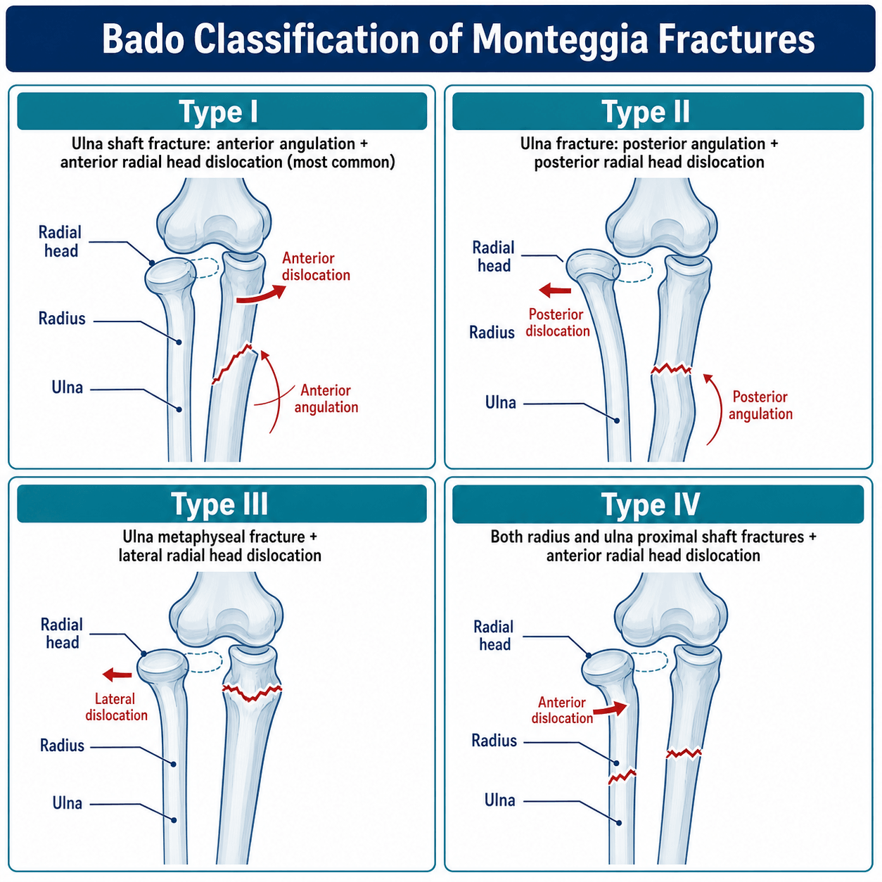
- RH Dislocation
- Anterior
- Ulna Pattern
- Anterior apex angulation
- Mechanism
- Fall on hyperextended arm
- Frequency Adult
- 15%
- RH Dislocation
- Posterior/Posterolateral
- Ulna Pattern
- Posterior apex angulation
- Mechanism
- Direct blow to flexed elbow
- Frequency Adult
- 70%
- RH Dislocation
- Lateral
- Ulna Pattern
- Metaphyseal fracture (valgus)
- Mechanism
- Varus force on extended arm
- Frequency Adult
- 10%
- RH Dislocation
- Anterior
- Ulna Pattern
- Both bone fractures (same level)
- Mechanism
- Hyperpronation injury
- Frequency Adult
- 5%
Type I in more detail. The force is a hyperpronation with hyperextension: a fall on the outstretched hand with the forearm pronated, the biceps pulling the radial head anteriorly. In children the ulna often fails as a greenstick fracture.
Type II in more detail. A direct blow to the posterior aspect of the flexed elbow, or an axial load on it, angulates the ulna posteriorly and drives the radial head posteriorly or posterolaterally.
Jupiter's subdivision of Type II. Jupiter subdivides the posterior lesion, and the subtype helps predict difficulty: proximal fractures near the coronoid (IIA) may require a different approach and have higher complication rates.
- Pattern
- Ulna fracture at coronoid level
- Implications
- Most common subtype
- Pattern
- Fracture distal to coronoid
- Implications
- Standard plating approach
- Pattern
- Fracture at diaphysis
- Implications
- May need longer plate
- Pattern
- Fracture at ulna diaphysis + radius fracture
- Implications
- Both bone fixation required
Clinical Assessment
Look for forearm deformity, swelling over the proximal forearm, and a radial head prominent laterally or posteriorly.
Feel and move. Palpate the ulnar fracture site and the position of the radial head, and check for tenderness over the DRUJ. Supination and pronation are limited and elbow movement is painful; assess grip strength.
The interosseous membrane is often disrupted in Monteggia injuries. This affects load transfer and forearm stability. Assess for tenderness along the entire interosseous space.
The nerve. The posterior interosseous nerve (PIN) is the nerve most at risk. Test finger extension (EDC, EIP) and thumb extension (EPL) specifically. Wrist extension is usually preserved, because ECRL and ECRB are innervated proximal to the PIN. The incidence, prognosis and management of the palsy are set out under Complications.
Investigations
Radiographs. Image the whole forearm and the elbow in its own right:
- AP and lateral forearm, which must include the elbow and wrist joints
- AP and lateral elbow, to confirm the relationship of the radial head
- Contralateral comparison if ulnar length is questionable
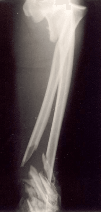
The radiocapitellar line. A line drawn through the centre of the radial shaft and head must pass through the centre of the capitellum on every view: AP, lateral and oblique. If it does not, the radial head is subluxed or dislocated.
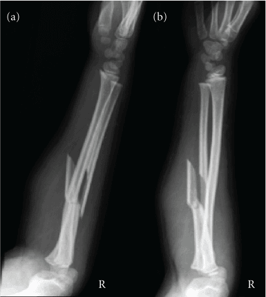
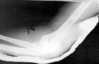
CT is indicated for assessing a coronoid fracture, for complex proximal ulna fractures, and in chronic Monteggia to assess changes in the shape of the radial head.
MRI is rarely indicated. It has a place where interosseous membrane disruption (the Essex-Lopresti variant) is suspected, for assessing cartilage integrity in chronic injuries, and for planning where soft tissue is interposed.
Differential Diagnosis
- Ulna
- Proximal/shaft fracture or plastic bowing
- Radial Head / PRUJ
- Dislocated (radiocapitellar line disrupted)
- Discriminating Feature
- Ulna fracture WITH radial head dislocation - check the line on every view
- Ulna
- Mid-shaft fracture, often transverse
- Radial Head / PRUJ
- Located normally (line intact)
- Discriminating Feature
- No radial head dislocation - the classic Monteggia trap if the line is not checked
- Ulna
- Normal, no fracture
- Radial Head / PRUJ
- Chronically dislocated; convex/dome-shaped radial head, hypoplastic capitellum
- Discriminating Feature
- Bilateral, no trauma, dysmorphic radial head - do NOT mistake for acute injury
- Ulna
- Fracture
- Radial Head / PRUJ
- Radius fractured but radial head located
- Discriminating Feature
- Both bones broken without PRUJ dislocation (vs Bado IV which adds RH dislocation)
- Ulna
- Intact
- Radial Head / PRUJ
- Radial HEAD fracture + IOM + DRUJ disruption
- Discriminating Feature
- Longitudinal forearm instability and DRUJ disruption, not proximal RU dislocation
- Ulna
- Olecranon fracture
- Radial Head / PRUJ
- PRUJ intact; ulnohumeral subluxation
- Discriminating Feature
- Forearm translates with the proximal ulna; radiocapitellar relationship preserved
A pulled or angulated ulna shaft fracture is a Monteggia until proven otherwise. The single discriminator is the radiocapitellar line - a normal-looking "isolated ulna fracture" with a disrupted line IS a Monteggia.
Management Algorithm
The principle. Fix the ulna anatomically, restoring its length and alignment, and in an acute injury the radial head typically reduces spontaneously. If it does not, suspect interposed tissue: the annular ligament, capsule or biceps.
Acute or chronic. The dividing line is 4 weeks. An injury presenting later, missed or delayed, is chronic and is reconstructed. Treatment principles also differ significantly between adults and children.
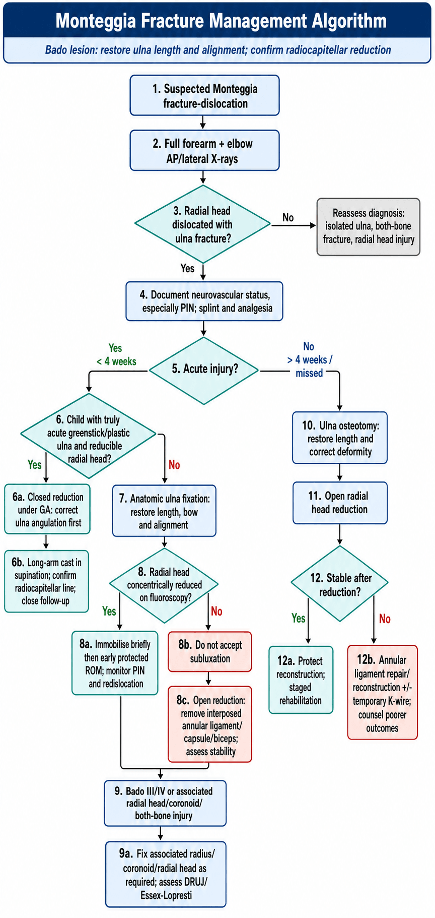
An anterior (Type I) or posterior (Type II) radial head dislocation presenting within 4 weeks of injury is the most common scenario in both children and adults. After a closed reduction attempt (see the timeline under Surgical Technique):
- Anatomic ulnar ORIF, restoring length and alignment; compare with the contralateral side for length
- Intraoperative fluoroscopy to confirm the radiocapitellar line
- The radial head reduces spontaneously in 95% of cases; if it does not, explore for interposed tissue
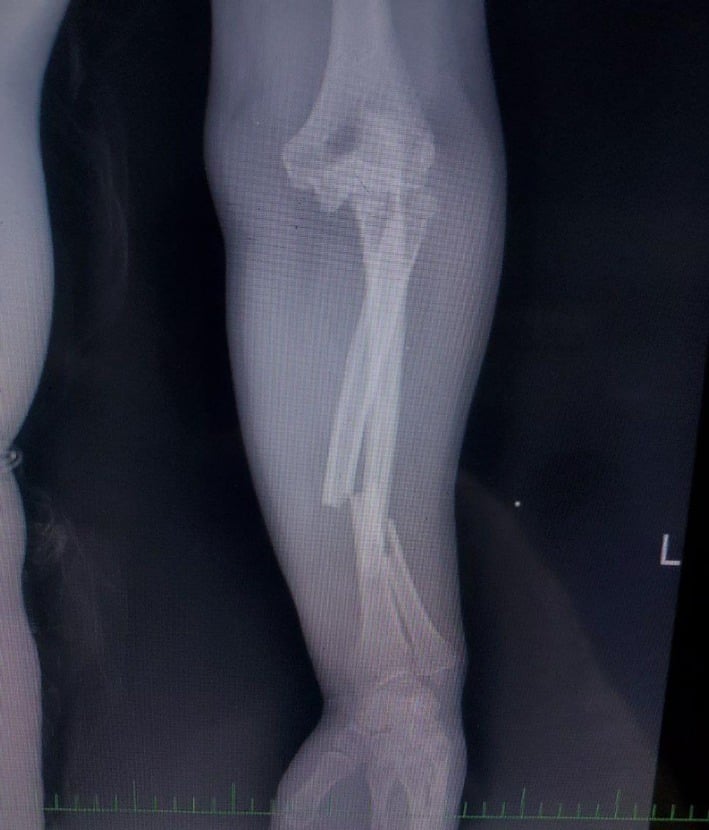
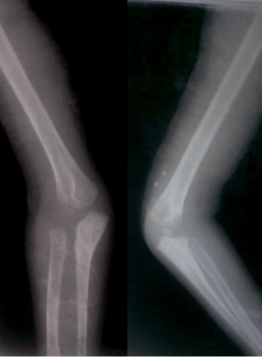
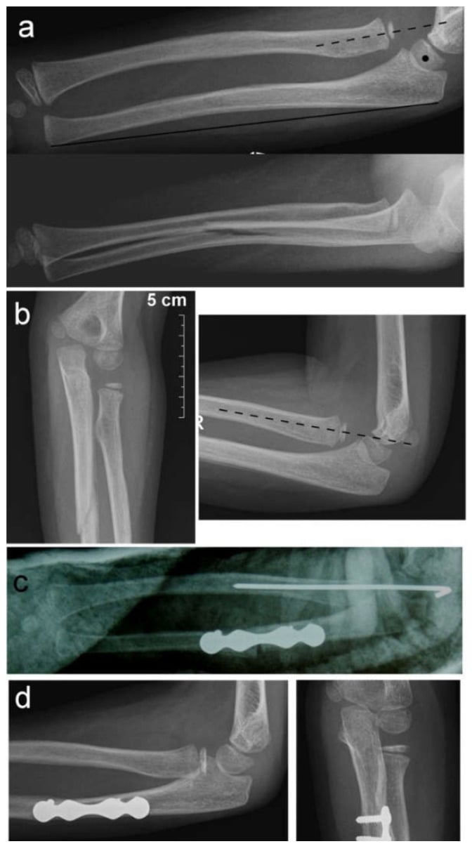
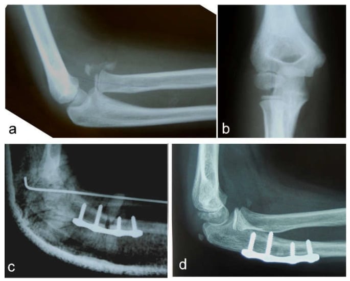
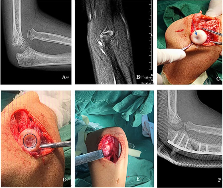
The decision table below summarises the tabs.
- Key Feature
- Anterior ulna angulation
- Radial Head Status
- Anterior RH dislocation
- Treatment
- Anatomic ulna ORIF - RH reduces
- Key Feature
- Posterior ulna angulation - most common adult
- Radial Head Status
- Posterior RH dislocation
- Treatment
- Plate fixation ulna - check RH reduction
- Key Feature
- Ulna metaphyseal fracture
- Radial Head Status
- Lateral RH dislocation
- Treatment
- Ulna fixation - may need RH ORIF
- Key Feature
- Both bone fractures
- Radial Head Status
- Anterior RH dislocation
- Treatment
- Fix both radius and ulna
- Key Feature
- Missed or delayed presentation
- Radial Head Status
- RH remains dislocated
- Treatment
- Open reduction + annular ligament reconstruction
- Key Feature
- Plastic deformation possible
- Radial Head Status
- Check radiocapitellar line
- Treatment
- Closed reduction if acute, open if chronic
Surgical Technique
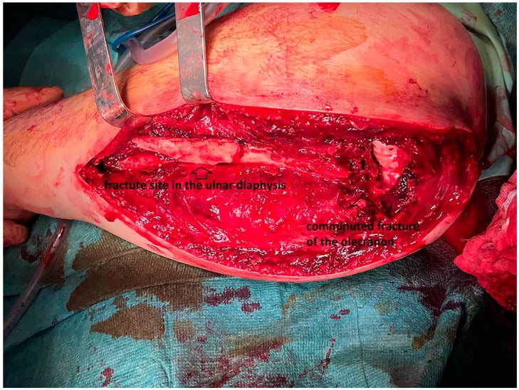
Approach. Direct posterior (Boyd) approach along the subcutaneous border of the ulna.
- Position supine, arm across the chest or on a table
- Posterior incision along the subcutaneous border
- Reduce the fracture, restoring length and alignment
- Plate fixation: 3.5mm LCP, 6-8 holes minimum
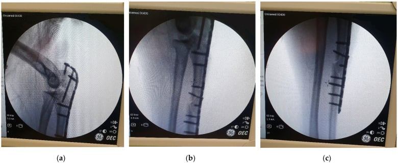
- Check the radiocapitellar relationship under fluoroscopy
- If the radial head does not reduce, explore for interposition
Length and bow. Ulnar length must be restored, compared with the contralateral side. The slight bow of the ulna is important for rotation, and the plate sits on the tension side (posterior/lateral).
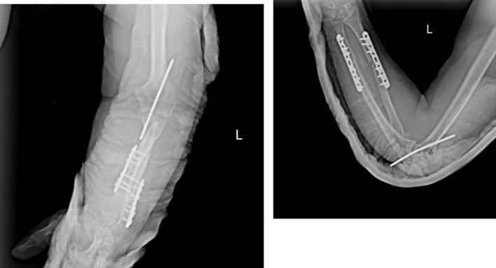
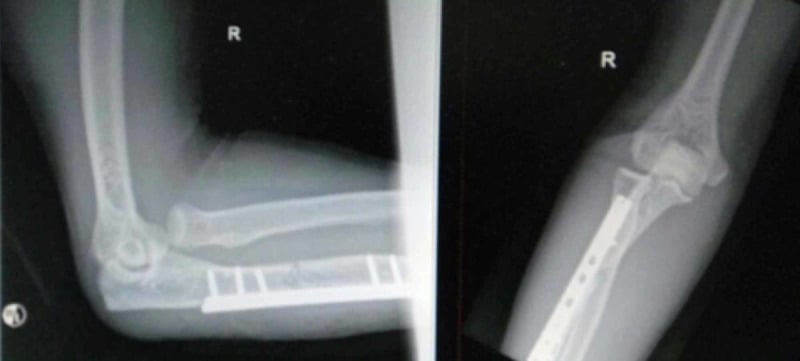
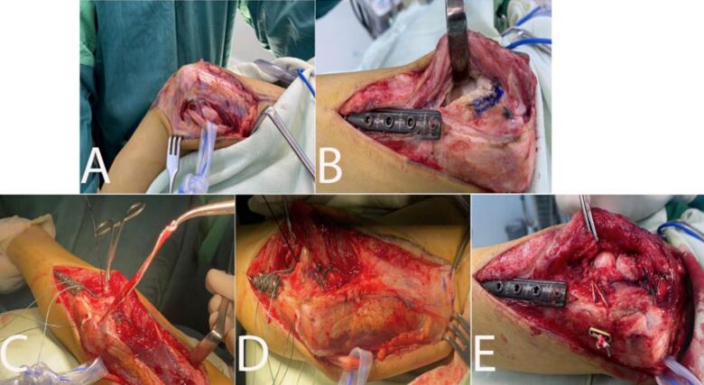
The straight-plate pitfall. A straight or generically pre-contoured plate on the dorsal proximal ulna flattens the normal PUDA, subtly shortening and malaligning the proximal ulna, which then levers the radial head into subluxation or redislocation even when the shaft looks reduced. This is the mechanism behind the malaligned posterior Monteggia fractures that Ring salvaged with a dorsal contoured plate.
Avoiding it. Template the PUDA against the contralateral uninjured ulna, contour the plate (or use a dedicated pre-contoured proximal ulna plate) to the patient's own geometry, and confirm a concentric radiocapitellar line on fluoroscopy before leaving theatre. Anatomic means restoring the ulna's length and its native sagittal bow, not making it straight.
Complications and Management
- Incidence
- ~11% (8/73 population-based); 10-20% quoted
- Prevention/Management
- Usually neurapraxia - observe; explore if no recovery by 3-4 months
- Incidence
- 5-10%
- Prevention/Management
- Anatomic ulna reduction, check intraoperative fluoro
- Incidence
- 2-5%
- Prevention/Management
- Single incision, careful soft tissue handling
- Incidence
- 10-15%
- Prevention/Management
- Early ROM, static progressive splinting
- Incidence
- Under 5%
- Prevention/Management
- Compression plating, bone graft if needed
- Incidence
- 5%
- Prevention/Management
- Gentle surgery, consider prophylaxis if prior HO
Nerve injury. The PIN is injured by stretch as the radial head dislocates, or by direct compression, and the association is strongest with Bado Type III. Most palsies recover spontaneously by 3-4 months; if there is no clinical recovery, obtain an EMG at 6 weeks. Explore the nerve for:
- No recovery by 3-4 months
- An open fracture with suspected nerve injury
- Worsening after closed reduction
Stiffness. Prevention is early motion, stable fixation, avoiding prolonged immobilisation, and aggressive physiotherapy from week 2. For established contracture, static progressive splinting (low-load, prolonged stretch) is more effective than dynamic splinting. If therapy reaches a plateau, consider open or arthroscopic capsular release at 6-12 months.
Heterotopic ossification. Prophylaxis for a patient with prior HO is indomethacin or radiation. Established heterotopic bone is resected when mature, at 12-18 months.
Postoperative Care and Rehabilitation
Principles. Move early if fixation is stable. Avoid forced supination for the first 4 weeks, which protects the annular ligament, and protect against varus stress if the LCL has been repaired. Rehabilitation works on forearm rotation as well as elbow flexion and extension.
Return to activities is staged:
- Desk work: 2-4 weeks
- Manual labour: 3-6 months
- Contact sports: 6 months minimum
- Full unrestricted activity: when strength and range of motion have normalised
Outcomes and Prognosis
The ranges in this section are conventional teaching estimates unless a cited series is named; the verified outcome data on this page are Ring 1998 (83% good/excellent in 48 adults), Zheng 2020 (75% congruent at 19 months in missed paediatric cases) and Zilliacus 2024 (all 60 followed children with full motion).
- Expected ROM
- Most of contralateral ROM
- Union Rate
- Union expected with anatomic plate fixation
- Functional Outcome
- Good to excellent in 83% (Ring 1998)
- Expected ROM
- 70-85% of contralateral ROM
- Union Rate
- 90-95%
- Functional Outcome
- Good to excellent in 70-80%
- Expected ROM
- 60-70% of contralateral ROM
- Union Rate
- N/A (osteotomy)
- Functional Outcome
- Fair to good in 50-60%
- Expected ROM
- Near-normal ROM
- Union Rate
- Union near-universal
- Functional Outcome
- All 60 followed children had full motion (Zilliacus 2024)
- Expected ROM
- Flexion improved 108 to 140 degrees (Zheng 2020)
- Union Rate
- N/A
- Functional Outcome
- Radiocapitellar alignment in 39 of 52 (75%) at 19 months (Zheng 2020)
What predicts a good result. An acute injury (under 4 weeks), an anatomic ulnar reduction, stable fixation, early mobilisation and a Type I or II pattern. Children have better outcomes overall because of their remodelling potential; adults require an anatomic reduction for a good outcome.
What predicts a poor one. A chronic injury (over 4 weeks), a comminuted proximal ulna, an associated coronoid fracture, a PIN palsy that is not recovering, a Type III or IV pattern, and an open wound.
The most important prognostic factor is timing of diagnosis and treatment. Acute injuries treated promptly do well (83% good/excellent in Ring's adult series). Chronic injuries fare worse even with reconstruction (75% congruent alignment in Zheng's missed-injury series).
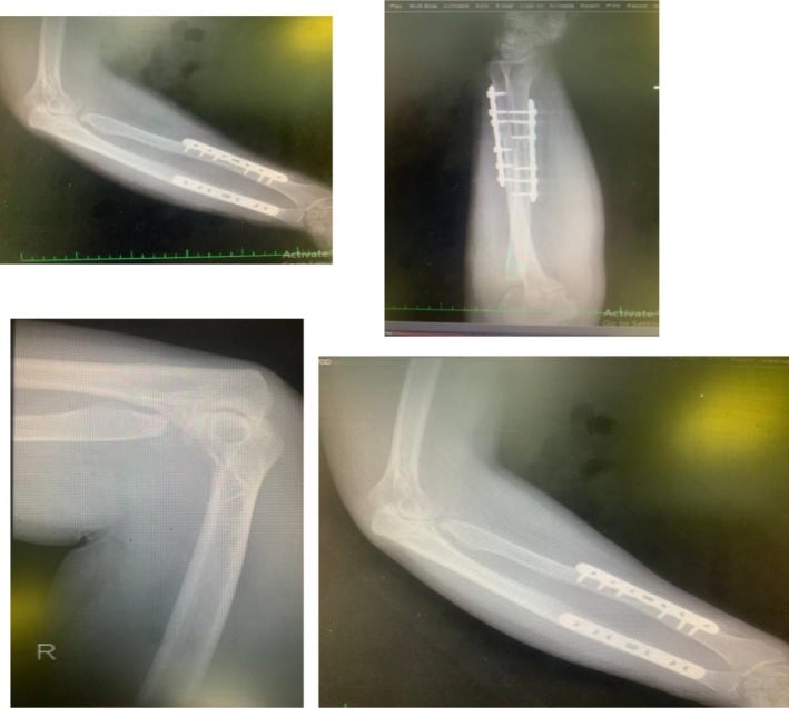
The long term. After acute anatomic fixation, ulnar union and a stable, concentrically reduced radial head are the expectation, with most forearm rotation recovered. Mild residual extension loss is common and rarely functionally limiting; heterotopic ossification and persistent PIN weakness are uncommon. Even malalignment is salvageable: re-fixation in Ring's series of 17 malaligned posterior injuries achieved union and a concentric ulnohumeral reduction in all of them. But 9 of those 17 had radiographic ulnohumeral arthrosis at a mean of 59 months, and late degeneration is the honest long-term caveat.
Counsel patients that while union and stability are reliably achieved, 10-15 degrees of motion loss is common and some loss of grip strength may persist. Most return to full activities by 6 months.
Guidelines, Registries & Global Practice
Global Epidemiology
- Monteggia lesions are uncommon: roughly 1-2% of all forearm fractures and under 5% of paediatric elbow/forearm fractures across published series worldwide.
- Bimodal distribution: paediatric peak around 4-10 years (predominantly Bado type I) and an adult peak in middle age and the elderly (predominantly type II).
- There is no single-country pattern - the adult type II predominance and paediatric type I predominance are consistent across North American, European and Asian cohorts.
Guidelines & Society Positions (Side by Side)
- Core Position
- Anatomic reduction and stable plate fixation of the ulna; radial head reduces with correct ulna alignment
- Practical Emphasis
- Restore ulna length, alignment and rotation; intra-op fluoroscopic check of radiocapitellar line
- Core Position
- Treat as a fracture-dislocation - urgent assessment, documented neurovascular exam, early definitive fixation
- Practical Emphasis
- Open injuries follow standard open-fracture pathways; senior-led decision-making
- Core Position
- Operative fixation is standard in adults; emphasis on detecting associated radial head and coronoid injury
- Practical Emphasis
- Address coronoid and radial head fractures at the index procedure
- Core Position
- Closed reduction acceptable for acute, stable (incomplete) paediatric ulna patterns; operate if unstable or chronic
- Practical Emphasis
- Low threshold to fix unstable complete ulna fractures; never excise the radial head in a child
There is no dedicated randomised guideline for Monteggia fractures - guidance is consensus/expert based. The internationally shared principle across AO, BOA, AAOS and paediatric bodies is identical: fix the ulna anatomically and the radial head follows.
Registry & Resource-Setting Variation
- No national arthroplasty/implant registry tracks Monteggia outcomes specifically; evidence is from single-centre and tertiary trauma-centre series rather than registry data.
- High-resource settings: routine fluoroscopy, locking plates, on-table radial head implants, and ready paediatric subspecialty referral.
- Limited-resource settings: missed and chronic (neglected) Monteggia is proportionally far more common because the radiocapitellar line is not checked on the initial film; reconstruction (ulna osteotomy with indirect or open radial head reduction) is therefore a more frequent presentation.
Related pages: Monteggia and Galeazzi Fractures sets the two forearm fracture-dislocations side by side if you want the comparison rather than this depth; Monteggia Fractures in Children and Paediatric Forearm Fractures for the plastically deformed ulna that hides the injury; Galeazzi Fractures for the mirror-image lesion at the wrist. For the injuries that decide a Bado II outcome see Coronoid Fractures and Radial Head Fractures, with Olecranon Fractures for the proximal ulna differential, Radial Head Dislocations for the isolated and congenital mimics, and Radioulnar Synostosis for the complication that costs rotation.
Monteggia fractures are among the most commonly missed injuries.
- Document radiocapitellar line assessment on ALL views
- Note PIN function pre- and post-operatively
- Record stability of radial head after fixation
- Missed diagnosis (most common)
- Delayed treatment leading to chronic Monteggia
- Failure to document pre-existing PIN palsy
- Poor outcomes from chronic reconstruction
Controversies & Areas of Uncertainty
Routine annular ligament reconstruction? In chronic paediatric cases, ulnar osteotomy with indirect reduction can stabilise the radial head without formal reconstruction (Song 2012). When the annular ligament is addressed, repair of the native ligament outperformed reconstruction (Zheng 2020). Many now reserve formal Bell Tawse reconstruction for cases that remain unstable intra-operatively.
Open or indirect (ulna-based) reduction in chronic cases. Whether to open the radiocapitellar joint or rely on over-correction of the ulna (angulation-translation osteotomy) remains debated. Both can succeed, and the choice is often driven by chronicity, radial head shape and intra-operative stability.
The temporary transarticular K-wire. Trans-capitellar or transarticular K-wires can hold a reduced radial head but carry a real risk of wire breakage and migration. Use is selective, kept short, and avoided where stable fixation alone is sufficient.
Managing the PIN palsy. Most PIN palsies are neurapraxic and recover, so early exploration is generally not indicated. The grey zone is the new, complete post-reduction palsy where nerve entrapment cannot be excluded; the timing of exploration and the role of early EMG or nerve ultrasound are not standardised.
MCQ Practice Points
Q: What radiographic line must be checked on every elbow X-ray to avoid missing a Monteggia fracture?
A: The radiocapitellar line. A line drawn through the center of the radial neck must bisect the center of the capitellum on ALL views (AP, lateral, oblique). Disruption indicates radial head dislocation.
Q: What is the most common Bado type in adults vs children?
A: Adults: Type II - posterior radial head dislocation with posterior ulna angulation. Conventionally quoted at ~70%; Ring's tertiary-centre series found 38 of 48 (79%), which is a referral pattern as much as an epidemiological one. Children: Type I - anterior radial head dislocation. Note that in the one population-based paediatric series, Bado I and Bado III together accounted for all but 3 of 73 fractures, so type III is the second commonest childhood pattern, not a rarity.
Q: After anatomic ulna fixation, the radial head does not reduce. What is your next step?
A: The radial head should reduce spontaneously after anatomic ulna fixation. If it doesn't: 1) Confirm ulna reduction is truly anatomic (length and alignment). 2) If still subluxed, explore through Kocher approach for interposed tissue (annular ligament, capsule, or biceps).
Q: A patient develops finger drop after Monteggia ORIF. What is the likely diagnosis and prognosis?
A: PIN (Posterior Interosseous Nerve) palsy. Conventionally quoted at 10-20%; the one population-based series gives associated nerve injury in 8 of 73 children (11%) (Zilliacus 2024), so the lower end is better supported. Usually a neurapraxia from traction during injury (not iatrogenic). Excellent prognosis - most recover spontaneously within 3-4 months. Observe unless new post-op or concern for entrapment, and remember the association is strongest with Bado type III.
Q: What is the treatment for a Monteggia fracture diagnosed 6 weeks after injury?
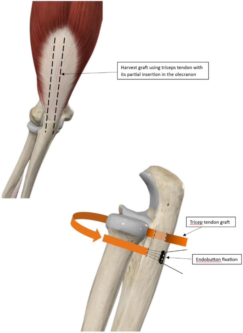
A: Chronic Monteggia (greater than 4 weeks) has poor outcomes with simple reduction. Requires: ulna osteotomy (to restore length), open reduction of radial head, and annular ligament reconstruction (Bell Tawse technique using triceps tendon strip). Results are inferior to acute treatment.
Clinical Decision Scenarios
Practise clinical reasoning and management decisions out loud
“A 45-year-old construction worker presents after falling from scaffolding. X-rays show proximal ulna fracture with posterior radial head dislocation.”
“A 7-year-old child presents 6 weeks after a fall. Parents were told the 'wrist fracture healed well' at another hospital. You notice limited forearm rotation and prominent radial head.”
“You fix an acute Type II Monteggia fracture with anatomic ulna reduction and the radial head reduces concentrically. Post-operatively, the patient cannot extend fingers at MCPJs.”
Key Stats
- 1-2% of forearm fractures
- Type II (posterior) = 70% in adults
- Type I (anterior) = most common in children
- Nerve injury ~11% (8/73, Zilliacus 2024); conventionally quoted as 10-20%
- Missed at first consultation in 20-50% (Zivanovic 2022); 22% delayed 1-8 days in a population series
Bado Classification
- Type I - Anterior RH dislocation, anterior ulna angulation
- Type II - Posterior RH dislocation, posterior ulna angulation
- Type III - Lateral RH dislocation, ulna metaphyseal fracture
- Type IV - Anterior RH dislocation, both bone fractures
Key Principles
- Radiocapitellar line MUST bisect capitellum on ALL views
- Fix ulna anatomically - RH reduces spontaneously
- If RH doesn't reduce - explore for interposed tissue
- Chronic (more than 4 weeks) - annular ligament reconstruction needed
- PIN palsy - usually recovers, observe 3-4 months
Surgical Steps
- Position supine, arm on table
- Boyd posterior approach to ulna
- Anatomic reduction - restore length/alignment
- 3.5mm LCP plate fixation (6-8 holes)
- Check radiocapitellar line on fluoro
- If RH subluxed - Kocher approach to explore
Must Know for Exam
- Most missed injury - ALWAYS check radiocapitellar line
- Adult vs pediatric types differ (II vs I)
- PIN palsy is neurapraxia - observe
- Chronic has poor outcomes - emphasizes early diagnosis
- Bell Tawse technique for annular ligament reconstruction
Evidence Base and Literature
Key Studies and Papers
Bado Original Classification
- Defined the Monteggia lesion and four types by direction of radial head dislocation and ulna deformity
Annular Ligament Reconstruction (Bell Tawse)
- Described use of a triceps fascia strip to create a neo-annular ligament for chronic radial head dislocation
Monteggia Fractures in Adults
- 48 adults; 38 (79%) were Bado type II; 68% of type II had an associated radial head fracture
- Excellent or good Broberg-Morrey result in 40 of 48 patients (83%) after stable anatomic plate fixation of the ulna
- 6 of 8 unsatisfactory results were type II with concomitant radial head/coronoid fractures
Monteggia Fractures in Children and Adults (Principle Review)
- Stable anatomic reduction of the ulna fracture restores anatomic reduction of the radial head
- Character of the ulna fracture (not the direction of dislocation) guides treatment
- Good non-operative results in children reflect prevalence of incomplete (stable) ulna fractures