The Dually Innervated Adductor and the Roof of the Medial Hip
- Pectineus is the classic dually innervated muscle of the lower limb: the anterior division of the obturator nerve (L2, L3, L4) plus a branch from the femoral nerve, which is why it is sometimes grouped with the anterior compartment.
- Origin is the pectineal line (pecten pubis) on the superior pubic ramus and the adjacent pubic surface; insertion is the pectineal line of the femur on the posterior aspect below the lesser trochanter.
- It forms the medial portion of the floor of the femoral triangle, alongside the iliopsoas laterally and the adductor longus medially.
- Its actions are hip adduction and flexion with a contribution to internal rotation in the flexed hip; it is the most anterior and most superior of the adductor group and lies in the same plane as the hip capsule.
- In the medial (Ludloff) approach to the hip, the medial circumflex femoral artery lies immediately deep and posterior to the pectineus and adductor brevis — the vessel whose injury causes avascular necrosis of the femoral head.
- “Ask what happens to adduction after a pure obturator nerve injury: it is weakened but not abolished, partly because the pectineus retains its femoral nerve supply and the ischiocondylar part of adductor magnus retains its sciatic supply.
- “The obturator nerve anterior division lies anterior to adductor brevis and posterior to pectineus and adductor longus — the key plane to state when asked how the divisions are separated.
- “The medial circumflex femoral artery passes between pectineus and psoas anteriorly, then runs posteriorly between the obturator externus and the quadratus femoris — the anatomical route explaining why it is at risk in every medial hip approach.
- “Pectineus is anatomically an adductor compartment muscle by origin and action but has an anterior compartment nerve as well; this hybrid status is exactly why examiners like it.
Overview
The pectineus is a flat, quadrilateral muscle in the uppermost part of the medial thigh. It sits at the junction between the anterior (femoral) compartment and the medial (adductor) compartment, and this hybrid position is the source of both its anatomical interest and its surgical importance. It contributes to the floor of the femoral triangle, lies directly on the anteromedial hip capsule, and separates the femoral vessels in front from the medial circumflex femoral artery and the obturator nerve behind.
Because it is the most anterior and most superior of the adductor group, the pectineus is the muscle encountered first when approaching the hip from the medial side and the muscle that must be released last (and most carefully) when a spastic adducted hip will not abduct despite adductor longus and brevis release.
The pectineus is the textbook dually innervated muscle of the lower limb.
- Anterior division of the obturator nerve — its adductor compartment supply.
- A branch of the femoral nerve — usually arising directly from the femoral nerve or from the nerve to iliopsoas, entering the anterior surface of the muscle. Accessory obturator nerve, when present, is another source.
- Root values are L2, L3, L4 whichever route you follow.
Why the examiner asks it
- It explains why adduction is never abolished by an obturator nerve lesion. Two adductor group muscles have a non-obturator supply: the pectineus (femoral nerve) and the ischiocondylar (hamstring) part of adductor magnus (tibial division of the sciatic nerve). Adduction is therefore weakened, not lost.
- It explains the embryology. The pectineus lies at the boundary of the pre-axial territory and receives supply from both plexus divisions — a developmental fossil.
- It explains a clinical pattern. In a pure femoral nerve lesion, hip adduction is largely preserved; in a pure obturator lesion, adduction is weak with a characteristic sensory patch on the medial thigh. A patient with weak adduction plus quadriceps weakness plus medial thigh numbness has a lesion at or above the lumbar plexus, not an isolated peripheral nerve injury.
The other dually innervated muscles worth pairing it with: adductor magnus (obturator posterior division plus tibial division of sciatic), biceps femoris (tibial division for the long head, common peroneal division for the short head), and flexor digitorum profundus in the upper limb (median and ulnar).
P-L-B-M-GAdductor Compartment — Superficial to Deep, Anterior to Posterior
Hook:Pectineus and magnus are the two dually innervated ones; brevis is the landmark between the obturator divisions.
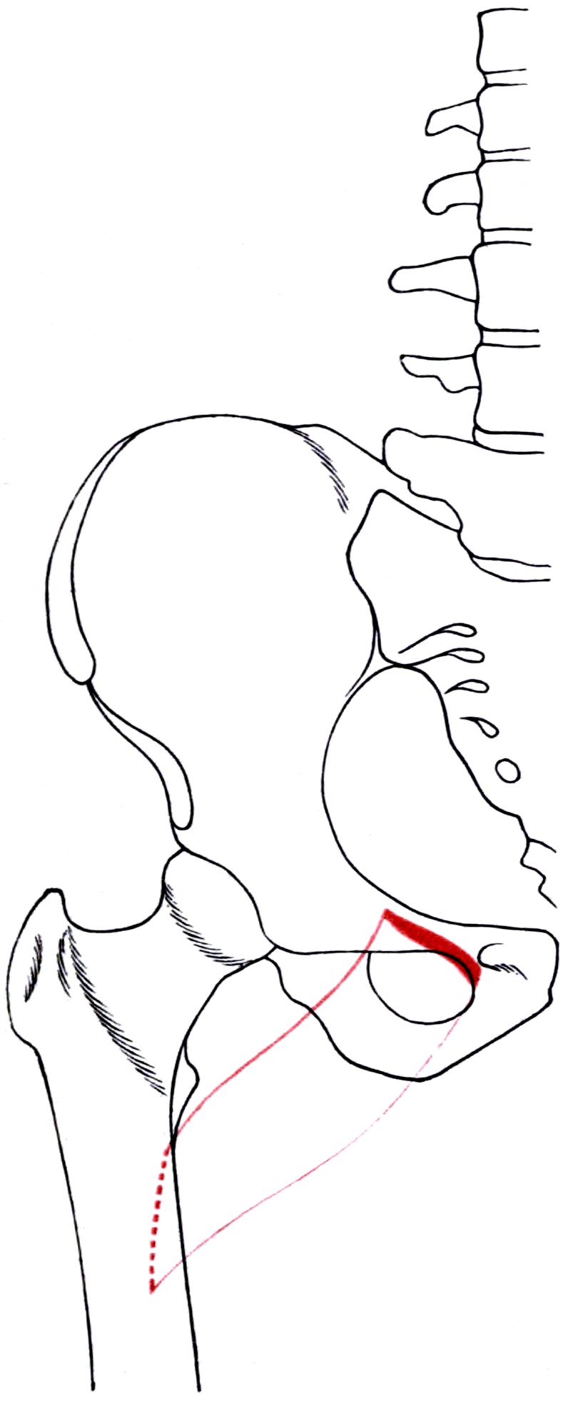
Attachments, Innervation and Relations
Origin
- Pectineal line (pecten pubis) on the superior surface of the superior pubic ramus — the sharp ridge running from the pubic tubercle to the iliopubic eminence.
- The adjacent bone anterior to the pectineal line, and the pectineal fascia.
- The pectineal (Cooper's) ligament — the thickened periosteum along the pectineal line — is closely related and is the structure used for fixation in some hernia repairs.
Insertion
- The pectineal line of the femur, a vertical ridge on the posterior surface of the femur running from the lesser trochanter to the upper end of the linea aspera.
- The muscle therefore passes posteriorly and laterally from the pubis to the back of the proximal femur, wrapping around the anteromedial aspect of the hip.
Shape and plane
- Flat and quadrilateral, roughly 5-8 cm wide in its middle portion.
- Occupies the most anterior and most superior position of the adductor group, in the same coronal plane as (and immediately anterior to) the hip joint capsule.
- Its medial border abuts the adductor longus; its lateral border abuts the iliopsoas at the iliopectineal groove, which is the point where the femoral vessels lie.
Named landmarks and measurements
- Relationship
- Origin; runs from the pubic tubercle to the iliopubic eminence
- Practical use
- Landmark for the pectineal ligament, femoral ring and hernia repair fixation
- Relationship
- Medial end of the pectineal line, just lateral to the adductor longus origin
- Practical use
- Palpable landmark for the medial approach and for adductor tenotomy
- Relationship
- Insertion, on the posterior femur from the lesser trochanter to the linea aspera
- Practical use
- Explains the posteriorly directed line of pull and the internal rotation component in flexion
- Relationship
- Between the pectineus medially and the iliopsoas laterally, on the floor of the femoral triangle
- Practical use
- The femoral artery and vein lie in this groove — the danger zone
- Relationship
- Approximately 2-3 cm inferolateral to the pubic tubercle, above the obturator membrane
- Practical use
- Site of the obturator nerve emergence; target for obturator nerve block; site of obturator hernia
- Relationship
- Just proximal and lateral to the femoral insertion
- Practical use
- Reference for the depth of a medial approach and for iliopsoas tenotomy
The vessel that determines whether the femoral head survives.
- The MCFA passes posteriorly between pectineus and psoas and continues between obturator externus and quadratus femoris.
- It is the dominant blood supply to the adult femoral head.
- Consequence: in a medial approach to the hip, dissection carried posteriorly behind the pectineus and adductor brevis risks the MCFA and therefore avascular necrosis of the femoral head — a reported complication of medial open reduction for developmental dysplasia.
- Answer: define the adductor longus and brevis, work anterior to adductor brevis, keep the obturator nerve anterior division in view, and never dissect blindly posteriorly.
Because pectineus keeps its femoral supply.
- The pectineus retains a femoral nerve branch; the ischiocondylar part of adductor magnus retains a tibial (sciatic) supply.
- Consequence: obturator nerve block or neurectomy weakens adduction but never abolishes it, and a residual adductor twitch during transurethral resection may persist despite an apparently successful block.
- Answer: explain this to the patient before neurectomy, and to the anaesthetist and urologist before relying on a block to abolish the adductor jerk.
Action and Biomechanics
Actions by plane
- Action
- Hip adduction
- Position of greatest efficiency
- Hip abducted
- Note
- Contributes to the adductor group; not the strongest member
- Action
- Hip flexion
- Position of greatest efficiency
- Hip extended
- Note
- Significant flexion component because the muscle lies anterior to the joint axis
- Action
- Internal rotation in the flexed hip; contributes to external rotation in some positions
- Position of greatest efficiency
- Depends on hip flexion angle
- Note
- The insertion on the posterior femur means the rotational effect reverses with hip position
- Action
- Stabilises the pelvis in single-leg stance and controls contralateral pelvic drop with the other adductors
- Position of greatest efficiency
- Weight-bearing
- Note
- The adductors are more important stance-phase stabilisers than their name suggests
The rotational paradox worth knowing
- Because the pectineus inserts on the posterior surface of the femur but originates anterior to the joint, its rotational moment depends on hip position. With the hip extended it contributes to external rotation; with the hip flexed the line of pull shifts and it contributes to internal rotation. The same reversal applies to the other adductors and is the reason the adductor group is described inconsistently across textbooks.
- The safe exam answer: the pectineus is an adductor and flexor of the hip with a position-dependent rotational component. State the mechanism rather than committing to a single direction.
Force contribution and architecture
- The adductor group as a whole is substantial — comparable in mass to the quadriceps in some estimates — and the pectineus is a modest contributor within it, being the smallest and most proximal.
- Its short fibres and proximal position give it a short moment arm for adduction but a useful flexion moment.
Length-tension and gait
- The adductors as a group are active in early stance and in pre-swing, contributing to pelvic stabilisation and to the transition between stance and swing rather than to gross adduction.
- In spastic hip disease the adductors are overactive throughout, producing a scissoring gait and, over time, progressive hip displacement as the femoral head is levered out of a shallow acetabulum.
Synergists and antagonists
- Adduction synergists: adductor longus, brevis and magnus, gracilis, and to a small extent the inferior fibres of gluteus maximus and the quadratus femoris.
- Flexion synergists: iliopsoas (dominant), rectus femoris, sartorius, tensor fascia lata.
- Antagonists: gluteus medius and minimus, tensor fascia lata (abduction); gluteus maximus and hamstrings (extension).
What happens when it fails
- Obturator nerve lesion: weak but not absent adduction, a numb patch on the medial thigh, and a subtle circumducting or wide-based gait. The pectineus retains its femoral supply.
- Adductor group overactivity (spasticity): scissoring gait, difficulty with perineal hygiene and catheterisation, and progressive hip displacement in cerebral palsy.
- Adductor group contracture: fixed adduction deformity, apparent limb shortening, and difficulty positioning for hip surgery.
The spastic hip in cerebral palsy displaces because of an imbalance of forces on a hip that is dysplastic from the outset.
- Adductor and iliopsoas overactivity with relatively weak abductors and extensors produces a flexed, adducted, internally rotated hip.
- The resulting persistent coxa valga and femoral anteversion, combined with a shallow, deficient posterosuperior acetabulum, allows the head to migrate superolaterally and posteriorly.
- Migration percentage (Reimers index) quantifies it: the proportion of the femoral head width lying lateral to Perkins' line. Displacement of greater than 30 percent is generally the trigger for intervention, and greater than 50 percent usually indicates that a soft-tissue release alone will not suffice.
- Hip surveillance programmes (for example the Australian and Scandinavian programmes and the UK equivalents) mandate radiographic monitoring by GMFCS level, because early displacement is asymptomatic and painless dislocation is the endpoint that must be prevented.
- Soft-tissue release — adductor longus and gracilis tenotomy, with adductor brevis and pectineus added if abduction remains restricted, plus iliopsoas lengthening at the pelvic brim — is effective in the young child with a migration percentage of roughly 30-50 percent and preserved acetabular morphology.
- Once the acetabulum is deficient or the migration percentage is high, reconstruction is required: varus derotation femoral osteotomy plus pelvic osteotomy (Dega, San Diego, Pemberton) with open reduction as needed.
- The pectineus is the deepest and most anterior of the adductors and is released last, with the obturator nerve and the medial circumflex femoral artery immediately behind it — which is why an extended adductor release is not a trivial operation.
Surface Anatomy and Examination
Palpation and positioning
- Pubic tubercle: the key landmark. Palpable at the medial end of the inguinal ligament, roughly 2-3 cm lateral to the symphysis. The adductor longus tendon is the prominent cord arising just below and medial to it, and the pectineus lies immediately lateral and deeper.
- Pectineus itself is not reliably palpable as a discrete muscle — it lies deep to the femoral vessels and the fascia in the femoral triangle. It can sometimes be appreciated as a deep resistance lateral to the adductor longus tendon with the hip flexed and abducted.
- Femoral pulse: lies at the mid-inguinal point, in the iliopectineal groove on the floor formed by the iliopsoas laterally and the pectineus medially. Feeling the pulse orientates you to the danger plane.
- Obturator canal: approximately 2-3 cm inferolateral to the pubic tubercle; the target for an obturator nerve block, palpable only as a soft depression in a slim patient.
Named tests and their interpretation
- How to perform
- Supine, hips at 0, 45 or 90 degrees of flexion; the patient squeezes the examiner's hand or a dynamometer between the knees
- Positive finding
- Pain or weakness reproduced
- What it means
- Adductor-related groin pain, adductor injury, or adductor weakness
- False positives
- Pubic or hip joint pathology, osteitis pubis, inguinal-related groin pain
- How to perform
- Supine, pelvis level; abduct with the knee extended, then with the knee flexed
- Positive finding
- Abduction improves markedly with the knee flexed
- What it means
- The limiting structure crosses the knee — that is the gracilis, the only biarticular adductor; unchanged abduction implicates the monoarticular adductors including pectineus
- How to perform
- Compare adductor power and passive range in hip flexion and extension
- Positive finding
- Difference between positions
- What it means
- Reflects the position-dependent line of pull of the proximal adductors, including pectineus
- False positives
- Pain inhibition; hip joint pathology limiting flexion
- How to perform
- Palpate the adductor longus origin at the pubis, the pubic symphysis, the inguinal canal and the hip joint line separately
- Positive finding
- Localised tenderness at one site
- What it means
- Distinguishes adductor-related from pubic-related, inguinal-related and hip-related groin pain per the Doha classification
- False positives
- Overlapping tenderness in multi-entity groin pain, which is common
- How to perform
- Test light touch over the distal medial thigh
- Positive finding
- Reduced sensation
- What it means
- Obturator nerve cutaneous branch involvement — a small but specific patch
- False positives
- Overlap with the medial cutaneous nerve of the thigh from the femoral nerve
- How to perform
- Standard hip impingement and hip/sacroiliac provocation tests
- Positive finding
- Groin pain reproduced
- What it means
- Hip-related groin pain, which must be excluded before attributing symptoms to the adductors
- False positives
- Pain from any anterior structure; FABER is not specific
- How to perform
- Supine with the pelvis square, measure the maximum passive abduction on each side
- Positive finding
- Reduced abduction with a hard end-feel
- What it means
- Fixed adductor contracture, relevant to positioning for hip surgery and to spastic hip management
- False positives
- Pelvic obliquity not controlled; pain-limited guarding
Pitfalls in examination
- Localise groin pain by entity, not by region. The Doha agreement framework requires the examiner to separate adductor-related, iliopsoas-related, inguinal-related, pubic-related and hip-related groin pain, because more than one entity coexists in a large proportion of athletes.
- Always square the pelvis before measuring abduction, or a pelvic obliquity will masquerade as an adductor contracture.
- Test with the knee both flexed and extended to separate the biarticular gracilis from the monoarticular adductors.
- Do not forget the hip joint. Referred pain from hip pathology, and an articular branch of the obturator nerve supplying the hip, mean that hip disease can present as medial thigh and knee pain.
- Consider referred and systemic causes: an obturator hernia (Howship-Romberg sign — medial thigh pain on hip extension, adduction and internal rotation, in an elderly thin woman with bowel obstruction), a retroperitoneal mass or haematoma, and pelvic malignancy.
Complications
- Mechanism
- Injury to or spasm of the medial circumflex femoral artery deep to pectineus and adductor brevis; excessive traction; forceful reduction; extreme abduction in a spica
- Avoidance / management
- Work anterior to adductor brevis; limit posterior dissection; avoid forced reduction; use the human position in the spica
- Mechanism
- Dissection anterior to the pectineal fascia in the iliopectineal groove
- Avoidance / management
- Identify the adductor longus first and work medially; never dissect anteriorly toward the pulse
- Mechanism
- Anastomotic vessel across the superior pubic ramus divided during anterior pelvic exposure
- Avoidance / management
- Inspect the ramus within 3-6 cm of the symphysis and ligate deliberately before plating
- Mechanism
- Division during adductor release or pelvic surgery; retraction injury
- Avoidance / management
- Identify the anterior division on adductor brevis; deliberate rather than incidental neurectomy
- Mechanism
- Complete obturator neurectomy combined with an extensive adductor release
- Avoidance / management
- Avoid or limit neurectomy; release only what is needed to reach about 40-45 degrees of abduction per side
- Mechanism
- Pectineus retains its femoral nerve supply; ischiocondylar adductor magnus retains its sciatic supply
- Avoidance / management
- Counsel that adduction is weakened rather than abolished; plan additional tenotomy if a complete effect is needed
- Mechanism
- Release performed too late, with an already deficient acetabulum or a high migration percentage
- Avoidance / management
- Adhere to surveillance schedules; add femoral and pelvic osteotomy when the migration percentage exceeds about 50-60 percent
- Mechanism
- Disruption of deep inguinal nodes and lymphatics anterior to the pectineus
- Avoidance / management
- Careful haemostasis and lymphostasis; avoid unnecessary nodal dissection
- Mechanism
- Extensive muscle dissection, acetabular fracture surgery, head or spinal cord injury
- Avoidance / management
- Gentle handling, haemostasis, prophylaxis in high-risk patients; excise only when mature
- Mechanism
- Moist, contaminated area, particularly in a child in a spica or a patient with poor continence
- Avoidance / management
- Meticulous closure, careful cast padding and hygiene planning, early review
- Mechanism
- Medial thigh and knee pain attributed to musculoskeletal causes in an elderly thin woman
- Avoidance / management
- Consider the Howship-Romberg and Hannington-Kiff signs; CT if bowel symptoms coexist
Denervation pattern
- Isolated pectineus denervation is not clinically detectable because of its dual supply and small contribution.
- Obturator nerve lesion: adduction is weakened by roughly the loss of adductor longus, brevis, gracilis and the adductor part of magnus, with a small patch of medial thigh sensory loss. The retained supplies to pectineus (femoral) and to the ischiocondylar adductor magnus (sciatic) preserve some adduction.
- On MRI, obturator neuropathy produces denervation oedema and later fatty atrophy in adductor longus, brevis, gracilis and the adductor portion of magnus, with relative sparing of the pectineus and of the ischiocondylar magnus — a pattern that localises the lesion.
Clinical Relevance
The medial (Ludloff) approach to the hip
Purpose: direct access to the inferomedial hip capsule, the transverse acetabular ligament and the psoas tendon — the structures blocking reduction in a dislocated hip. Principally used for open reduction of developmental dysplasia of the hip in the infant, and occasionally for psoas release, biopsy of a medial lesion, or removal of a medially placed foreign body.
Positioning and incision
- Supine with the hip flexed, abducted and externally rotated (the frog-leg position) to bring the medial structures forward.
- Incision: a transverse or longitudinal incision over the adductor longus, beginning roughly 1-2 cm distal to the pubic tubercle and extending distally and posteriorly for 4-6 cm.
Interval — the part that matters
- Ludloff's original interval: between adductor longus and gracilis superficially, then between adductor brevis and adductor magnus deeply. This is the classic description.
- The Ferguson modification: between adductor longus and adductor brevis, that is anterior to the adductor brevis.
- Why the distinction matters: the anterior division of the obturator nerve lies anterior to adductor brevis, and the posterior division lies posterior to it. The medial circumflex femoral artery lies posteriorly, deep to the pectineus and adductor brevis. Working anterior to adductor brevis keeps the surgeon away from the posterior division of the obturator nerve and, critically, further from the medial circumflex femoral artery.
- The pectineus is the anterior boundary of the working corridor; it is retracted anteriorly (or partially released) and it protects the femoral vessels lying on its anterior surface.
Sequence
- Identify and, if required, tenotomise the adductor longus close to its origin.
- Develop the interval anterior to the adductor brevis.
- Identify the anterior division of the obturator nerve on the anterior surface of adductor brevis and protect it.
- Retract the pectineus anteriorly, protecting the femoral vessels.
- Identify and release the iliopsoas tendon at the level of the lesser trochanter — a major block to reduction.
- Perform the capsulotomy, excise the ligamentum teres and the pulvinar, and divide the transverse acetabular ligament to allow reduction.
Advantages and disadvantages
- Medial (Ludloff)
- Direct access to the psoas, ligamentum teres, pulvinar and transverse acetabular ligament
- Anterior (Smith-Petersen)
- Good access, plus access to the acetabulum and capsule for capsulorrhaphy
- Medial (Ludloff)
- Not possible — a recognised limitation
- Anterior (Smith-Petersen)
- Possible, and considered a major advantage
- Medial (Ludloff)
- Not possible through this approach
- Anterior (Smith-Petersen)
- Possible in the same exposure
- Medial (Ludloff)
- Small, hidden in the groin crease
- Anterior (Smith-Petersen)
- Longer, more visible
- Medial (Ludloff)
- Minimal
- Anterior (Smith-Petersen)
- Greater
- Medial (Ludloff)
- The concern that has limited its use — the medial circumflex femoral artery lies immediately posterior
- Anterior (Smith-Petersen)
- Lower reported rates in some comparisons, though the literature is heterogeneous
- Medial (Ludloff)
- The very young infant (commonly quoted under about 12-18 months) with a hip that needs only soft-tissue release
- Anterior (Smith-Petersen)
- Older infants and children, and any hip needing capsulorrhaphy or a bony procedure
Reported rates of avascular necrosis after medial open reduction for developmental dysplasia vary widely between series, and injury to the medial circumflex femoral artery — which passes immediately deep and posterior to the pectineus and adductor brevis, and is the dominant blood supply to the femoral head — is the mechanism implicated. Practical safeguards: work anterior to the adductor brevis, do not dissect posteriorly beyond what is required, avoid excessive traction and forceful reduction, avoid extreme abduction in the postoperative spica (aim for roughly 100 degrees of flexion and 45-55 degrees of abduction, the human position), and confirm a concentric reduction rather than forcing one.
Surgical Relevance
Procedure-by-procedure summary
- How pectineus is involved
- Retracted anteriorly (or partially released) as the anterior boundary of the corridor
- Structures at risk with location
- Medial circumflex femoral artery immediately deep and posterior; femoral vessels on its anterior surface; obturator nerve anterior division on adductor brevis
- How pectineus is involved
- Released last, after adductor longus, gracilis and brevis, if abduction remains restricted
- Structures at risk with location
- Obturator nerve branches on its deep surface; medial circumflex femoral artery deeper still
- How pectineus is involved
- The anterior division lies deep to it; the femoral branch is NOT blocked
- Structures at risk with location
- Femoral vessels anteriorly; obturator vessels adjacent to the nerve in the canal
- How pectineus is involved
- Its origin is elevated from the pectineal line and the superior pubic ramus
- Structures at risk with location
- Corona mortis crossing the ramus within about 3-6 cm of the symphysis; external iliac and obturator vessels
- How pectineus is involved
- Forms the floor of the iliopectineal groove on which the vessels lie
- Structures at risk with location
- Femoral artery and vein; deep inguinal nodes and lymphatics (lymphocele)
- How pectineus is involved
- The pectineal ligament along its origin is the fixation point
- Structures at risk with location
- Femoral vein laterally; obturator vessels and a possible corona mortis
- How pectineus is involved
- The pectineus insertion is adjacent on the posterior femur
- Structures at risk with location
- Medial circumflex femoral artery; hip capsule; femoral nerve if approached anteriorly
- How pectineus is involved
- Origin elevated for plate placement along the pectineal line
- Structures at risk with location
- Corona mortis; obturator nerve and vessels; external iliac vessels
Distances and landmarks worth quoting
- Pubic tubercle: the reference point. The adductor longus tendon arises just below and medial to it; the pectineus is immediately lateral and deeper.
- Obturator canal: approximately 2-3 cm inferolateral to the pubic tubercle, at the upper part of the obturator foramen above the obturator membrane.
- Corona mortis: crosses the superior pubic ramus typically within 3-6 cm lateral to the symphysis pubis — the zone to inspect and ligate before plating.
- Saphenofemoral junction: approximately 3-4 cm inferolateral to the pubic tubercle, on the anterior surface of the region.
- Medial circumflex femoral artery: passes between the pectineus and psoas major anteriorly, then between obturator externus and quadratus femoris posteriorly to reach the posterosuperior head-neck junction.
- Ludloff incision: commencing roughly 1-2 cm distal to the pubic tubercle over the adductor longus, extending 4-6 cm distally and posteriorly.
- Spica position after medial open reduction: approximately 100 degrees of hip flexion and 45-55 degrees of abduction (the human position), specifically to avoid the extreme abduction associated with avascular necrosis.
When asked how you protect the femoral head's blood supply in a medial approach to the hip, give a plane, not a platitude.
- The anterior division of the obturator nerve lies anterior to adductor brevis; the posterior division lies posterior to it. The medial circumflex femoral artery lies posteriorly, deep to the pectineus and adductor brevis.
- Therefore: work anterior to the adductor brevis. This is the Ferguson modification of the Ludloff approach, and it keeps you away from both the posterior division of the obturator nerve and the medial circumflex femoral artery.
- Additional safeguards: identify and protect the anterior division of the obturator nerve as your marker for the correct plane; retract the pectineus anteriorly to shield the femoral vessels; release the psoas at the lesser trochanter under direct vision; do not dissect posteriorly beyond what the reduction requires; and avoid extreme abduction in the postoperative cast.
Guidelines, Registries & Global Practice
Variation and prevalence
- The femoral nerve contribution to the pectineus is present in the majority of individuals but its exact origin varies — sometimes directly from the femoral trunk, sometimes from the nerve to iliopsoas. An accessory obturator nerve (crossing over the superior pubic ramus rather than through the obturator canal) is present in a minority and supplies the pectineus and sometimes the hip joint; it is a recognised reason an obturator nerve block may be incomplete.
- Corona mortis is present in a substantial proportion of individuals, with venous connections more frequent than arterial. Its prevalence and calibre vary between anatomical series, which is why the practical instruction is always to inspect and ligate rather than to rely on an expected absence.
- The level at which the obturator nerve divides varies from within the obturator canal to several centimetres distal, which is relevant to the reliability of a selective anterior branch block or neurectomy.
Differences in described technique
- Emphasis relating to pectineus and the adductor compartment
- Explicit warning about the corona mortis crossing the superior pubic ramus in the Stoppa and ilioinguinal approaches, and about the obturator neurovascular bundle during anterior column plating along the pectineal line.
- Emphasis relating to pectineus and the adductor compartment
- Medial open reduction generally reserved for the young infant; many surgeons prefer the anterior approach because it permits capsulorrhaphy and a concurrent pelvic procedure, citing the avascular necrosis concern with the medial approach.
- Emphasis relating to pectineus and the adductor compartment
- Similar caution regarding medial open reduction; strong emphasis on early detection through newborn examination and selective ultrasound screening so that open reduction is rarely required.
- Emphasis relating to pectineus and the adductor compartment
- Mandate stratified radiographic surveillance by GMFCS level with migration percentage as the measurement, on the explicit basis that displacement is painless until late.
- Emphasis relating to pectineus and the adductor compartment
- Requires anatomical classification of groin pain into defined clinical entities and warns that multiple entities coexist; adductor-related pain requires both tenderness at the origin and pain on resisted adduction.
- Emphasis relating to pectineus and the adductor compartment
- Obturator nerve block used to prevent the adductor jerk during transurethral bladder resection; incomplete block attributed to the femoral contribution to pectineus and to an accessory obturator nerve.
Registry and outcome signals
- National hip surveillance registries and programmes for cerebral palsy have demonstrated a measurable reduction in the rate of hip dislocation where surveillance is implemented systematically, which is the strongest argument for programme-based rather than opportunistic radiographic review.
- Developmental dysplasia registries and national screening programmes report that the requirement for open reduction is strongly dependent on the age at diagnosis, so the effectiveness of newborn examination and selective ultrasound screening determines how often a medial or anterior open reduction is needed at all.
- Elite football injury surveillance consistently identifies the adductors as one of the four dominant muscle-injury groups, with adductor injury recurrence rates that support criteria-based rather than calendar-based return to play.
High- versus limited-resource practice
- Well-resourced settings: ultrasound-guided obturator nerve blocks, MRI and electromyography for suspected obturator neuropathy, 3D CT for acetabular fracture planning with corona mortis identification, stratified hip surveillance with digital radiographic measurement, and arthrography before open reduction.
- Limited-resource settings: the medial approach is attractive precisely because it is small, quick and needs no special equipment, but it demands anatomical discipline — work anterior to adductor brevis and avoid the medial circumflex femoral artery. Hip surveillance in cerebral palsy needs only a properly positioned AP pelvis radiograph and a ruler to calculate a migration percentage, which makes it one of the highest-value low-cost interventions in paediatric orthopaedics. Adductor release requires no implants.
- Universal principles: in any medial hip exposure, define the adductor longus and brevis first and stay anterior to adductor brevis; inspect for a corona mortis before plating the superior pubic ramus; measure migration percentage rather than waiting for pain; and never promise that obturator neurectomy will abolish adduction.
MCQ Practice Points
Q: Which nerves supply the pectineus? A: The anterior division of the obturator nerve AND a branch of the femoral nerve (L2, L3, L4). It is the classic dually innervated muscle of the lower limb. An accessory obturator nerve, when present, is a further source.
Q: Give the origin and insertion of the pectineus. A: Origin — the pectineal line (pecten pubis) on the superior pubic ramus. Insertion — the pectineal line of the femur, on the posterior surface running from the lesser trochanter to the linea aspera.
Q: What forms the floor of the femoral triangle? A: Iliopsoas laterally, pectineus medially, and adductor longus most medially. The femoral vessels lie in the iliopectineal groove between iliopsoas and pectineus.
Q: What separates the anterior and posterior divisions of the obturator nerve in the thigh? A: The adductor brevis. The anterior division lies anterior to it (and posterior to pectineus and adductor longus); the posterior division lies posterior to it.
Q: Which artery lies immediately deep and posterior to the pectineus, and why does it matter? A: The medial circumflex femoral artery, the dominant blood supply to the adult femoral head. Its injury during a medial approach to the hip causes avascular necrosis.
Q: Describe the interval of the medial (Ludloff) approach and its safer modification. A: Classically between adductor longus and gracilis superficially, then adductor brevis and adductor magnus deeply. The Ferguson modification works anterior to the adductor brevis, keeping the surgeon away from the posterior division of the obturator nerve and the medial circumflex femoral artery.
Q: Why is adduction weakened but not abolished after a complete obturator nerve lesion? A: Pectineus retains a femoral nerve branch and the ischiocondylar (hamstring) part of adductor magnus is supplied by the tibial division of the sciatic nerve.
Q: What and where is the corona mortis? A: An anastomotic vessel (more often venous) between the obturator and external iliac or inferior epigastric systems, crossing the superior pubic ramus typically within 3-6 cm lateral to the symphysis pubis — behind the pectineus origin. Identify and ligate it in anterior pelvic approaches.
Q: What is the migration percentage and what threshold triggers intervention in spastic hip disease? A: The proportion of the femoral head width lying lateral to Perkins' line on a standardised AP pelvis. Greater than 30 percent generally triggers intervention; above about 50-60 percent a soft-tissue release alone will not suffice.
Q: In what order are the adductors released for spastic hip displacement, and what is the target? A: Adductor longus, then gracilis, then adductor brevis, then pectineus if needed. Target approximately 40-45 degrees of abduction per side. Avoid a complete obturator neurectomy, which risks an abduction contracture.
Q: What is the Howship-Romberg sign? A: Medial thigh pain provoked by hip extension, adduction and internal rotation, caused by obturator nerve compression in an obturator hernia. The Hannington-Kiff sign is an absent adductor reflex with a preserved patellar reflex.
Exam Viva Scenarios
Practise clinical reasoning and management decisions out loud
“A 10-month-old girl has a dislocated left hip that has failed Pavlik harness treatment and remains irreducible under anaesthesia on arthrography. You plan a medial open reduction. Describe the anatomy of the approach and how you avoid avascular necrosis.”
“A 5-year-old boy with spastic quadriplegic cerebral palsy, GMFCS level IV, has a migration percentage of 40 percent on the right on surveillance radiographs. Passive abduction is 20 degrees. He has no pain. What do you do?”
“A 24-year-old footballer describes deep medial thigh pain that begins a few minutes into training, is associated with a feeling of weakness, and resolves with rest. Resisted adduction is mildly weak but not especially painful. Adductor origin palpation is unremarkable. What is your differential and how do you investigate?”
Anatomy
- Origin: pectineal line (pecten pubis), superior pubic ramus
- Insert: pectineal line of femur, posterior surface below lesser trochanter
- Flat quadrilateral; most anterior and superior adductor
- Nerve: obturator anterior division PLUS femoral nerve branch, L2-L4
- Supply: medial circumflex femoral and obturator arteries
Femoral Triangle
- Floor: iliopsoas laterally, pectineus medially, adductor longus most medial
- Femoral vessels lie in the iliopectineal groove
- Deep inguinal nodes anterior to pectineus
- Pectineal (Cooper's) ligament along the origin
Obturator Nerve
- L2-L4 anterior divisions; obturator canal 2-3 cm inferolateral to pubic tubercle
- Anterior division: anterior to adductor brevis
- Posterior division: posterior to adductor brevis
- Adductor brevis SEPARATES the divisions
- Cutaneous patch on distal medial thigh
Medial Approach
- Ludloff: adductor longus/gracilis then brevis/magnus
- Ferguson modification: ANTERIOR to adductor brevis (safer)
- MCFA lies deep and posterior — femoral head blood supply
- Release psoas at lesser trochanter, ligamentum teres, pulvinar, transverse acetabular ligament
- No capsulorrhaphy possible
- Spica: 100 degrees flexion, 45-55 degrees abduction
Clinical
- Obturator block does NOT abolish adduction (pectineus keeps femoral supply)
- Migration percentage over 30 percent triggers release; over 50-60 percent needs bony surgery
- Release order: longus, gracilis, brevis, pectineus; target 40-45 degrees
- Avoid complete obturator neurectomy — abduction contracture
- Corona mortis 3-6 cm lateral to symphysis
- Howship-Romberg sign in obturator hernia
Evidence Base
Read this section knowing what it is. Pectineus has almost no primary literature of its own: there is no trial of pectineus release, no cadaveric series devoted to it, no outcome study naming it. Every paper below is therefore a context paper - it defines the vessel the muscle guards, the approach that divides it, the operation it is added to, or the nerve it shares. Each card's limitation opens by saying what that paper does not contain. That is a fair basis for teaching a landmark muscle; it is not a basis for claiming any of these studies measured anything about pectineus itself.
Anatomy of the Medial Femoral Circumflex Artery and Its Surgical Implications
- The deep branch of the MFCA is stated in the paper's opening line to be THE PRIMARY SOURCE of blood supply to the femoral head
- 24 cadaver hips injected with neoprene-latex; the extracapsular course of the deep branch of the MFCA was CONSTANT in every specimen
- A trochanteric branch arises at the proximal border of quadratus femoris in all cases, and marks the level of the obturator externus tendon - which the deep branch crosses POSTERIORLY
- Travelling superiorly the deep branch crosses ANTERIOR to the conjoint tendon of gemellus inferior, obturator internus and gemellus superior, then perforates the capsule at the level of gemellus superior
- THE HEADLINE FINDING: obturator externus PROTECTED the deep branch from disruption or stretch during dislocation of the hip in ANY direction, after serial release of every other soft-tissue attachment including a complete circumferential capsulotomy
- Intracapsularly it runs along the posterosuperior neck and divides into two to four subsynovial retinacular vessels
Open Reduction Through a Medial Approach for Congenital Dislocation of the Hip: A Critical Review of the Ludloff Approach in Sixty-Six Hips
- 66 hips in 63 children, mean age at operation 12 months (range 2-63), mean follow-up 6 years
- Avascular necrosis appeared POSTOPERATIVELY in 7 of 66 hips (11 per cent); a further 2 hips (3 per cent) had AVN before any surgery
- AVN correlated with AGE: the prevalence rose in children operated after 24 MONTHS - which is the paper's actual selection rule
- Acetabular index fell from a mean of 38 to 16 degrees, but dysplasia persisted in 33 PER CENT and those hips went on to pelvic osteotomy
- One redislocation and two subluxations were found at the first cast change four weeks postoperatively
- Stated advantages: direct access to the iliopsoas, the transverse acetabular ligament and the constricted capsule, minimal blood loss, acceptable scar
Soft-Tissue Releases to Treat Spastic Hip Subluxation in Children with Cerebral Palsy
- 65 children (129 hips) treated by open adductor tenotomy with psoas recession or iliopsoas tenotomy, followed for a mean of 10.8 years
- 49% good, 17% fair, 4% poor and 30% OUTRIGHT FAILURE; release prevented dislocation long term in 67%
- NEITHER preoperative migration percentage NOR age at surgery significantly affected outcome - the two variables usually quoted as the selection criteria
- What did predict success: ability to WALK preoperatively and a spastic diplegic pattern (p=0.01)
- The migration percentage at ONE YEAR AFTER SURGERY was the strongest predictor of final outcome (p=0.001)
Doha Agreement Meeting on Terminology and Definitions in Groin Pain in Athletes
- International consensus establishing a clinical classification of groin pain in athletes
- Three major categories: defined clinical entities (adductor-related, iliopsoas-related, inguinal-related, pubic-related), hip-related, and other causes
- Diagnosis based on clinical examination findings including palpation and resisted testing
- Recognised that multiple entities frequently coexist in the same athlete
Obturator Nerve Entrapment: A Cause of Groin Pain in Athletes
- Series of athletes with exercise-induced medial thigh pain due to obturator nerve entrapment
- Pain began after minutes of exercise and was associated with adductor weakness and medial thigh paraesthesia
- Electromyography performed after exercise provocation was the most useful diagnostic test
- Surgical release of the fascia over the short adductor at the obturator canal exit allowed return to sport
Hip Displacement in Cerebral Palsy
- Inception cohort from the Victorian Cerebral Palsy Register, birth years 1990-1992; full data on 323 of 374 children (86 per cent), mean follow-up 11 years 8 months
- Overall incidence of hip displacement (migration percentage greater than 30 per cent) was 35 PER CENT
- The relationship with gross motor function was LINEAR: 0 per cent at GMFCS I rising to 90 PER CENT at GMFCS V
- Against GMFCS II, the relative risks at levels III, IV and V were 2.7, 4.6 and 5.9
- Displacement tracked gross motor function rather than clinical subtype - the finding that made GMFCS, not diplegia-versus-quadriplegia, the basis of surveillance