Two Heads, Two Laminae, One Predictable Failure
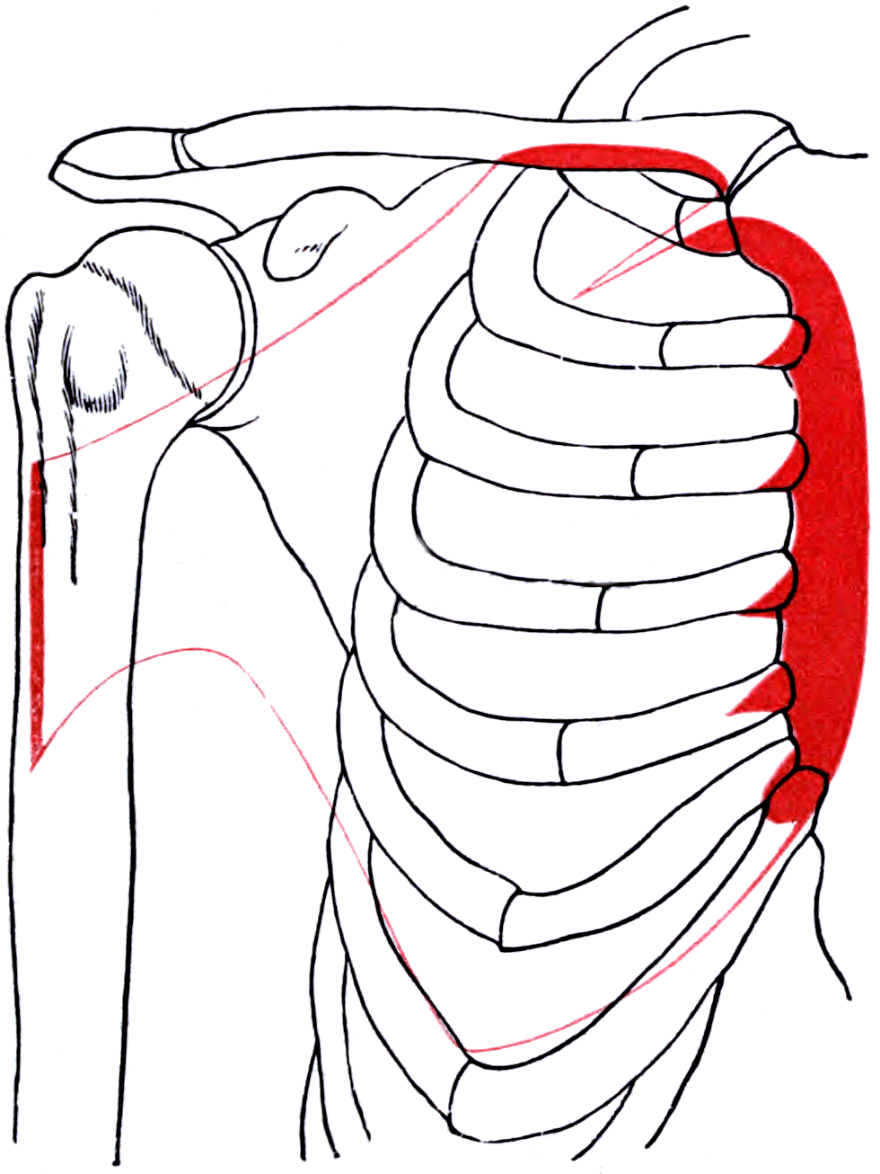
- Origin: medial half of the anterior clavicle (clavicular head); sternum, upper six costal cartilages and the external oblique aponeurosis (sternocostal head).
- Insertion: lateral lip of the intertubercular (bicipital) groove by a bilaminar tendon roughly 5 cm high and 5 mm thick.
- The tendon twists approximately 180 degrees, so the most inferior fibres insert most superiorly.
- Innervation: lateral pectoral nerve (C5, C6, C7) to the clavicular head; medial pectoral nerve (C8, T1) to the sternocostal head, piercing pectoralis minor.
- The dominant pedicle is the pectoral branch of the thoracoacromial trunk on the deep surface.
- “The inferior sternocostal fibres are maximally stretched in the abducted, externally rotated, eccentrically loaded position — the bottom of a bench press — which is why the inferior lamina tears first.
- “The medial pectoral nerve is lateral in position and pierces pectoralis minor; the lateral pectoral nerve is medial in position. The names refer to their cords of origin.
- “The 5 cm musculocutaneous safe zone is unreliable: the nerve entered coracobrachialis within 5 cm of the coracoid in a third of cadaveric specimens, and in 92 per cent once its twigs were counted.
- “Poland syndrome is the congenital absence of the sternocostal head, with ipsilateral hand hypoplasia.
Overview
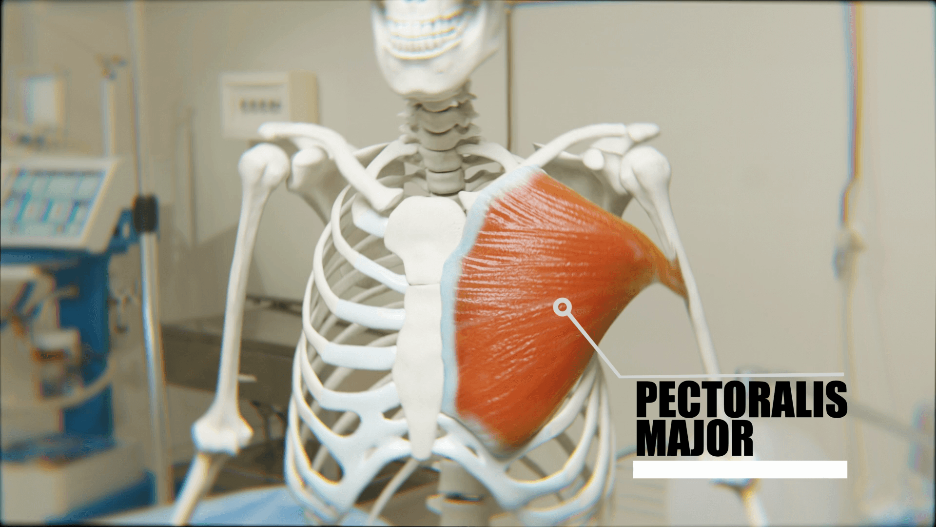
The pectoralis major is a large fan-shaped muscle covering the anterior chest wall. It arises from the clavicle, sternum, costal cartilages and external oblique aponeurosis, and converges on a short flat tendon at the lateral lip of the bicipital groove.
Why it matters surgically. Its surgical importance is concentrated in four areas:
- It forms the medial wall of the deltopectoral interval
- It ruptures in a characteristic and increasingly common pattern in weight-training athletes
- It is the standard transfer for irreparable subscapularis insufficiency
- It is the muscle congenitally absent in Poland syndrome
The concept that unlocks it. The anatomy behind all four is the same: a bilaminar tendon that twists, and it is the single anatomical concept the examiners are testing. Knowing which fibres end up where at the insertion explains why the inferior lamina fails first, why a detached sternal lamina can hide behind an intact clavicular head on examination, why the sternal head is chosen for transfer, and why the repair must reconstruct a footprint rather than simply reattach a stump.
Attachments and the Twisted Insertion
Origin. The clavicular head arises from the anterior surface of the medial half of the clavicle, and its fibres run downward and laterally. The sternocostal head arises from the anterior surface of the sternum down to the level of the sixth or seventh costal cartilage, from the upper six costal cartilages, and from the aponeurosis of the external oblique, which provides the abdominal fibres. Its abdominal fibres run upward and laterally, its sternal fibres horizontally.
The cleft between the heads. A small cleft is often visible between the two heads on the chest wall. It is the plane exploited when the sternal head alone is harvested for transfer.
Insertion. The tendon inserts into the lateral lip of the intertubercular (bicipital) groove of the humerus. It is flat, approximately 5 cm in vertical dimension and about 5 mm thick, and fibrous expansions blend with the deltoid insertion laterally and with the brachial fascia distally.
The twist. The tendon has two laminae. The clavicular head forms the anterior lamina and inserts more distally; the sternocostal head forms the posterior lamina and inserts more proximally. The tendon therefore twists through approximately 180 degrees, so the most inferior (abdominal) fibres of the sternocostal head insert most superiorly on the lateral lip.
Neighbours at the insertion. The insertion sits lateral to latissimus dorsi in the floor of the groove and lateral to teres major on the medial lip. The long head of biceps lies immediately deep to the pectoralis major tendon within the groove, the relationship exploited in subpectoral biceps tenodesis.
Innervation
Two nerves, one for each head. The lateral pectoral nerve (C5, C6, C7) comes from the lateral cord. It passes anterior to the axillary artery, pierces the clavipectoral fascia, and supplies the clavicular head primarily. The medial pectoral nerve (C8, T1) comes from the medial cord, pierces pectoralis minor, supplying it, and continues to supply the sternocostal head.
Named for their cords. The names describe the cord of origin, not where you find the nerve: the lateral pectoral nerve lies medially in position and the medial pectoral nerve lies laterally. "Medial pierces minor" fixes which is which.
The ansa pectoralis. A communicating loop joins the two nerves anterior to the axillary artery. Because of this loop and the overlapping territories, complete denervation of pectoralis major requires an injury proximal to both nerves. Denervation of the sternal head alone produces a characteristic loss of the inferior chest contour with preservation of the upper fibres.
Surgical consequences. Splitting the muscle between the heads during a sternal head transfer preserves the innervation of both, because each head has its own nerve. Each nerve has its own operative hazard:
- The medial pectoral nerve is at risk during pectoralis minor tenotomy, since it pierces that muscle
- The lateral pectoral nerve is encountered in the clavipectoral fascia during the deltopectoral approach and can be injured by aggressive medial retraction
Blood Supply and Flap Anatomy
The dominant pedicle. The pectoral branch of the thoracoacromial trunk enters the deep surface of the muscle. The thoracoacromial trunk arises from the second part of the axillary artery deep to pectoralis minor, and its pectoral branch descends on the deep surface roughly along a line from the acromion toward the xiphoid, just lateral to a line dropped from the mid-clavicle.
The other vessels. The lateral thoracic artery supplies the lateral and inferior part of the muscle. Internal thoracic (internal mammary) perforators through the second to fourth intercostal spaces supply the medial part segmentally, and intercostal perforators contribute inferiorly.
Flap anatomy. Pectoralis major is a Mathes-Nahai Type V muscle: one dominant pedicle, the thoracoacromial pectoral branch, with secondary segmental pedicles from the internal thoracic perforators. Like the latissimus, it can therefore be raised in two directions:
- As a pedicled myocutaneous flap on the thoracoacromial pedicle, a workhorse for head and neck reconstruction, reaching the oral cavity, pharynx and neck
- As a turnover flap on the internal thoracic perforators, used to cover sternal wounds after median sternotomy, one of the most valuable applications in cardiothoracic practice
The deltopectoral (Bakamjian) flap is a fasciocutaneous flap based on the same internal thoracic perforators over the muscle.
Relations
Superficial. Skin, subcutaneous fat and the breast lie over the muscle. It forms the bed of the breast, which is why its fascia is the plane of a subpectoral implant and the deep margin of a mastectomy.
Deep. Beneath it lie the clavipectoral fascia and pectoralis minor, the axillary vessels and brachial plexus in the axilla, and the ribs and intercostal muscles medially. The axillary vessels and plexus are at risk deep to pectoralis minor, particularly during a subcoracoid transfer.
Lateral border. The lateral border forms the anterior axillary fold, whose loss is the visible sign of rupture.
The deltopectoral groove. The interval between pectoralis major and the deltoid contains the cephalic vein, at risk in the groove, and the deltoid branch of the thoracoacromial artery.
The clavipectoral fascia. It encloses subclavius and pectoralis minor, and the structures that pierce it are a classic examination list:
- Lateral pectoral nerve
- Thoracoacromial artery
- Cephalic vein
- Lymphatics
Action and Biomechanics
- Primary actions
- Forward flexion of the humerus
- Secondary actions
- Horizontal adduction, internal rotation, assists adduction
- Primary actions
- Adduction and internal rotation
- Secondary actions
- Extension of the flexed humerus back to the side, depression of the shoulder girdle
- Primary actions
- Powerful adduction and depression
- Secondary actions
- The fibres most stretched in the abducted externally rotated position
The whole muscle. Adduction and internal rotation are the dominant actions, working with latissimus dorsi, teres major and subscapularis. With the humerus fixed it depresses the shoulder girdle, contributing to trunk elevation in climbing and to crutch walking. With the arms fixed it elevates the ribs as an accessory muscle of respiration, the reason a breathless patient braces the arms on a table.
Two heads that oppose each other. From a position of full flexion the sternocostal head extends the arm back to the side, while the clavicular head flexes it. The two heads therefore oppose one another through part of the arc, which is why they can be separately tested.
Synergists and antagonists. Besides the adductors and internal rotators above, anterior deltoid shares flexion and horizontal adduction, and coracobrachialis is a synergist. The antagonists are deltoid and supraspinatus for abduction, infraspinatus and teres minor for external rotation, and trapezius and serratus anterior for girdle elevation.
Where the strain falls. Peak tendon strain occurs in the abducted, extended and externally rotated position under eccentric load. The descent of a bench press reproduces this exactly, which is why bench pressing accounts for the majority of ruptures. The fibres under the greatest stretch are the inferiormost sternocostal fibres, because their spiral has placed them at the top of the insertion and given them the longest excursion. Load is not distributed evenly across the tendon; it concentrates on the inferior lamina, which tears from inferior to superior at the tendon-bone junction.
What is lost. Loss of the pectoralis major reduces adduction and internal rotation power by roughly a quarter to a half in objective testing, with the greater deficits at higher speeds and in the horizontal adduction vector.
Surface Anatomy and Examination
The anterior axillary fold. The fold is the lateral border of pectoralis major and the structure to inspect and palpate. Ask the patient to press the palms together in front of the chest, or to push the hands into the hips, and compare the two folds.
Inspect in three positions. Look from the front with the arms at the side, then with the arms elevated, and then during resisted adduction. A partial sternal head rupture may be visible only in one of these positions.
The two signs together. Look for the medially retracted muscle bulge and the thinning of the axillary fold. The two together are more reliable than either alone.
- How to perform
- Compare fold contour with the arms at the side and during resisted adduction
- Positive finding
- Loss of the normal fold with a step or hollow
- What it means
- Tendon avulsion or complete rupture
- False positives and pitfalls
- Obesity and swelling obscure the sign acutely; re-examine at two weeks
- How to perform
- Hands into the hips, press inward while palpating the fold
- Positive finding
- Weakness and a palpable defect; the muscle bunches medially
- What it means
- Loss of tendon continuity
- False positives and pitfalls
- Pain inhibition acutely; compare with the opposite side
- How to perform
- Resist forward flexion at 90 degrees
- Positive finding
- Weakness
- What it means
- Clavicular head or lateral pectoral nerve involvement
- False positives and pitfalls
- Anterior deltoid substitutes; palpate the head directly
- How to perform
- Resist extension of the flexed arm back toward the side, and resisted adduction from abduction
- Positive finding
- Weakness
- What it means
- Sternocostal head involvement, the head that usually ruptures
- False positives and pitfalls
- Latissimus dorsi and teres major substitute for adduction
- How to perform
- Inspect the medial arm and chest wall at 3 to 7 days
- Positive finding
- Extensive ecchymosis tracking down the medial arm
- What it means
- Supports an acute tendon rupture
- False positives and pitfalls
- Absent in muscle belly strains and in some chronic tears
- How to perform
- Standard subscapularis tests
- Positive finding
- Weakness or lag
- What it means
- Subscapularis insufficiency, the indication for a pectoralis major transfer
- False positives and pitfalls
- Stiffness confounds the lift-off test; use belly-press if the hand cannot reach the back
Because the tendon is bilaminar and fails from inferior to superior, an intact clavicular head can preserve a reasonable anterior axillary fold and much of the flexion strength while the entire sternal lamina is detached. Two habits prevent the miss: test the heads separately, and re-examine at two weeks once swelling and pain inhibition have settled, when the contour asymmetry becomes obvious.
Pectoralis Major Rupture
Who. Men aged roughly 20 to 40, weight training, injured most often during the eccentric (descent) phase of the bench press. Anabolic steroid use is a recognised association, and the injury also occurs in wrestling, rugby and water sports.
Where it fails. Most commonly at the tendon or tendon-bone junction, less often at the musculotendinous junction, and rarely at the muscle belly or the origin. Bony avulsion is uncommon.
Presentation. An audible or felt tearing sensation and immediate pain, then ecchymosis tracking down the medial arm over the following days, loss of the anterior axillary fold and a medially retracted muscle bulge.
Imaging. MRI is the standard, and imaging the arm in abduction and external rotation improves visualisation of a retracted lamina. Ultrasound is a reasonable alternative in experienced hands.
Treatment. In an active patient, surgical repair gives better strength and functional outcomes than non-operative treatment, and acute repair, generally within six weeks, outperforms delayed repair. Non-operative treatment is reasonable for low-demand patients, muscle belly tears and partial tears with preserved strength.
Exposure. A deltopectoral incision, or a limited anterior axillary incision along the fold, gives direct access to the lateral lip footprint and to the retracted stump. The retracted tendon lies medially, often adherent within a haematoma, and the intact clavicular lamina guides you to the sternal lamina. The two laminae must be identified separately to restore anatomy.
The footprint. Decorticate the lateral lip of the bicipital groove lightly to bleeding bone over the 5 cm footprint, which restores a broad tendon-bone contact area rather than a point of fixation. Unicortical buttons, suture anchors, or a bone trough with transosseous sutures all perform acceptably, and buttons and anchors avoid a large cortical defect. Avoid large bicortical drill holes at this level (see Complications).
Restoring the twist. The retracted inferior lamina belongs at the superior part of the footprint, not simply wherever it most easily reaches. Reducing it there restores the spiral anatomically; otherwise the repair is tensioned incorrectly.
Chronic tears. Retraction and scarring prevent direct reattachment beyond a few months, and a chronic tear may require allograft or autograft interposition (Achilles, hamstring, fascia lata).
Rehabilitation. The protocol protects the repair through the position of maximum strain:
- Sling with limited external rotation for about six weeks
- Then progressive motion
- Resisted work at three months
- Full return at four to six months
Poland Syndrome and Other Conditions
Poland syndrome. A congenital, almost always unilateral and sporadic condition, classically defined by the absence of the sternocostal head of pectoralis major with ipsilateral hand hypoplasia. The anterior axillary fold is absent from birth.
The full spectrum. Absence of the sternocostal head is the defining feature, and the rest varies:
- Often absence of pectoralis minor
- Hypoplasia or absence of the breast and nipple
- Deficiency of subcutaneous tissue
- Rib and costal cartilage deficiency
- Ipsilateral symbrachydactyly, with short webbed digits and hypoplastic middle phalanges
Epidemiology and cause. It is uncommon, with a reported incidence in the order of one in 20,000 to 30,000 births, more common in males, and right-sided in the majority of cases. The prevailing theory is an interruption of the embryonic subclavian artery blood supply during the sixth week of gestation, the subclavian artery supply disruption sequence, which accounts for the unilateral and variable presentation.
The hand. The hand is what the orthopaedic surgeon usually treats. Symbrachydactyly management ranges from observation to syndactyly release, distraction lengthening, and toe-to-hand transfer for severe deficiency.
The chest wall. Reconstruction is usually undertaken for contour and, in the presence of significant rib deficiency, for protection of the underlying viscera, using latissimus dorsi transfer, custom implants and fat grafting. The latissimus is the usual donor, and the surgeon must confirm it is present, because it is occasionally deficient in the same patient.
Function. Despite the missing muscle, functional deficit is usually modest, because latissimus dorsi, teres major and subscapularis duplicate its actions.
Other conditions involving the muscle are compared below.
- Presentation
- Restricted abduction and external rotation with a taut anterior band
- Key finding
- Clinical diagnosis
- Management
- Release, Z-plasty or flap reconstruction
- Presentation
- Dehiscence and infection after cardiac surgery
- Key finding
- Exposed sternum
- Management
- Pectoralis major turnover flap on internal thoracic perforators
- Presentation
- Incidental palpable parasternal mass or an unexpected finding on mammography
- Key finding
- A longitudinal muscle slip anterior to the sternal origin of pectoralis major
- Management
- Recognition only; no treatment required
Transfer for Subscapularis Insufficiency
Indication. An irreparable subscapularis tear with functional deficit in a patient too young for arthroplasty. The transfer restores the anterior arm of the transverse force couple.
Why the sternal head. The subscapularis pulls from the anterior scapula, medially and slightly inferiorly, to the lesser tuberosity, and it both internally rotates and compresses the head. The sternal head of pectoralis major originates low and medially on the chest wall, so its line of pull is the closest available approximation to that vector. The clavicular head pulls from above and in front, producing flexion rather than the required internal rotation and compression, and it lies too superficial to be routed correctly.
The donor. The sternal head is split from the clavicular head, or the upper two thirds of the sternal head is taken.
The limitation. Even the sternal head pulls from anterior to the chest wall, whereas the subscapularis pulls from behind the coracoid, on the anterior scapula, so no pectoralis transfer truly reproduces the vector. That is the rationale for routing the transfer beneath the conjoined tendon, and for the alternative anterior latissimus dorsi transfer, which comes from genuinely behind the chest wall and reproduces the subscapularis vector more faithfully than any pectoralis route.
The route. The choice trades vector against the musculocutaneous nerve:
- The subcoracoid route (Resch) passes the transfer deep to the conjoined tendon, so that it turns the corner at the coracoid and approaches the lesser tuberosity from a more medial direction. It is the best available reproduction of the subscapularis vector, but it places the musculocutaneous nerve directly at risk.
- The superficial route (Wirth and Rockwood) passes it superficial to the conjoined tendon. It is technically simpler and avoids the nerve, but the vector is less anatomic.
Fixation and rehabilitation. The tendon is fixed to the lesser tuberosity with anchors or through a bone trough, restoring a footprint rather than a point. A sling in internal rotation, with external rotation restricted for about six weeks, then progressive motion and motor re-education, protects the transfer through the position that would stretch it.
Prognosis. Pain relief and improvement in activities of daily living are reliable, but results are markedly worse if the supraspinatus is also irreparable. Jost, Gerber and colleagues showed a large difference in Constant score between isolated and combined irreparable tears.
The musculocutaneous nerve arises from the lateral cord and enters coracobrachialis a mean of roughly 5 cm distal to the tip of the coracoid, but the mean is the least useful number here. In Singh and colleagues' in situ cadaveric study the main trunk entered anywhere from 17 to 72 mm from the coracoid, and the most proximal motor twigs entered a mean of 34 mm and as close as 22 mm. The nerve entered within 5 cm of the coracoid in a third of specimens, and in 92 per cent once twigs were counted, so the conventional 5 cm safe zone is not reliable, and a 3 cm working limit is the safer teaching. The nerve is also tethered proximally, so traction rather than laceration is the usual injury.
- Identify the nerve on the deep surface of the conjoined tendon before creating any tunnel beneath it.
- Create the tunnel bluntly with a finger and confirm it admits the tendon freely before pulling anything through. A transfer forced through a tight subcoracoid window compresses the nerve permanently.
- Do not retract the conjoined tendon forcefully medially; retract gently and intermittently. Forceful medial retraction is the mechanism of musculocutaneous palsy in the deltopectoral approach generally, including in the Latarjet procedure.
- If the anatomy is unfavourable or the window tight, use the superficial route and accept a less anatomic vector. A functioning transfer with a suboptimal vector is a far better outcome than an anatomic transfer with a paralysed biceps and a numb forearm.
- 1Confirm the deficit is structuralWeak internal rotation, positive belly-press and lift-off tests, increased passive external rotation compared with the other side, and Goutallier 3 to 4 subscapularis on the sagittal MRI, with retraction to the glenoid.
- 2Assess the rest of the cuffAn irreparable supraspinatus alongside an irreparable subscapularis predicts a substantially worse outcome from any transfer, and pushes the decision toward arthroplasty.
- 3Assess age, arthritis and escapeAnterosuperior escape and established arthritis in an older patient point to reverse arthroplasty. A younger patient with a preserved joint is a transfer candidate.
- 4Choose the donor and routeSternal head of pectoralis major, subcoracoid route where the musculocutaneous nerve anatomy permits, superficial route otherwise; anterior latissimus dorsi transfer where the more faithful vector is preferred.
Approaches and Landmarks
The deltopectoral interval. Pectoralis major (medial and lateral pectoral nerves) medially and deltoid (axillary nerve) laterally form a true internervous plane, the workhorse anterior approach to the shoulder. The incision runs from just lateral to the coracoid toward the deltoid insertion.
The cephalic vein. It marks the interval and is usually retracted laterally with the deltoid, since most of its tributaries come from the deltoid side.
Going deeper. The clavipectoral fascia is incised lateral to the conjoined tendon, and the conjoined tendon is retracted medially, gently, to protect the musculocutaneous nerve. The upper 1 to 2 cm of the pectoralis major insertion may be released to improve exposure of the humeral shaft and is repaired at closure. The three sisters, the anterior circumflex humeral vessels, mark the inferior border of subscapularis and are ligated in an extensile exposure.
Subpectoral biceps tenodesis. The inferior border of pectoralis major is the landmark, and the tenodesis is performed just proximal to it in the bicipital groove, where the biceps tendon lies deep to the pectoralis tendon. Cadaveric work places the musculocutaneous nerve roughly 3 cm medial to the tenodesis site and the radial nerve a similar distance distal and posterior, and the axillary nerve lies proximally. Use unicortical fixation or a small-diameter implant, for the fracture risk described under Complications.
Complications
Musculocutaneous nerve injury. The mechanism is a transfer forced through a tight subcoracoid tunnel, forceful medial retraction of the conjoined tendon, or dissection deep to the coracoid without identifying the nerve. The cost is loss of elbow flexion power and numbness over the lateral forearm, a poor exchange for improved internal rotation.
Failure of a rupture repair. Re-rupture is uncommon after acute anatomic repair but rises steeply in chronic tears requiring graft reconstruction and in patients who return to heavy bench pressing too early. Fixing the tendon as a point, instead of restoring the 5 cm footprint, gives weaker fixation and poorer strength recovery.
Humeral shaft fracture. Large bicortical drill holes at the level of the pectoralis insertion create a stress riser, and fracture through them is a recognised complication, either at rupture repair or at subpectoral biceps tenodesis. Avoid it with unicortical fixation, small-diameter implants, and by not placing multiple closely spaced holes.
Denervation of the muscle. Because of the ansa pectoralis and the separate supply to each head, partial denervation is more common than complete, producing a segmental contour defect.
After a transfer. Internal rotation power is not always restored, because the pectoralis vector is a compromise; patients should be counselled that pain relief and improvement in activities of daily living are more reliable than restoration of power. Anterior overtightening limits external rotation, and re-education can fail, since the patient must learn to fire the transfer in a new role.
Flap complications. The flap can bring bulk and tethering to a head and neck reconstruction, and a visible chest wall contour defect and loss of the anterior axillary fold, in women in particular. Distal flap necrosis is the risk where the skin paddle is placed beyond the reliable territory of the thoracoacromial pedicle, and once the muscle has been used it is lost as a future donor.
Guidelines, Registries & Global Practice
Anatomical variation across populations
- The sternalis muscle, a longitudinal slip lying anterior to the sternal origin of pectoralis major, is present in a minority of individuals with reported prevalence differences across populations and between the sexes. It is clinically irrelevant except as a cause of confusion on mammography and as an unexpected finding at surgery.
- The abdominal head derived from the external oblique aponeurosis is variably developed, which affects the amount of low-origin muscle available for transfer.
- The entry point of the musculocutaneous nerve into coracobrachialis is genuinely variable, and a proportion of specimens have proximal branches close to the coracoid. This variation, not a measurement error, is why the safe zone is a guide rather than a rule.
- Poland syndrome occurs sporadically worldwide with a reported incidence in the order of one in 20,000 to 30,000 births and a consistent right-sided and male predominance across reported series.
Side-by-side guidance
- Position Relevant to Pectoralis Major
- Supports surgical repair of complete pectoralis major tendon ruptures in active patients, and recognises tendon transfer for irreparable subscapularis insufficiency in younger patients.
- Position Relevant to Pectoralis Major
- Recommend early specialist referral for suspected pectoralis major rupture so that repair can occur within the acute window, and structured non-operative care for muscle belly injuries.
- Position Relevant to Pectoralis Major
- Emphasises the deltopectoral internervous plane, gentle retraction of the conjoined tendon to protect the musculocutaneous nerve, and avoidance of large bicortical drill holes at the pectoralis insertion.
- Position Relevant to Pectoralis Major
- Treat the pedicled pectoralis major myocutaneous flap as a reliable workhorse where free tissue transfer is unavailable or has failed, and the turnover flap as standard for sternal wound coverage.
Registry and outcome signals
- Pectoralis major rupture repair is not captured by joint registries. The evidence base is pooled case series and systematic reviews, and the consistent signal across them is that operative treatment outperforms non-operative treatment in active patients and that acute repair outperforms delayed repair.
- Reported incidence of rupture has risen over recent decades in parallel with recreational and competitive weight training, and this is a consistent international trend rather than a local one.
- Tendon transfers for subscapularis insufficiency are low-volume procedures without registry coverage; institutional series consistently show reliable pain relief, variable strength recovery, and a strong negative effect of concomitant irreparable supraspinatus disease.
- Reverse arthroplasty volumes continue to grow in every national registry, and in older patients a reverse has largely displaced transfer for anterosuperior cuff insufficiency.
High-resource versus limited-resource practice
- Well-resourced settings: MRI in the abducted externally rotated position for rupture characterisation, suture button and anchor fixation, arthroscopically assisted transfer, and free tissue transfer as the reconstructive first choice.
- Limited-resource settings: transosseous bone trough repair of a ruptured tendon achieves comparable results with no implant cost, and the pedicled pectoralis major flap remains one of the most valuable reconstructive operations available anywhere, requiring no microsurgery.
- The universally applicable clinical skill is separate testing of the two heads, which costs nothing and prevents the commonest diagnostic error — missing a sternal lamina rupture behind an intact clavicular head.
MCQ Practice Points
Q: Which head forms the anterior lamina of the pectoralis major tendon, and which inserts more proximally? A: The clavicular head forms the anterior lamina and inserts more distally; the sternocostal head forms the posterior lamina and inserts more proximally, with its lowest fibres reaching highest.
Q: Why does the inferior lamina rupture first? A: The tendon twists 180 degrees, so the inferior sternocostal fibres insert most superiorly and have the longest excursion. In abduction, extension and external rotation under eccentric load they are maximally stretched, and the tear propagates inferior to superior.
Q: Which nerve supplies each head of pectoralis major? A: Lateral pectoral nerve (C5, C6, C7) from the lateral cord to the clavicular head; medial pectoral nerve (C8, T1) from the medial cord to the sternocostal head. The medial pectoral nerve pierces pectoralis minor.
Q: Which four structures pierce the clavipectoral fascia? A: The lateral pectoral nerve, the thoracoacromial artery, the cephalic vein and lymphatics.
Q: How far distal to the coracoid does the musculocutaneous nerve enter coracobrachialis? A: The main trunk enters a mean of roughly 5 cm distal to the coracoid, but the range runs from 17 to 72 mm and the most proximal twigs enter as close as 22 mm. The nerve was within 5 cm in a third of cadaveric specimens and in 92 per cent once twigs were counted, so the 5 cm safe zone is unreliable — work within 3 cm and identify the nerve.
Q: Which head of pectoralis major is used for transfer in subscapularis insufficiency and why? A: The sternal head, because its low medial origin gives the closest available approximation to the subscapularis vector. The clavicular head would produce flexion instead.
Q: What Mathes-Nahai type is the pectoralis major and what does it permit? A: Type V — one dominant pedicle (pectoral branch of the thoracoacromial trunk) plus secondary segmental pedicles (internal thoracic perforators). It permits both a conventional flap and a turnover flap for sternal wound coverage.
Q: What defines Poland syndrome? A: Congenital absence of the sternocostal head of pectoralis major with ipsilateral hand hypoplasia, typically symbrachydactyly. Unilateral, sporadic, more common in males and usually right-sided.
Q: What is the accepted threshold between an acute and a chronic pectoralis major rupture, and why does it matter? A: Approximately six weeks. Acute repair outperforms delayed repair, and beyond this window retraction and scarring may require graft reconstruction rather than direct reattachment.
Q: Which structure lies immediately deep to the pectoralis major tendon at the bicipital groove? A: The long head of biceps tendon. The inferior border of pectoralis major is therefore the landmark for subpectoral biceps tenodesis, where large bicortical drill holes risk humeral shaft fracture.
Exam Viva Scenarios
Practise clinical reasoning and management decisions out loud
“A 31-year-old man felt a tearing sensation in his right chest while lowering a heavy bench press bar four days ago. He has ecchymosis down the medial arm and a normal-looking anterior axillary fold at rest. How do you assess and manage him?”
“A 52-year-old man has an irreparable subscapularis tear, Goutallier 4, with a positive belly-press test and increased passive external rotation, but a well-preserved supraspinatus and no arthritis. You plan a pectoralis major transfer. Which part do you take, how do you route it, and what will you not do?”
“A 6-year-old is referred with a hypoplastic right hand. On examination the right anterior axillary fold is absent and the nipple is higher and smaller than the left. What is the diagnosis and what else do you examine and plan?”
Attachments
- Clavicular head: medial half of the anterior clavicle
- Sternocostal head: sternum, upper six costal cartilages, external oblique aponeurosis
- Insertion: lateral lip of the bicipital groove, about 5 cm high, 5 mm thick
- Clavicular head is the anterior lamina, inserts distally
- Sternocostal head is the posterior lamina, inserts proximally
- Tendon twists about 180 degrees
Nerves and Vessels
- Lateral pectoral nerve C5-C7, lateral cord, clavicular head
- Medial pectoral nerve C8-T1, medial cord, pierces pectoralis minor, sternocostal head
- Ansa pectoralis connects the two
- Dominant pedicle: pectoral branch of the thoracoacromial trunk
- Secondary: internal thoracic perforators, lateral thoracic artery
- Mathes-Nahai Type V
Rupture
- Bench press eccentric descent, abduction, extension, external rotation
- Inferior lamina fails first, tears inferior to superior
- Commonest site is the tendon or tendon-bone junction
- ElMaraghy and Devereaux: timing, extent, anatomic site
- Acute repair within six weeks outperforms delayed repair
- Restore the full footprint and the tendon twist
Transfer
- Sternal head only, split preserves both nerves
- Subcoracoid route (Resch) is more anatomic, superficial route (Wirth and Rockwood) is safer
- Musculocutaneous 5 cm safe zone unreliable; twigs as close as 22 mm, work within 3 cm
- Poor results with a concomitant irreparable supraspinatus
- Anterior latissimus transfer reproduces the vector better
Other
- Deltopectoral internervous plane: pectoral nerves versus axillary nerve
- Clavipectoral fascia pierced by lateral pectoral nerve, thoracoacromial artery, cephalic vein, lymphatics
- Subpectoral biceps tenodesis: avoid large bicortical drill holes
- Poland syndrome: absent sternocostal head plus ipsilateral hand hypoplasia
- Turnover flap on internal thoracic perforators for sternal wounds
Evidence Base
A Systematic Review and Comprehensive Classification of Pectoralis Major Tears
- 365 published cases of pectoralis major injury from 1822 to 2010 analysed; 75 per cent were reported in the last 20 years
- 83 per cent resulted from indirect trauma and 48 per cent occurred during weight-training activities
- Existing reports classified injuries inconsistently in timing, location and extent, particularly regarding which muscle segments of the bilaminar tendon were affected
- A three-part classification is proposed: timing (acute versus chronic), location (muscle origin or belly, at or between the musculotendinous junction and the tendinous insertion, or bony avulsion), and standardised terminology for tear extent
Rupture of the Pectoralis Major Muscle: A Meta-Analysis of 112 Cases
- 108 published cases with sufficient data plus 4 of the authors' own, giving 112 analysed cases; every reported patient was male
- Rupture occurs most commonly in sport during weight training, weight lifting or wrestling, with the arm externally rotated and abducted
- Most reported ruptures are complete and located at the insertion to the humerus; work-related injuries occur more often at the musculotendinous junction
- Prognosis was related neither to patient age nor to the location of the rupture; surgical treatment, preferably within the first 8 weeks, gave better results
Outcome of Pectoralis Major Transfer for the Treatment of Irreparable Subscapularis Tears
- 30 consecutive pectoralis major transfers in 28 patients, mean age 53, for irreparable subscapularis tears (12 isolated, 18 with an associated supraspinatus or supraspinatus and infraspinatus tear)
- Mean relative Constant score improved from 47 to 70 per cent at a mean of 32 months
- Constant subscores for pain and activities of daily living, forward flexion and abduction strength all improved; subjective shoulder value rose from 23 to 55 per cent
- THE SELECTION RULE, WITH ITS NUMBER: where the supraspinatus was also irreparable the mean relative Constant score was 49 per cent, against 79 per cent where it was reparable (p = 0.002)
- The authors conclude that with an associated IRREPARABLE supraspinatus tear, pectoralis major transfer MAY NOT BE WARRANTED
Transfer of the Pectoralis Major Muscle for the Treatment of Irreparable Rupture of the Subscapularis Tendon
- 12 patients with irreparable subscapularis tears; the superior one-half to two-thirds of the pectoralis major tendon was used as a substitute
- To match the orientation of the transfer to that of the subscapularis it was routed BEHIND the conjoined tendon of coracobrachialis and the short head of biceps, to the lesser tuberosity
- At a mean of 28 months, 9 of 12 rated the result excellent or good, 3 fair and none poor; Constant score rose from 26.9 to 67.1 per cent of normal
- All four shoulders that were unstable pre-operatively were stable at final follow-up
Classification of the Vascular Anatomy of Muscles: Experimental and Clinical Correlation
- Describes five patterns of muscle circulation derived from studies of muscle vascular anatomy
- Validated against arc of rotation, skin territory, distally based flaps, microvascular transplantation and muscle-delay models
- Designed to assist the surgeon in both the choice and the design of muscle and musculocutaneous flaps
- Within this system pectoralis major is a Type V muscle — one dominant pedicle (the pectoral branch of the thoracoacromial trunk) plus secondary segmental pedicles from the internal thoracic perforators
Relationship of the Musculocutaneous Nerve and Its Twigs to the Coracoid Process: An Operative Exposure
- Twenty-four fresh-frozen cadaveric shoulders dissected after coracoid osteotomy, measuring the nerve and its twigs in situ
- The main trunk entered coracobrachialis a mean of 51.1 mm from the coracoid, but with a range from 16.7 to 71.9 mm
- The most proximal twigs entered a mean of 33.5 mm from the coracoid, as close as 21.9 mm; twigs were present in 70.8 per cent of specimens
- The nerve entered within 5 cm of the coracoid in 33.3 per cent of specimens, rising to 91.7 per cent once twigs were counted