The intermediate-grade, cartilage-forming surface osteosarcoma of the diaphysis
- Periosteal osteosarcoma is a chondroblastic osteosarcoma that arises on the surface of the DIAPHYSIS and sits on a thickened, saucerised cortex with perpendicular periosteal spiculation and Codman triangles at the margins.
- The medullary canal is almost never truly invaded. Convincing marrow tumour on MRI should force reconsideration of high-grade surface osteosarcoma or a dedifferentiated lesion.
- Matrix is predominantly cartilage and largely unmineralised — low attenuation on CT, lobulated bright T2 on MRI. It can be mistaken for periosteal chondroma or chondrosarcoma.
- Diagnosis rests on lace-like tumour osteoid produced directly by malignant spindled cells at the periphery of atypical cartilage lobules — a chondrosarcoma cannot do this.
- Staging is whole-bone MRI (skip lesions, true marrow status) plus CT chest. Bone scan or PET completes systemic survey.
- Treatment is wide en-bloc resection, usually diaphyseal intercalary resection with allograft, vascularised fibula, or the combined Capanna construct.
- Adequacy of surgical margin is the dominant prognostic variable; intralesional or marginal excision drives local recurrence and downstream mortality.
- “Diaphysis plus hair-on-end spiculation plus unmineralised chondroid mass equals periosteal osteosarcoma until proven otherwise.
- “Parosteal is metaphyseal, dense and low grade; periosteal is diaphyseal, lucent and intermediate grade — the names are counterintuitive, learn them as a pair.
- “Never call it chondrosarcoma without excluding lace-like osteoid; in a teenage diaphysis, surface chondrosarcoma is essentially non-existent.
- “Biopsy the soft-tissue component in line with the definitive incision, planned by the resecting surgeon.
- “Chemotherapy has never shown a clear survival benefit in retrospective periosteal series but is frequently used in young, fit patients.
A predominantly chondroid surface lesion in a teenage diaphysis is periosteal osteosarcoma, not periosteal chondroma or chondrosarcoma. Underdiagnosis leads to marginal excision and local recurrence. Insist on adequate tissue and specialist bone pathology review.
True medullary invasion is exceptional. If MRI shows unequivocal marrow tumour (not just reactive oedema), reclassify toward high-grade surface osteosarcoma and treat with neoadjuvant chemotherapy plus resection.
A badly placed diaphyseal biopsy contaminates compartments and can convert an intercalary resection into an amputation or a joint sacrifice. Biopsy in the referral sarcoma centre, along the planned approach, single tract, no transverse incisions.
Local recurrence after inadequate margin correlates strongly with metastasis and death. Plan the resection on MRI with the periosteal reaction and reactive rim included, and take an uninvolved cuff of muscle over the tumour surface.
Definition, Biology and Where It Sits
Periosteal osteosarcoma is a malignant bone-forming tumour arising from the deep layer of the periosteum, growing outward on the cortical surface, with a predominantly chondroblastic phenotype and intermediate (grade 2) histological grade. It is the middle child of the surface osteosarcoma family.
- Cell of origin: cambium layer of periosteum; the tumour therefore sits on, and erodes into, the outer cortex rather than arising within the medulla.
- Site: diaphysis or metadiaphysis of femur (approximately half) and tibia (approximately one third); humerus, ulna, ribs and jaw are described. Circumferential involvement is uncommon — the lesion typically occupies one aspect of the shaft.
- Age: peak in the second and third decades, essentially overlapping conventional osteosarcoma, and a decade younger than parosteal.
- Genetics: unlike parosteal osteosarcoma, periosteal lesions do not show consistent MDM2/CDK4 amplification. A negative MDM2 FISH therefore supports periosteal over parosteal when the imaging is ambiguous, but does not exclude conventional osteosarcoma.
- Behaviour: metastasises to lung in a minority; regional nodal spread is not a feature. Local behaviour is dominated by the soft-tissue component.
Pathology in Plain Terms
a lobulated, glistening, blue-grey cartilaginous mass sitting on the cortex, with gritty bone spicules extending into it from the cortical surface and a firm reactive base. The medulla, when sectioned, is normal.
- Lobules of atypical cartilage forming most of the tumour volume — hypercellular hyaline cartilage with binucleate cells and nuclear enlargement, corresponding to the low-density areas on CT.
- Peripheral spindling — at the edge of each cartilage lobule the cells become spindled and more atypical, blending into a sarcomatous stroma. This is where the diagnosis is made.
- Fine, lace-like tumour osteoid deposited directly by those malignant spindled cells between the lobules and along the spicules. Osteoid produced by tumour cells (not entrapped reactive bone) is what makes this an osteosarcoma rather than a chondrosarcoma.
intermediate (grade 2 of 3). Mitoses present but not frenzied; necrosis limited.
no specific marker. MDM2 and CDK4 amplification absent — useful against parosteal osteosarcoma and against dedifferentiated parosteal lesions. IDH1/IDH2 mutation is a feature of conventional cartilage tumours and its absence supports osteosarcoma in equivocal chondroid lesions.
"Malignant cartilage with peripheral spindling that makes lace-like osteoid" — that sentence separates periosteal osteosarcoma from every cartilage tumour on the differential. Reactive periosteal bone does not have malignant osteoblasts sitting in its lacunae.
Differentiating the Surface Osteosarcomas
- Parosteal
- Low grade (1)
- Periosteal
- Intermediate grade (2)
- High-grade surface
- High grade (3)
- Parosteal
- Third to fourth decade
- Periosteal
- Second to third decade
- High-grade surface
- Second to third decade
- Parosteal
- Posterior distal femoral metaphysis
- Periosteal
- Femoral or tibial diaphysis
- High-grade surface
- Femoral diaphysis, any surface
- Parosteal
- Dense ossified lobulated 'cauliflower' mass with radiolucent string sign cleft
- Periosteal
- Broad-based surface mass, perpendicular hair-on-end spicules, Codman triangles, saucerised sclerotic cortex
- High-grade surface
- Bulky mass, variable mineralisation, aggressive cortical destruction
- Parosteal
- Heavily ossified
- Periosteal
- Predominantly unmineralised chondroid matrix
- High-grade surface
- Osteoid, variably mineralised
- Parosteal
- Well-formed parallel bony trabeculae in bland spindle stroma
- Periosteal
- Lobules of intermediate-grade cartilage with peripheral osteoid
- High-grade surface
- Frankly high-grade sarcoma, as conventional osteosarcoma
- Parosteal
- Late, in about 20% — confers worse prognosis
- Periosteal
- Essentially absent; canal intact — invasion should prompt rethinking the diagnosis
- High-grade surface
- Common
- Parosteal
- MDM2 and CDK4 amplified
- Periosteal
- MDM2 negative
- High-grade surface
- MDM2 negative
- Parosteal
- Wide resection alone; chemotherapy not indicated
- Periosteal
- Wide resection; chemotherapy debated, often given to young patients
- High-grade surface
- Wide resection plus chemotherapy, as conventional osteosarcoma
- Parosteal
- Under 10%
- Periosteal
- 10 to 15%
- High-grade surface
- Comparable to conventional osteosarcoma
- Parosteal
- Greater than 90%
- Periosteal
- 85 to 90% with clear margins
- High-grade surface
- Approximately 50 to 70%
- Parosteal
- Low (1)
- Periosteal
- Intermediate (2)
- High-grade surface
- High (3)
- Parosteal
- Third to fourth decade
- Periosteal
- Second to third decade
- High-grade surface
- Second to third decade
- Parosteal
- Posterior distal femoral metaphysis
- Periosteal
- Femoral or tibial diaphysis
- High-grade surface
- Femoral diaphysis, any surface
- Parosteal
- Dense ossified lobulated mass, string sign
- Periosteal
- Broad-based mass, hair-on-end spicules, Codman triangles
- High-grade surface
- Bulky mass, variable mineralisation, aggressive destruction
- Parosteal
- Heavily ossified
- Periosteal
- Predominantly unmineralised cartilage
- High-grade surface
- Osteoid, variably mineralised
- Parosteal
- Late, in about 20% (worse prognosis)
- Periosteal
- Essentially absent — invasion rethinks the diagnosis
- High-grade surface
- Common
- Parosteal
- MDM2 / CDK4 amplified
- Periosteal
- MDM2 negative
- High-grade surface
- MDM2 negative
- Parosteal
- Not indicated
- Periosteal
- Debated; often given to young patients
- High-grade surface
- Indicated, as conventional osteosarcoma
- Parosteal
- Under 10%
- Periosteal
- 10 to 15%
- High-grade surface
- Comparable to conventional osteosarcoma
- Parosteal
- Greater than 90%
- Periosteal
- 85 to 90% with clear margins
- High-grade surface
- Approximately 50 to 70%
PARA-META / PERI-DIATelling the two apart
Hook:The denser the tumour looks, the lower the grade — parosteal is the most ossified and the most benign-behaving of the three.
SCRAMRadiographic features of periosteal osteosarcoma
Hook:SCRAM the diaphysis — the tumour scrapes the surface but never gets inside.
Staging and Multidisciplinary Decision-Making
- 1Step 1 — Presentation
Diaphyseal surface mass on a long bone, broad-based against cortex with a soft-tissue component.
Local extent and metastatic status defined ahead of biopsy.
- 2Step 2 — The marrow rule
Distinguish confluent T1 marrow replacement from reactive marrow oedema alone.
Reactive oedema alone supports a surface lesion; confluent T1 replacement means medullary involvement and a conventional high-grade osteosarcoma with surface extension until proven otherwise.
- 3Step 3 — Biopsy
Tissue must sample the deep, less ossified periphery, not the mature ossified base.
Lace-like chondroid matrix with intervening malignant osteoid confirms periosteal osteosarcoma (intermediate to high grade).
- 4Step 4 — Staging assignment
Extraosseous soft-tissue mass with no metastasis.
State explicitly in a viva that Enneking's binary grading forces this intermediate lesion into the high-grade box.
- 5Step 5 — MDT items to settle before theatre
Grade confirmation on adequate tissue; true marrow status; osteotomy levels; reconstruction strategy.
A single agreed plan covering margin, reconstruction and systemic therapy.
- 6Step 6 — Fertility and growth
Chemotherapy planned in an adolescent, or reconstruction in a skeletally immature patient.
Irreversible harms avoided before treatment begins.
- 7Step 7 — Resection and surveillance
Wide en bloc excision with the biopsy tract, osteotomies planned from MRI marrow extent.
Clear margins give excellent prognosis; marrow-positive or intralesional disease shifts management towards conventional high-grade osteosarcoma protocols and closer surveillance.
- Enneking (MSTS) surgical staging: periosteal osteosarcoma with an extraosseous soft-tissue mass and no metastasis is typically Stage IIB (high grade by Enneking's two-tier system, extracompartmental, M0). Enneking's binary grading forces intermediate lesions into the high-grade box — say this explicitly in a viva rather than pretending the system is finer than it is.
- AJCC: T-stage by size (8 cm threshold), G by grade, M by site of metastasis (lung versus other).
- MDT decisions to settle before theatre: grade confirmation on adequate tissue; true marrow status; osteotomy levels; reconstruction strategy; whether chemotherapy will be offered and, if so, whether neoadjuvantly (which allows assessment of necrosis) or adjuvantly.
- Fertility and growth: if chemotherapy is planned in an adolescent, arrange fertility counselling and gamete/ovarian tissue preservation before the first cycle. Consider remaining growth in the reconstruction plan for skeletally immature patients.
Clinical Presentation and Examination
- Duration: typically several months (commonly 3 to 12) of symptoms — slower than conventional osteosarcoma, faster than parosteal, which may smoulder for years.
- Pain: dull, activity-related mid-shaft thigh or shin pain; night pain is variable and its absence does not reassure.
- Swelling: a firm mass fixed to bone, often first noticed by the patient or after minor trauma. Trauma is a red herring that delays referral.
- Systemic features: absent. Weight loss, fever or malaise should prompt thought of Ewing sarcoma or osteomyelitis.
- Function: near normal until the mass is large; pathological fracture is rare because the cortex is thickened, not destroyed.
Imaging — The Diagnostic Signature
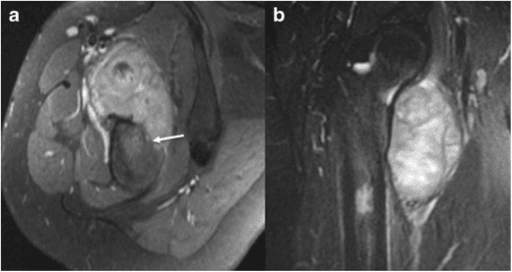
Orthogonal views of the whole bone including both adjacent joints.
The classic constellation:
- Broad-based soft-tissue mass applied to the diaphyseal cortex, usually occupying one third to one half of the shaft length.
- Cortical thickening and saucerisation — the outer cortex is scalloped and simultaneously sclerotic and reactive beneath the tumour.
- Perpendicular "hair-on-end" or "sunburst" spiculation radiating from the cortex into the mass — the single most characteristic feature.
- Codman triangles at the proximal and distal margins where periosteum is lifted.
- Sparse, faint matrix mineralisation within the mass — much less dense than parosteal osteosarcoma. The mass often looks surprisingly lucent for an osteosarcoma.
- Intact medullary canal — no endosteal destruction, no intramedullary sclerosis.
Periosteal osteosarcoma should not truly invade the medullary canal. If MRI shows unequivocal confluent T1 marrow replacement, the working diagnosis becomes high-grade surface osteosarcoma (or a periosteal lesion with an aggressive component), and the treatment algorithm shifts to neoadjuvant chemotherapy plus wide resection. Discuss every such case at the sarcoma multidisciplinary meeting before committing.
Biopsy — Doing It Once, Doing It Right
- Where it happens
- Pathology bench, after core biopsy
- Mechanism
- Periosteal osteosarcoma has an abundant chondroid matrix; a core taken from the cartilaginous lobules alone reads as a low-grade cartilage tumour, and the ossified periphery yields only reactive bone
- Avoid by
- Image-guided cores (multiple 14G to 16G) of the non-mineralised soft-tissue component, avoiding the densely ossified rim and necrotic centres; retain material for MDM2/CDK4 FISH if parosteal osteosarcoma is in the differential
- If it happens
- Treat as unrepresentative sampling, not as wrong imaging. A report of low-grade cartilage tumour against a spiculated diaphyseal surface mass in a teenager mandates MDM review and re-biopsy
- Where it happens
- MRI reporting
- Mechanism
- Reactive signal beneath a surface lesion is read as intramedullary extension, escalating the planned resection
- Avoid by
- Correlate MRI with the radiographic surface pattern and discuss at the MDM before committing to resection length
- If it happens
- Re-review imaging with the resecting surgeon and radiologist; do not extend the resection on oedema alone
- Where it happens
- Referring unit, before specialist review
- Mechanism
- Biopsy performed outside the centre that will resect contaminates planes that were never going to be excised — the commonest avoidable cause of a compromised reconstruction
- Avoid by
- Refer before biopsy; the resecting surgeon marks the tract on imaging so it lies within the skin ellipse and muscle cuff to be excised
- If it happens
- Excise the whole tract with the specimen; accept a larger soft-tissue defect, flap cover or, in the worst case, loss of a limb-salvage option
- Where it happens
- Definitive resection
- Mechanism
- Transverse incision, or a tract crossing an intermuscular plane or neurovascular bundle, forces a marginal excision
- Avoid by
- Longitudinal incision only, never transverse, never crossing an intermuscular plane or neurovascular bundle; plan the cuff at the same sitting as the biopsy
- If it happens
- Re-resection to negative margins where anatomy allows; radiology, pathology and surgery must agree before any further treatment
Biopsy is performed at the centre that will resect. Unplanned biopsy elsewhere is the commonest avoidable cause of a compromised reconstruction.
The resecting surgeon marks the tract so that it lies within the skin ellipse and muscle cuff that will be excised. Longitudinal incision only, never transverse, never crossing an intermuscular plane or neurovascular bundle.
Image-guided core needle biopsy (multiple 14G to 16G cores) of the non-mineralised soft-tissue component. Avoid the densely ossified periphery, which yields only reactive bone, and avoid necrotic centres.
Fresh tissue to pathology; send material for culture if any doubt about infection. Retain material for MDM2/CDK4 FISH if parosteal is in the differential.
Radiology, pathology and surgery must agree. A pathology report of low-grade cartilage tumour against radiology of a spiculated diaphyseal surface mass in a teenager means the biopsy is unrepresentative, not that the imaging is wrong.
Surgical Management
- 1Hemicortical resection and hemicortical allograftOnly where MRI proves circumferential cortical sparing and the tumour is truly limited to one aspect; preserves continuity. Mostly reserved for parosteal lesions — be more conservative in periosteal disease.
- 2Intercalary allograft with plate or nailRestores bone stock and matches host diameter. Compress at host-graft interfaces and supplement both junctions with iliac crest cancellous autograft. Risks: junctional nonunion, allograft fracture, infection; union commonly 6 to 12 months.
- 3Vascularised free fibulaBiologically active, hypertrophies with load, excellent in the tibia and in irradiated or chemotherapy-exposed hosts. Slow to bear load initially; needs microvascular set, second team and contralateral leg prepped.
- 4Capanna construct (vascularised fibula inside a hollowed intercalary allograft)Combines immediate structural strength of allograft with biological union and hypertrophy of the fibula, and is widely favoured for long femoral and tibial defects — but the best comparative series does NOT support it as the default. Errani's 46 children (25 composite against 21 allograft alone) found reconstruction survival 84 against 87 per cent (p = 0.89), near-identical complications, MORE infections with the fibula (4 against 1), and a 10-hour rather than 4-hour operation; those authors reconstruct with allograft alone and add the fibula to SALVAGE a fracture or nonunion. Note the counter-argument in their own data: the fibula was chosen for the longer resections, so equivalence may understate it.
- 5Segmental endoprosthesis (intercalary spacer)Immediate stability and rapid rehabilitation when adjuvant therapy or patient factors make a prolonged union unattractive; long-term stem loosening and aseptic failure risk.
- 6Distraction osteogenesis / bone transportBiologically ideal and avoids graft entirely, but demands prolonged frame time and high patient compliance.
- 7AmputationReserved for encasement of the neurovascular bundle beyond reconstruction, extensive contamination from unplanned surgery, or fungation and infection.
Principles
- Wide en-bloc resection is the treatment. Because the medulla is spared, most tumours are amenable to diaphyseal (intercalary) segmental resection preserving both adjacent joints — a major functional advantage over conventional osteosarcoma.
- The critical margins are the soft-tissue cuff over the mass (take an uninvolved layer of muscle or fascia across the entire surface, never dissect on tumour) and the bone cuts (2 to 3 cm of normal marrow beyond MRI-defined extent, confirmed with intraoperative frozen section of marrow from each cut end).
- If a portion of cortex is uninvolved circumferentially and the tumour is truly limited to one aspect, a hemicortical resection with hemicortical allograft is described and preserves continuity — but only where MRI proves circumferential sparing and margins are achievable. Most surgeons reserve this for parosteal lesions and are more conservative with periosteal disease.
- Amputation is reserved for tumours encasing the neurovascular bundle beyond reconstruction, extensive contamination from unplanned surgery, or fungation/infection.
PIPADRAW — Intercalary femoral diaphyseal resection with intercalary reconstruction
Because periosteal osteosarcoma is diaphyseal and spares the medulla, a young patient can often keep both native joints. Long-term function after intercalary reconstruction substantially exceeds that after a growing-child megaprosthesis. Preserving the epiphysis and physis is a legitimate operative goal — do not resect a joint reflexively because the word "osteosarcoma" appears on the report.
Chemotherapy — The Genuine Uncertainty
conventional osteosarcoma chemotherapy protocols (doxorubicin, cisplatin, high-dose methotrexate, with or without ifosfamide) were validated in high-grade disease. Periosteal osteosarcoma is intermediate grade, rare, and has never been the subject of a randomised trial.
large retrospective and cooperative-group analyses of periosteal osteosarcoma have not demonstrated a clear survival advantage for chemotherapy, and pathological necrosis response rates are generally poorer than in conventional osteosarcoma. Series consistently identify surgical margin adequacy, not chemotherapy, as the dominant determinant of outcome.
- Argues for chemotherapy
- Focal grade 3 areas identified
- Argues against
- Uniform intermediate grade
- Argues for chemotherapy
- Any true medullary invasion
- Argues against
- Canal clearly spared
- Argues for chemotherapy
- Large, rapidly enlarging mass
- Argues against
- Modest, indolent lesion
- Argues for chemotherapy
- Marginal margin anticipated or obtained
- Argues against
- Wide margin achieved with confidence
- Argues for chemotherapy
- Pulmonary metastases present
- Argues against
- M0 disease
- Argues for chemotherapy
- Young, fit, fertility preserved, accepts toxicity
- Argues against
- Comorbidity, cardiac or renal limitation, patient declines
A defensible position for a viva: "There is no randomised evidence that chemotherapy improves survival in periosteal osteosarcoma, and retrospective series have not shown a consistent benefit. Surgery with a wide margin is the mainstay. Many centres nonetheless offer conventional osteosarcoma chemotherapy to young patients, particularly where the tumour is large, the grade is heterogeneous, or the margin is compromised. I would take the case to the sarcoma multidisciplinary meeting and consent the patient with an honest account of the uncertainty."
Radiotherapy has no established role in primary management; it is reserved for unresectable disease, positive margins where re-excision is impossible, or palliation.
Outcomes, Surveillance and Complications
Prognosis
- Long-term survival 85 to 90% with a wide, negative margin — intermediate between parosteal (greater than 90%) and high-grade surface or conventional osteosarcoma.
- Pulmonary metastases in 10 to 15%, typically within the first 3 years but late events occur.
- Adverse factors: inadequate margin, local recurrence, large tumour volume, high-grade foci, true medullary invasion, metastasis at presentation.
- Resectable pulmonary metastases should be considered for metastasectomy — prolonged survival after complete resection of limited lung disease is well documented across osteosarcoma generally.
Surveillance schedule
- Local imaging
- Clinical review plus limb radiograph every 3 months (MRI if symptoms or equivocal)
- Systemic
- CT chest every 3 to 4 months
- Local imaging
- Every 6 months
- Systemic
- CT chest every 6 months
- Local imaging
- Annually to at least 10 years
- Systemic
- Annual chest imaging; also monitor reconstruction integrity for life
Complications of treatment
- Approximate risk
- Low with wide margin; substantial after marginal or intralesional excision
- Prevention
- Preoperative MRI planning, excise biopsy tract, muscle cuff over tumour
- Management
- Re-resection to wide margins; amputation if unsalvageable; restage systemically
- Approximate risk
- Common, often reported in the range of one in five to one in four
- Prevention
- Compression, long-span fixation, autograft at junctions, vascularised augmentation
- Management
- Revision fixation plus autograft, or conversion to vascularised fibula
- Approximate risk
- Recognised late complication of massive allografts
- Prevention
- Protected loading, avoid stress risers, adequate plate span
- Management
- Plate and graft, or graft exchange
- Approximate risk
- Higher with long operative time, large implants, prior chemotherapy
- Prevention
- Antibiotic prophylaxis, meticulous soft-tissue cover, flap coverage of the tibia
- Management
- Staged debridement, spacer, reconstruction or amputation
- Approximate risk
- Small but catastrophic
- Prevention
- Two-team approach, careful pedicle handling, flap monitoring
- Management
- Urgent re-exploration; salvage with allograft or transport
- Approximate risk
- Site dependent
- Prevention
- Early identification and protection, avoid excessive traction
- Management
- Orthosis, tendon transfer if permanent
- Approximate risk
- Cardiac, renal, ototoxic, infertility, secondary malignancy
- Prevention
- Echocardiography, renal and audiometric monitoring, fertility preservation
- Management
- Dose modification, long-term survivorship follow-up
Guidelines, Registries & Global Practice
Global epidemiology. Osteosarcoma incidence is broadly similar worldwide (roughly 3 to 4 per million per year across all subtypes). Estimates of the periosteal share vary with the denominator and the era: this page quotes 1 to 2%, while the SEER-based survey cited above puts it at 1 to 6% — quote a range and say which source you are using rather than a single figure. Because absolute numbers are tiny either way — that SEER query found just 54 patients in the whole United States registry — no single centre accumulates a large series and evidence is inevitably retrospective and multi-institutional. Outcome differences between regions are driven far more by time to referral and access to reconstruction than by tumour biology.
Society and network guidance (where it genuinely addresses this topic):
- Relevant position
- Suspected primary bone sarcoma should be referred untouched to a reference centre; biopsy and definitive surgery in the same institution; management by a bone sarcoma multidisciplinary team
- Relevant position
- Centralised sarcoma services with defined diagnostic pathways and specialist pathology review; supports the referral-before-biopsy principle
- Relevant position
- Diagnostic pathway for suspicious bone lesions, imaging before biopsy, specialist centre biopsy, and defined follow-up
- Relevant position
- Recognises surface osteosarcoma variants separately; low-grade parosteal treated surgically alone, high-grade surface treated as conventional osteosarcoma, periosteal handled as an individualised multidisciplinary decision
- Relevant position
- Defines periosteal osteosarcoma as an intermediate-grade chondroblastic surface osteosarcoma of the diaphysis — the reference definition used above
Registry and network evidence. There are no arthroplasty-style implant registries for this disease. The relevant datasets are national and supranational bone sarcoma registries and cooperative group databases (European Musculoskeletal Oncology Society registries, cooperative osteosarcoma study groups, national cancer registries), which pool rare variants to generate the survival and chemotherapy-response figures quoted above. Where segmental endoprostheses are used, national implant and tumour-prosthesis audits contribute long-term revision data.
Practice variation by resource setting.
- High-resource centres: MRI-planned intercalary resection, allograft banking, microvascular reconstruction, PET staging, fertility preservation, protocol chemotherapy where chosen.
- Intermediate-resource settings: intercalary resection with autograft, non-vascularised fibula, or bone transport where allograft banks and microsurgery are unavailable; CT chest available but PET is not.
- Low-resource settings: late presentation with large masses, limited pathology expertise (risk of misdiagnosis as a cartilage tumour), and higher amputation rates. The single highest-yield intervention in these settings is early referral and correct biopsy, not access to chemotherapy.
Controversies & Areas of Uncertainty
- Does chemotherapy help? The central unresolved question. No randomised data; retrospective series conflict and are underpowered. Practice ranges from surgery alone in all cases to routine conventional protocols in all young patients.
- What counts as medullary invasion? MRI reactive oedema is over-called. Some argue any T1 marrow change reclassifies the tumour; others require confluent replacement plus histological confirmation. The threshold materially changes both grade assignment and chemotherapy decisions.
- Is periosteal osteosarcoma a distinct entity or a spectrum? Some pathologists regard it as one end of a continuum with high-grade surface osteosarcoma, with the grade determined by sampling. Inadequate biopsy sampling can misgrade either way.
- Hemicortical versus segmental resection. Hemicortical excision preserves continuity and function but the oncological safety margin in an intermediate-grade tumour with periosteal spread is less secure than in parosteal disease. Most units resect segmentally.
- Best intercalary reconstruction. Allograft, vascularised fibula, Capanna construct, intercalary endoprosthesis and bone transport all have advocates; comparative data are small-series and centre-dependent. Choice is driven by defect length, host biology, chemotherapy exposure and local microsurgical capability.
- Duration of surveillance. Late metastasis and late reconstruction failure both occur; whether chest imaging should continue beyond 10 years is not settled.
MEDSReasons to doubt a diagnosis of periosteal osteosarcoma
Hook:If any of MEDS is present, take the case back to the multidisciplinary meeting before you cut.
MCQ Practice Points
Q: Which combination is characteristic of periosteal osteosarcoma?
A: Diaphyseal location, broad-based surface mass, perpendicular hair-on-end spiculation, largely unmineralised chondroid matrix, and an intact medullary canal. Parosteal osteosarcoma is metaphyseal and densely ossified; high-grade surface osteosarcoma frequently invades the medulla.
Q: What is the histological grade of periosteal osteosarcoma, and why does that cause trouble at staging?
A: Intermediate — grade 2 of 3. Parosteal is grade 1 and high-grade surface is grade 3. The trouble is that the Enneking system has only TWO grades, so a periosteal lesion is forced into the high-grade category and is commonly staged IIB. Say so explicitly, because IIB here does not carry the meaning it carries in a conventional osteosarcoma.
Q: Which molecular finding argues against periosteal osteosarcoma?
A: MDM2 and CDK4 amplification. That is the signature of parosteal osteosarcoma and of low-grade central osteosarcoma, and it is absent in periosteal osteosarcoma.
Q: A periosteal-type surface tumour shows unequivocal confluent T1 marrow replacement. What is the implication?
A: Reconsider the diagnosis in favour of high-grade surface osteosarcoma. True medullary invasion is essentially not a feature of periosteal osteosarcoma; if it is present the patient is managed with neoadjuvant chemotherapy plus wide resection as for conventional osteosarcoma.
Q: What is the single strongest determinant of outcome, and what is the role of chemotherapy?
A: Adequacy of the surgical margin — wide negative margins are associated with long-term survival of roughly 85 to 90 per cent, while inadequate margins drive local recurrence and subsequent metastatic death. On chemotherapy, be precise: Tsukamoto's systematic review of 13 studies and 291 patients found mortality 11.3 against 16.3 per cent, pooled odds ratio 0.89, p = 0.800, with local recurrence and distant metastasis odds ratios pointing the other way and every p value above 0.4. State it as NOT PROVEN BENEFICIAL rather than proven useless — all the data are retrospective and confounded by indication.
Q: Which histological feature confirms osteosarcoma rather than a cartilage tumour in a predominantly chondroid surface lesion?
A: Fine lace-like osteoid produced directly by malignant spindled cells at the periphery of the cartilage lobules. Reactive periosteal bone and entrapped host bone do not qualify — the osteoid must be tumour-derived.
Exam Viva Scenarios
Practise clinical reasoning and management decisions out loud
“An 18-year-old presents with 6 months of dull mid-thigh pain and a firm, deep, immobile mass. Radiographs show a broad-based soft-tissue mass on the anterolateral femoral diaphysis with a thickened, saucerised cortex, perpendicular spiculation and Codman triangles at each end. There is faint matrix mineralisation. How do you proceed?”
“The core biopsy from that patient is reported by a general pathologist as a low to intermediate grade cartilaginous neoplasm, favouring chondrosarcoma. The oncology team asks whether chemotherapy is needed. What is your response?”
“Diagnosis is confirmed as periosteal osteosarcoma of the mid-femoral diaphysis, 9 cm in length, with a soft-tissue mass abutting but not encasing the superficial femoral vessels, and a clearly normal medullary canal. Take me through your operative plan.”
“Two years after wide resection with clear margins, surveillance CT chest shows two peripheral pulmonary nodules, each about 8 mm, in the right lower lobe. The limb is disease free. What now?”
Identity
- Intermediate-grade (2 of 3) chondroblastic surface osteosarcoma
- Femoral or tibial DIAPHYSIS; second and third decades
- 1 to 2% of all osteosarcomas; MDM2/CDK4 negative
- Arises from cambium layer of periosteum, grows outward on the cortex
Imaging signature (SCRAM)
- Spiculation — perpendicular hair-on-end bone into the mass
- Codman triangles at proximal and distal margins
- Reactive cortex — thickened, sclerotic, saucerised
- Adjacent diaphysis — joint and physis spared
- Medulla spared — CT low-density chondroid matrix, MRI lobulated bright T2 with septal enhancement
Contrast with the other surface tumours
- Parosteal — metaphyseal, dense, low grade, MDM2 amplified, surgery alone, survival greater than 90%
- Periosteal — diaphyseal, lucent chondroid, intermediate grade, surgery plus debated chemotherapy, survival 85 to 90%
- High-grade surface — bulky, marrow invasion common, grade 3, chemotherapy plus surgery, survival like conventional osteosarcoma
Pathology
- Atypical hyaline cartilage lobules forming the bulk of the tumour
- Peripheral spindling of lobules into sarcomatous stroma
- Fine lace-like tumour osteoid made by malignant cells — the diagnostic feature
- MDM2/CDK4 negative; IDH wild type
Staging
- Whole-bone MRI with adjacent joint — extent, skip lesions, true marrow status
- CT of the lesion for matrix and cortex; CT chest for metastases
- Bone scintigraphy or PET-CT for skeletal survey
- Typically Enneking Stage IIB
Biopsy rules
- Refer before biopsy; biopsy in the resecting centre
- Longitudinal single tract planned by the resecting surgeon, excised at resection
- Core the non-mineralised soft-tissue component, multiple cores
- Radiology-pathology discordance means repeat or review, not proceed
Surgery
- Wide en-bloc intercalary diaphyseal resection preserving both joints
- Osteotomies 2 to 3 cm beyond MRI extent; frozen section marrow at both ends
- Muscle cuff over the tumour surface; never dissect on tumour
- Reconstruction: allograft, vascularised fibula, Capanna construct, intercalary endoprosthesis or bone transport
- Long-span fixation with compression and autograft at junctions; flap cover for the subcutaneous tibia
Systemic therapy
- No randomised evidence of chemotherapy benefit in periosteal osteosarcoma
- Necrosis response generally poorer than conventional osteosarcoma
- Often offered to young patients with large tumours, heterogeneous grade or compromised margins
- Radiotherapy reserved for unresectable disease, positive margins or palliation
Outcome and follow-up
- Long-term survival 85 to 90% with wide margins; pulmonary metastases in 10 to 15%
- Adverse: inadequate margin, local recurrence, large size, high-grade foci, marrow invasion
- Consider metastasectomy for resectable pulmonary disease
- Surveillance 3-monthly for 2 years, 6-monthly to 5 years, annually to 10 years and beyond
Doubt the diagnosis if (MEDS)
- Medulla truly involved — think high-grade surface osteosarcoma
- Epicentre metaphyseal and posterior distal femur — think parosteal, test MDM2
- Dense ossification throughout — against a chondroid tumour
- Systemic symptoms — think Ewing sarcoma or osteomyelitis
Evidence Base
Demographic and Treatment Analysis of Periosteal Osteosarcoma
- The first SEER-based epidemiological survey of periosteal osteosarcoma in the United States; 54 patients identified (ICD-O-3 9193/3)
- Periosteal osteosarcoma accounts for between 1 and 6 PER CENT of osteosarcomas; median age at diagnosis was 20-24 years
- Surgery carried a 20-year cause-specific survival benefit (HR 0.08, p = 0.040); CHEMOTHERAPY DID NOT (p = 0.29)
- Mean survival was longer for appendicular disease (16.0 years) than axial disease (10.9 years), and younger patients more often had appendicular tumours
- Stage-stratified analysis found surgery alone NON-INFERIOR to surgery plus chemotherapy for both local (p = 0.37) and regional (p = 0.85) disease
Effect of Adjuvant Chemotherapy on Periosteal Osteosarcoma: A Systematic Review
- Systematic review of 13 studies and 291 patients with non-metastatic periosteal osteosarcoma, comparing surgery plus (neo)adjuvant chemotherapy against surgery alone
- Mortality 11.3 per cent (8/71) with chemotherapy against 16.3 per cent (16/98) without; pooled odds ratio 0.89, p = 0.800
- Local recurrence 12.1 per cent (8/66) against 17.6 per cent (13/74); pooled odds ratio 1.31, p = 0.601
- Distant metastasis 15.2 per cent (10/66) against 10.8 per cent (8/74); pooled odds ratio 1.51, p = 0.444
- Secondary malignancy 7.6 per cent (9/118) against 2.7 per cent (2/74); pooled odds ratio 2.29, p = 0.187
- The authors conclude adjuvant chemotherapy did not appear to improve prognosis, and found no association with secondary malignancy
A System for the Surgical Staging of Musculoskeletal Sarcoma
- Stratifies bone and soft-tissue sarcoma of any histogenesis by biological grade, anatomical setting and the presence of metastasis
- Three stages - I low grade, II high grade, III metastatic - each subdivided by whether the lesion is confined within a well-delineated compartment (A) or extends beyond it (B)
- Defines operative margins as intralesional, marginal, wide and radical, relating the margin to the lesion, its reactive zone and the compartment
- The stated purpose is to allow new treatment protocols to be compared against standard surgical treatment
The Hazards of the Biopsy, Revisited
- 597 patients from 21 institutions, repeating the Musculoskeletal Tumor Society's 1982 survey of 329 biopsies to see whether anything had improved in a decade
- IT HAD NOT: diagnostic error 17.8 per cent overall; a biopsy problem forced a different or more complex operation in 19.3 per cent (against 18 per cent in 1982)
- Outcome was changed - more complex resection with disability, loss of function, local recurrence or death - in 10.1 per cent, against 8.5 per cent previously
- EIGHTEEN patients underwent an unnecessary amputation because of the biopsy, against fifteen in the earlier study
- Errors, complications and changes in course were TWO TO TWELVE TIMES greater (p less than 0.001) when biopsy was done at the referring institution rather than the treatment centre
Does the Addition of a Vascularized Fibula Improve the Results of a Massive Bone Allograft Alone for Intercalary Femur Reconstruction of Malignant Bone Tumors in Children?
- 46 children with femoral sarcoma after intercalary resection: 25 reconstructed with massive allograft PLUS vascularised free fibula, 21 with massive allograft ALONE, followed a mean of 117 and 130 months
- NO DIFFERENCE in reconstruction survival: 84 per cent with the fibula against 87 per cent without (p = 0.89)
- Complications were near identical - 12 of 25 with the fibula against 12 of 21 without. Allograft fracture 7 against 5, NONUNION 4 against 6, INFECTION 4 against 1
- Surgery took 10 hours with the fibula against 4 hours without (p = 0.001)
- Functional scores were the same (26 against 27, p = 0.39)
- The authors' conclusion: reconstruct with ALLOGRAFT ALONE and add a vascularised fibula only to SALVAGE a fracture or nonunion - which would have spared about half their patients the more invasive procedure