A tiny, occult, usually benign mesenchymal tumour secreting FGF23, producing renal phosphate wasting, osteomalacia and multiple insufficiency fractures — curable by complete excision
- Biochemical signature: low serum phosphate, high renal phosphate loss (low TmP/GFR), inappropriately low or normal 1,25-dihydroxyvitamin D, NORMAL calcium and parathyroid hormone, raised alkaline phosphatase.
- The single most common misdiagnosis is osteoporosis or fibromyalgia — an adult with diffuse bone pain, proximal myopathy, height loss and multiple insufficiency fractures needs a serum phosphate.
- Phosphate is not on most routine biochemistry panels and must be requested specifically, on a fasting sample.
- Localisation is functional first: somatostatin receptor imaging (68-Ga DOTATATE PET/CT) has the highest yield, then anatomical MRI or CT of the flagged site.
- Do not fix insufficiency fractures blindly — unaddressed osteomalacia gives non-union, implant cut-out and new fractures elsewhere.
- Complete excision with a clear margin is curative; incomplete excision means persistent or recurrent disease.
- “Normal calcium plus normal PTH plus low phosphate is the discriminator from hyperparathyroidism and from nutritional osteomalacia.
- “FGF23 both wastes phosphate and suppresses 1-alpha-hydroxylase — hence the paradox of low phosphate with a low or 'inappropriately normal' 1,25-dihydroxyvitamin D.
- “Histology: bland spindle cells, prominent 'grungy' smudgy calcified matrix, haemangiopericytoma-like branching vessels, osteoclast-type giant cells; FN1-FGFR1 fusion is characteristic.
- “Selective venous sampling for FGF23 is reserved for equivocal or multiple candidate lesions.
- “Burosumab (anti-FGF23 monoclonal antibody) is the modern option when the tumour is unlocalised, unresectable or metastatic.
Multiple insufficiency fractures in an adult with normal calcium and normal PTH is TIO until proven otherwise. A fasting serum phosphate costs almost nothing and is the whole diagnosis. Most delays are because nobody ordered it.
Fixing a femoral neck or subtrochanteric insufficiency fracture in untreated osteomalacia risks non-union, screw cut-out and contralateral fracture. Correct the metabolic disease and, where possible, remove the tumour first or concurrently.
PMT recurs if shelled out. Plan a marginal-to-wide excision as for a soft tissue sarcoma of unknown grade, with pre-operative imaging-defined margins. Persistent hypophosphataemia at day 5 to 7 post-op means residual tumour.
Adult onset, no family history, no childhood bowing or short stature = acquired. Childhood onset with a positive family history = X-linked hypophosphataemia. Getting this wrong sends the patient to lifelong supplements instead of a curative operation.
Pathophysiology: what FGF23 actually does
Fibroblast growth factor 23 (FGF23) is a bone-derived hormone whose physiological job is to lower serum phosphate. A phosphaturic mesenchymal tumour secretes it autonomously and in excess.
Two coordinated effects, both of which are on the exam:
- Renal phosphate wasting. FGF23 acting through FGF receptor 1 with the co-receptor alpha-Klotho in the proximal tubule downregulates the sodium-phosphate co-transporters NaPi-IIa and NaPi-IIc in the brush border. Tubular reabsorption of phosphate falls, urinary phosphate loss rises, serum phosphate falls.
- Suppression of active vitamin D. FGF23 inhibits renal 1-alpha-hydroxylase and stimulates 24-hydroxylase. So 1,25-dihydroxyvitamin D is low or inappropriately normal. Normally, hypophosphataemia is a powerful stimulus to 1-alpha-hydroxylase, so a "normal" active vitamin D in the face of a low phosphate is pathological.
Because gut calcium absorption is not driven up, serum calcium stays normal and PTH stays normal (or is only mildly raised late, or after phosphate supplementation). Chronic hypophosphataemia impairs mineralisation of newly formed osteoid — osteomalacia, not osteoporosis. Unmineralised osteoid seams accumulate, bone becomes mechanically incompetent and painful, and insufficiency fractures and Looser zones appear.
Osteoporosis is too little normally mineralised bone. Osteomalacia is normal or increased osteoid that is inadequately mineralised. DXA may be low in both — a low T-score does not exclude TIO and often anchors the wrong diagnosis. The give-aways are bone pain, proximal myopathy, raised alkaline phosphatase and Looser zones.
Pathology
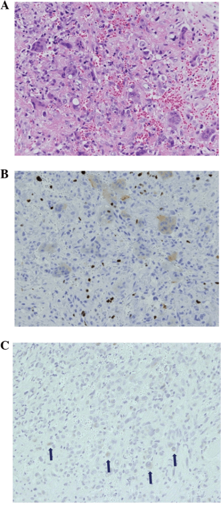
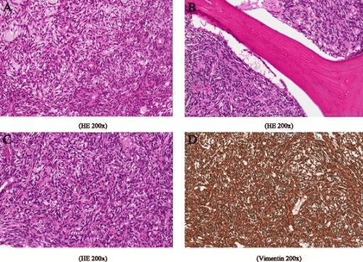
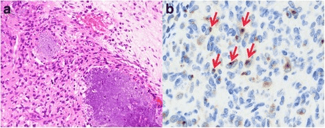
Macroscopic. Usually small (often 1 to 5 cm), well circumscribed, tan-grey to reddish-brown, sometimes gritty on sectioning because of the calcified matrix. May be intraosseous, intramuscular, subcutaneous or sinonasal.
Microscopic — the classic quartet:
- Bland spindle to stellate cells with low mitotic activity and minimal atypia, in a myxoid to fibrous stroma.
- "Grungy" or smudgy calcified matrix — flocculent, basophilic, poorly formed chondroid/osteoid-like material. This is the single most distinctive feature and the phrase examiners expect.
- Prominent branching, staghorn (haemangiopericytoma-like) vasculature, often with a rim of osteoclast-type multinucleated giant cells.
- Variable mature adipose tissue, microcysts and haemorrhage.
The original descriptive subtypes are of historical interest: mixed connective tissue variant (by far the commonest), osteoblastoma-like, non-ossifying fibroma-like and ossifying fibroma-like.
Malignant PMT is rare: infiltrative margins, marked nuclear pleomorphism, high mitotic count, necrosis, and capacity for local recurrence and pulmonary metastasis. It behaves as a sarcoma and demands sarcoma-standard oncological management.
Clinical presentation — the classic story
an adult in the fourth to sixth decade, previously well, with:
- Insidious diffuse bone pain over 2 to 5 years — axial and appendicular, worse on weight-bearing, poorly localised, often attributed to fibromyalgia, ankylosing spondylitis or "degenerative change".
- Progressive proximal muscle weakness — difficulty rising from a chair or climbing stairs, waddling gait. Ask specifically; patients call it "tiredness".
- Height loss from vertebral compression and kyphosis.
- Multiple low- or no-energy fractures in atypical sites: femoral neck, subtrochanteric femur, pubic rami, ribs, metatarsals, sacrum, calcaneus. A patient with three or more insufficiency fractures without a clear osteoporosis risk profile is a red flag.
- No childhood abnormality — normal growth, no bowing, no dental abscesses, no family history. This is the pivotal negative.
- Prior investigations often show a "low BMD", a normal calcium, and a failed course of bisphosphonate or denosumab with continued fractures — treatment failure is itself a diagnostic clue.
tenofovir, adefovir, ifosfamide, iron carboxymaltose infusions (a recognised cause of FGF23-mediated hypophosphataemia), coeliac disease, bariatric surgery, alcohol excess, and any renal tubular disorder.
Biochemistry — the signature every examiner wants
- TIO / PMT
- Low — the entry point
- Primary hyperparathyroidism
- Low or low-normal
- Nutritional osteomalacia
- Low-normal
- TIO / PMT
- NORMAL — the discriminator
- Primary hyperparathyroidism
- High
- Nutritional osteomalacia
- Low or low-normal
- TIO / PMT
- Normal (may rise once phosphate therapy starts)
- Primary hyperparathyroidism
- High (primary drive)
- Nutritional osteomalacia
- High (secondary)
- TIO / PMT
- Normal
- Primary hyperparathyroidism
- Normal
- Nutritional osteomalacia
- Low
- TIO / PMT
- Low or inappropriately normal (FGF23 blocks 1-alpha-hydroxylation)
- Primary hyperparathyroidism
- High
- Nutritional osteomalacia
- Low or normal
- TIO / PMT
- Raised — tracks activity and healing
- Primary hyperparathyroidism
- Raised
- Nutritional osteomalacia
- Raised
- TIO / PMT
- Low TmP/GFR; TRP not maximally raised despite hypophosphataemia — proves renal wasting
- Primary hyperparathyroidism
- Low
- Nutritional osteomalacia
- Normal or high — appropriate conservation
- TIO / PMT
- Raised or inappropriately normal
- Primary hyperparathyroidism
- Normal
- Nutritional osteomalacia
- Normal or low
- TIO / PMT
- Isolated phosphaturia, no glycosuria or aminoaciduria
- Primary hyperparathyroidism
- Hypercalciuria
- Nutritional osteomalacia
- Nil specific
- TIO / PMT
- Normal (raised suggests myositis, not TIO)
- Primary hyperparathyroidism
- Normal
- Nutritional osteomalacia
- Normal
- TIO / PMT
- Low
- Primary hyperparathyroidism
- Low or low-normal
- Nutritional osteomalacia
- Low-normal
- Renal Fanconi syndrome
- Low
- TIO / PMT
- Normal
- Primary hyperparathyroidism
- High
- Nutritional osteomalacia
- Low or low-normal
- Renal Fanconi syndrome
- Normal or low
- TIO / PMT
- Normal (may rise with therapy)
- Primary hyperparathyroidism
- High
- Nutritional osteomalacia
- High (secondary)
- Renal Fanconi syndrome
- Normal or high
- TIO / PMT
- Raised
- Primary hyperparathyroidism
- Raised
- Nutritional osteomalacia
- Raised
- Renal Fanconi syndrome
- Raised
- TIO / PMT
- Normal
- Primary hyperparathyroidism
- Normal
- Nutritional osteomalacia
- Low
- Renal Fanconi syndrome
- Normal
- TIO / PMT
- Low or inappropriately normal
- Primary hyperparathyroidism
- High
- Nutritional osteomalacia
- Low or normal
- Renal Fanconi syndrome
- Low
- TIO / PMT
- Low (renal phosphate wasting)
- Primary hyperparathyroidism
- Low
- Nutritional osteomalacia
- Normal or high (appropriate conservation)
- Renal Fanconi syndrome
- Low
- TIO / PMT
- Raised or inappropriately normal
- Primary hyperparathyroidism
- Normal
- Nutritional osteomalacia
- Normal or low
- Renal Fanconi syndrome
- Normal or low
- TIO / PMT
- Isolated phosphaturia
- Primary hyperparathyroidism
- Hypercalciuria
- Nutritional osteomalacia
- Nil specific
- Renal Fanconi syndrome
- Glycosuria, aminoaciduria, acidosis
How to prove renal phosphate wasting. Take a fasting morning serum phosphate and creatinine with a paired second-void urine phosphate and creatinine. Calculate the tubular reabsorption of phosphate (TRP):
TRP = 1 minus (urine phosphate multiplied by serum creatinine) divided by (serum phosphate multiplied by urine creatinine).
Then derive TmP/GFR from the Walton–Bijvoet nomogram or standard formula. In hypophosphataemia the kidney should reabsorb almost all filtered phosphate (TRP greater than 95 percent). A TRP that is not maximally raised, and a low TmP/GFR, prove inappropriate renal loss. If phosphate is being lost in the gut or shifted into cells instead, TRP will be appropriately high — that excludes TIO.
FGF23 assays. Intact and C-terminal assays exist and are not interchangeable; both are affected by iron deficiency and by iron carboxymaltose. A value that is raised, or simply not suppressed, in the presence of hypophosphataemia is supportive. A normal FGF23 does not exclude TIO if the clinical and TmP/GFR picture fits — a minority of tumours secrete other phosphatonins (secreted frizzled-related protein 4, MEPE, FGF7).
Serum phosphate falls after a carbohydrate meal and after insulin. Request a fasting sample. Phosphate is absent from many routine renal panels — request it explicitly. Also check creatine kinase (normal in TIO — raised suggests myositis), 25-OH vitamin D, PTH, calcium, albumin, magnesium, bicarbonate, glucose and urine dipstick for glycosuria.
PHOSPHATEThe TIO biochemical signature
Hook:Every letter is a box you must tick before saying 'tumour-induced osteomalacia' in a viva.
Localisation cascade — hunting a tumour the size of a pea
- 1Start
Biochemical diagnosis secure — renal phosphate wasting with inappropriately normal or raised FGF23. The tumour is typically only 1 to 2 cm and may lie in bone or soft tissue anywhere: sinonasal cavity and head and neck, femur, tibia, foot, hand, groin or thigh.
Proceed to functional imaging
- 2Step 1 — whole-body functional imaging
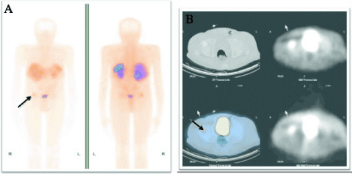
68-Ga DOTATATE (or DOTATOC / DOTANOC) PET/CT is first line: PMTs strongly express somatostatin receptor subtype 2, giving high tumour-to-background contrast and the best detection rate of any single modality.
Positive focus, multiple foci, or negative scan
- 3Step 1b — if PET unavailable
111-In-octreotide (Octreoscan) SPECT/CT is the fallback but has lower resolution and more false negatives. 18F-FDG PET/CT is a reasonable second functional study, though PMTs are often only mildly FDG-avid so a negative study does not exclude.
Any flagged site goes to anatomical confirmation
- 4Step 2 — anatomical confirmation
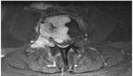
MRI of the flagged region: low to intermediate on T1, high on T2 and STIR, avid post-contrast enhancement; intraosseous lesions may be expansile with a sclerotic rim. CT is superior for craniofacial, sinonasal and small intraosseous lesions and shows the characteristic matrix mineralisation.
Single concordant lesion — plan resection
- 5Step 3 — selective venous sampling
Reserved for two or more candidate lesions, or a single equivocal focus. Catheter-directed selective venous sampling with regional FGF23 measurement; a step-up gradient of roughly two-fold or more over the systemic level localises the secreting lesion.
Culprit lesion identified
- 6Step 4 — biopsy with caution
Percutaneous biopsy confirms histology but is not always required when imaging and biochemistry are conclusive and excision is planned.
Never allow a non-oncological team to biopsy through an uninvolved compartment
- 7Step 5 — nothing found
Declare occult tumour-induced osteomalacia.
Tumours enlarge and become detectable; many patients localise on a repeat scan years later
- 8Two scans that mislead
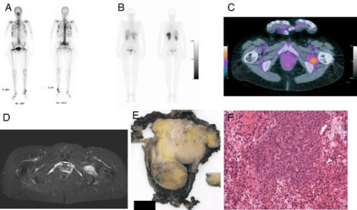
Bone scintigraphy in TIO shows multiple hot spots — these are the fractures, not the tumour. An incidental DOTATATE-avid rib focus may simply be a healing insufficiency fracture.
Never operate on functional imaging alone
The tumour may be anywhere: bone or soft tissue, head and neck sinuses, femur, tibia, foot, hand, groin, thigh, or the nasal cavity. It is often 1 to 2 cm and has usually been present, asymptomatic and unnoticed, for years.
68-Ga DOTATATE (or DOTATOC/DOTANOC) PET/CT is the modern first-line study. PMTs express somatostatin receptor subtype 2 strongly, giving high tumour-to-background contrast and the highest reported detection rate of any single modality. It is a whole-body study, which is exactly what an occult tumour of unknown site requires. Know the physiological uptake pitfalls: spleen, kidneys, adrenals, pituitary, uncinate process of the pancreas, and inflammatory or degenerative uptake at healing insufficiency fracture sites and joints.
111-In-octreotide (Octreoscan) SPECT/CT where DOTATATE PET is unavailable — lower resolution, more false negatives. 18F-FDG PET/CT is a reasonable second functional study; PMTs are often only mildly FDG-avid so a negative scan does not exclude. In resource-limited settings a structured whole-body MRI may be the only practical whole-body search.
MRI of the region: PMTs are typically low to intermediate on T1, high on T2 and STIR, with avid post-contrast enhancement; intraosseous lesions may be expansile with a sclerotic rim. CT is superior for craniofacial, sinonasal and small intraosseous lesions and demonstrates the characteristic matrix mineralisation. The anatomical study also defines the surgical margin and neurovascular relations.
When functional imaging shows two or more candidate lesions, or a single equivocal focus, catheter-directed selective venous sampling with regional FGF23 measurement is used. A step-up gradient of roughly two-fold or more over the systemic level localises the secreting lesion. It is invasive, technically demanding and available in few centres — it is a problem-solving tool, not a screening test.
Percutaneous biopsy confirms histology but is not always necessary if imaging and biochemistry are conclusive and excision is planned. If performed, the tract must be planned by the resecting surgeon and placed so it can be excised en bloc, exactly as for a soft tissue sarcoma. Never let a non-oncological team biopsy through an uninvolved compartment.
Declare it occult TIO, start medical therapy, and re-image every 1 to 2 years. Tumours enlarge and become detectable over time. Many patients localise on a repeat scan years later.
Bone scintigraphy in TIO shows multiple hot spots — these are the fractures, not the tumour. And an incidental DOTATATE-avid focus at a rib may simply be a healing insufficiency fracture. Always correlate functional uptake with anatomical imaging before committing to an operation.
Medical management when the tumour is not found or not resectable
- Oral phosphate plus active vitamin D
- Replaces losses; calcitriol/alfacalcidol bypasses blocked 1-alpha-hydroxylation
- Burosumab (anti-FGF23 monoclonal antibody)
- Binds and neutralises circulating FGF23, restoring tubular phosphate reabsorption and 1,25-dihydroxyvitamin D synthesis
- Oral phosphate plus active vitamin D
- Divided oral phosphate 3 to 5 times daily plus daily active vitamin D
- Burosumab (anti-FGF23 monoclonal antibody)
- Subcutaneous injection at intervals of several weeks
- Oral phosphate plus active vitamin D
- Poor — high pill burden and gastrointestinal upset
- Burosumab (anti-FGF23 monoclonal antibody)
- Good
- Oral phosphate plus active vitamin D
- Diarrhoea, secondary and tertiary hyperparathyroidism, nephrocalcinosis, hypercalciuria, renal impairment
- Burosumab (anti-FGF23 monoclonal antibody)
- Hyperphosphataemia if overdosed, ectopic mineralisation, injection site reactions; requires phosphate monitoring
- Oral phosphate plus active vitamin D
- Phosphate, calcium, PTH, ALP, urinary calcium, renal ultrasound for nephrocalcinosis
- Burosumab (anti-FGF23 monoclonal antibody)
- Fasting trough phosphate before each dose, ALP, renal imaging
- Oral phosphate plus active vitamin D
- First line where the antibody is unavailable or unfunded; bridging before surgery
- Burosumab (anti-FGF23 monoclonal antibody)
- Unlocalised, unresectable, recurrent or metastatic PMT; failure of or intolerance to conventional therapy
- Oral phosphate plus active vitamin D
- None
- Burosumab (anti-FGF23 monoclonal antibody)
- None — it treats the hormone, not the tumour; continue surveillance imaging
High-dose phosphate salts drive PTH secretion. Untreated, this progresses to tertiary hyperparathyroidism requiring parathyroidectomy, and active vitamin D plus phosphate together cause hypercalciuria, nephrocalcinosis and chronic kidney disease. Monitor urinary calcium and obtain periodic renal ultrasound. Never simply "keep escalating the phosphate".
Cinacalcet has been used adjunctively to blunt the compensatory rise in PTH and reduce phosphaturia, at the cost of hypocalcaemia risk and close monitoring. It is a specialist manoeuvre, not routine.
Surgical management — excision is the cure
Indication. A localised, resectable PMT in a patient with biochemically confirmed TIO. There is no role for watchful waiting once the tumour is found and resectable.
Contraindications. Unlocalised tumour; unresectable site with unacceptable functional cost where medical therapy is a reasonable alternative; disseminated malignant PMT (then treat as sarcoma with systemic and local strategies plus burosumab).
Why excision over anything else. It is the only treatment that returns FGF23, phosphate, vitamin D metabolism and bone mineralisation to normal and abolishes lifelong drug therapy and its renal risks.
P — Position. Dictated by site. Supine with a bump for anterior thigh/groin; lateral for buttock or proximal femur; prone for posterior compartments; supine with head extended and neurosurgical/ENT co-positioning for craniofacial and sinonasal lesions; tourniquet where the site allows for acral and distal limb tumours.
I — Imaging and equipment. Cross-sectional images on screen in theatre. Image intensifier for intraosseous lesions. Consider intra-operative gamma probe after a pre-operative radiolabelled somatostatin analogue dose for a deep or poorly palpable tumour, or wire/skin-marker localisation under ultrasound or CT the same morning. Bone curettes, high-speed burr and bone graft substitute if intraosseous. Have frozen section and fresh tissue handling for molecular studies arranged with pathology.
P — Preparation. Pre-operative optimisation: correct severe hypophosphataemia to reduce peri-operative respiratory and cardiac muscle weakness, but stop or reduce phosphate and calcitriol on the morning of surgery so that the post-operative phosphate trend is interpretable. Anaesthetic caution — profound hypophosphataemia impairs diaphragmatic function and delays weaning. Plan the biopsy tract for excision. Book the case with a sarcoma-capable team.
A — Approach. Longitudinal, extensile, compartmental — as for a soft tissue sarcoma of unknown grade. Avoid transverse incisions in a limb. Approach through a single compartment; do not cross uninvolved compartments or neurovascular sheaths.
D — Dissection. Do not enucleate. Dissect a cuff of normal tissue around the pseudocapsule. For intraosseous lesions in expendable sites, excise en bloc; in a load-bearing bone where en bloc excision would mandate major reconstruction, extended intralesional curettage with a burr and an adjuvant may be an accepted compromise — but counsel the patient about persistence.
R — Resection/Reconstruction. Confirm margins macroscopically; mark orientation for pathology. Reconstruct bone defects with graft or cement plus prophylactic fixation if the residual bone is mechanically compromised — remember the surrounding bone is osteomalacic and will not hold screws well until it remineralises.
A — At-risk structures. Site-specific: sciatic nerve and superior gluteal vessels in the buttock; femoral neurovascular bundle in the groin and thigh; common peroneal nerve at the fibular neck; posterior tibial neurovascular bundle in the foot; skull base, orbit, optic nerve and internal carotid for sinonasal/craniofacial tumours (joint ENT/skull-base approach mandatory).
W — Wound closure and aftercare. Layered closure over a drain; no dead space. Post-operatively check serum phosphate daily — it should rise within 2 to 5 days. Watch for hungry bone syndrome: with sudden withdrawal of FGF23, avid remineralisation can produce symptomatic hypocalcaemia and profound hypophosphataemia in the first two weeks. Have calcium and calcitriol ready, and monitor calcium, phosphate, magnesium and PTH.
Orthopaedic management of the fractures
Staged management of insufficiency fractures in tumour-induced osteomalacia
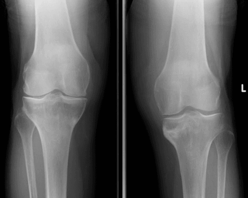
Bilateral, multiple or symmetrical insufficiency fractures — ribs, pubic rami, femoral necks — with a raised alkaline phosphatase should stop the fixation list. Do not fix a fracture in undiagnosed osteomalacia: unmineralised bone gives poor screw purchase, cut-out, non-union and periprosthetic fracture.
Fasting serum phosphate, calcium, PTH, alkaline phosphatase, 25-OH vitamin D and paired urine phosphate to demonstrate renal phosphate wasting. If the workup has not been done, do it before or at the time of any operative intervention. Withdraw bisphosphonates and denosumab once TIO is recognised — antiresorptives do not treat osteomalacia, may worsen the mineralisation defect and can cause severe hypocalcaemia in a mineral-deficient patient.
Correct the phosphate and restore mineralisation. Many lesions are metabolic pseudofractures rather than traumatic fractures: an undisplaced femoral neck insufficiency fracture in a metabolically untreated patient can unite with protected weight-bearing and metabolic correction alone. Falling alkaline phosphatase and rising phosphate are the signals that bone is remineralising.
Definitive treatment of the driver. Complete excision removes the source of phosphaturia and allows durable healing of the fracture and of the underlying mineralisation defect; without it, metabolic correction is only suppressive.
Reserve fixation for displaced or progressive fractures, and run metabolic therapy concurrently. Avoid arthroplasty as a reflex — osteomalacic bone gives poor cement interdigitation and a high early periprosthetic fracture risk. Where arthroplasty is unavoidable, defer until the phosphate is corrected if the clinical situation allows.
Proximal myopathy needs a graded strengthening and gait programme, with falls prevention maintained throughout the period of recovering bone quality.
This is where the orthopaedic surgeon most often meets the disease, and where the most damage is done.
Principles:
- Do not fix a fracture in undiagnosed osteomalacia. If the biochemistry is not yet done, do it before or at the time of surgery. Fixation into unmineralised bone gives poor screw purchase, cut-out, non-union and periprosthetic fracture.
- Suspect the pattern. Bilateral or multiple insufficiency fractures, especially symmetrical rib, pubic ramus and femoral neck lesions, in a patient with raised alkaline phosphatase.
- Undisplaced femoral neck insufficiency fracture in a metabolically untreated patient: protected weight-bearing and metabolic correction alone can achieve union, because these are metabolic pseudofractures rather than traumatic fractures. Displaced or progressive fractures need fixation, but with metabolic therapy running concurrently.
- Avoid arthroplasty as a reflex. Osteomalacic bone gives poor cement interdigitation and a high early periprosthetic fracture risk. Where arthroplasty is unavoidable, defer until the phosphate is corrected if the clinical situation allows.
- Antiresorptives are the wrong drug. Bisphosphonates and denosumab do not treat osteomalacia; they may worsen the mineralisation defect and cause severe hypocalcaemia in a mineral-deficient patient. Withdraw them once TIO is recognised.
- Rehabilitation. Proximal myopathy needs a graded strengthening and gait programme; falls prevention while bone quality is recovering.
"Before I fix this insufficiency fracture I would check a fasting serum phosphate, calcium, PTH, alkaline phosphatase, 25-OH vitamin D and paired urine phosphate to exclude a metabolic mineralisation defect, because fixation without correcting osteomalacia fails."
Complications
- Mechanism
- Years of unexplained bone pain and proximal myopathy mislabelled as osteoporosis, degenerative spine disease or a rheumatological disorder; futile antiresorptive therapy, unnecessary spine surgery, opioid dependence
- Prevention
- Measure fasting serum phosphate in any adult with unexplained fragility fractures, bone pain or proximal weakness; confirm renal phosphate wasting and elevated or inappropriately normal FGF23 before labelling the pain degenerative
- Salvage
- Functional localisation with Ga-68 DOTATATE PET/CT, then anatomical confirmation on MRI or CT; irreversible deformity may need deliberate corrective osteotomy or arthroplasty
- Mechanism
- Osteomalacic bone with wide unmineralised osteoid: multiple insufficiency fractures, non-union, kyphosis and height loss, chronic pain, falls, and rarely cord or root compromise from ligamentous ossification and vertebral deformity
- Prevention
- Early tumour localisation and excision; phosphate and active vitamin D replacement while the tumour is being sought so mineralisation is supported
- Salvage
- Fracture fixation only once mineralisation is restored; correct deformity after biochemical cure rather than before
Multiple insufficiency fractures, non-union, kyphosis and height loss, chronic pain, proximal myopathy with loss of independence, falls, and rarely spinal cord or nerve root compromise from ligamentous ossification and vertebral deformity in long-standing disease.
Years of futile antiresorptive therapy, unnecessary spine surgery for "degenerative" pain, rheumatological labelling, opioid dependence, and irreversible skeletal deformity. Diagnostic delay is the dominant morbidity of this condition.
Incomplete excision with persistent hypophosphataemia (the commonest failure); local recurrence; site-specific neurovascular injury; pathological fracture through an excision defect; hungry bone syndrome with symptomatic hypocalcaemia in the first two weeks.
Gastrointestinal intolerance, secondary and tertiary hyperparathyroidism, hypercalciuria, nephrocalcinosis and chronic kidney disease with conventional therapy; hyperphosphataemia and ectopic mineralisation with excessive anti-FGF23 dosing.
FIRSTDifferentiating adult hypophosphataemic osteomalacia
Hook:Exclude the first four, and the fifth is your diagnosis.
Guidelines, Registries and Global Practice
Global epidemiology. TIO is rare, reported worldwide with no clear ethnic predilection, affecting men and women roughly equally, most often presenting between the third and sixth decades. Reported case numbers have risen steeply since somatostatin receptor PET became widely available and since serum phosphate became a more frequently requested test — the true incidence is almost certainly higher than historical figures because of under-diagnosis. Paediatric cases occur but are distinctly uncommon and must be separated from inherited rickets.
Society and organisational guidance, where it genuinely applies:
- Endocrine and metabolic bone societies (including the American Society for Bone and Mineral Research and European endocrine bodies) provide the most directly applicable consensus: confirm FGF23-mediated hypophosphataemia biochemically, localise with functional then anatomical imaging, resect completely where possible, and use conventional or anti-FGF23 therapy when resection is not achievable.
- Sarcoma and musculoskeletal tumour guidance (ESMO soft tissue and bone sarcoma guidance, and national sarcoma network standards) applies to the surgical component: biopsy through a planned, excisable tract, referral to a specialist musculoskeletal tumour unit, and margin-directed excision with multidisciplinary review.
- WHO Classification of Soft Tissue and Bone Tumours lists phosphaturic mesenchymal tumour as a distinct entity of uncertain differentiation, most cases benign with a rare malignant variant.
- NICE and equivalent health technology bodies have appraised anti-FGF23 antibody therapy principally in the context of X-linked hypophosphataemia, with variable extension to TIO by jurisdiction — access remains the dominant determinant of practice.
- AAOS and BOA/BOAST documents do not address PMT specifically; their relevance is indirect, through fragility and insufficiency fracture pathways which should prompt metabolic bone screening.
Registries. There is no implant registry relevance here. Instead, the useful data sources are rare disease and metabolic bone registries and multicentre international case series, which have been the mechanism by which localisation strategies and outcomes have been defined. Contributing cases to such registries is the practical way a unit adds to the evidence base.
Practice variation by resource setting.
- Diagnosis
- Fasting phosphate, TmP/GFR, intact FGF23 assay
- Localisation
- 68-Ga DOTATATE PET/CT then targeted MRI; venous sampling if equivocal
- Treatment
- Sarcoma-unit excision; burosumab if unresectable
- Diagnosis
- Fasting phosphate, TRP calculation, FGF23 sent to reference laboratory
- Localisation
- Octreotide SPECT/CT or whole-body MRI, targeted CT
- Treatment
- Excision at a regional tumour unit; phosphate plus calcitriol
- Diagnosis
- Serum phosphate, calcium, PTH, alkaline phosphatase, urine phosphate ratio
- Localisation
- Careful whole-body clinical examination, plain films, ultrasound of soft tissue lumps, regional MRI
- Treatment
- Excision if a lesion is found; otherwise oral phosphate and alfacalcidol with clinical monitoring
Across all settings the single highest-value intervention is the cheapest: measuring serum phosphate in an adult with unexplained bone pain and fragility fractures.
Controversies and Areas of Uncertainty
- How hard, and how long, to hunt. There is no consensus on the optimal re-imaging interval in occult TIO. Practice ranges from annual to every three years. The argument for persistence is that cure remains available; the argument against is cost and radiation.
- Which functional tracer. Somatostatin receptor PET is first line, but head-to-head data with newer tracers are limited and FDG PET retains advocates for the minority of tumours with low receptor expression.
- Value of selective venous sampling. Some centres regard it as essential when multiple candidates exist; others consider it obsolete given modern PET resolution and the technical failure rate.
- Margin width. Whether a truly wide sarcoma-style margin is needed for a histologically benign tumour, or whether a marginal excision with a thin cuff suffices, is unresolved. The pragmatic position is that recurrence follows enucleation, so aim for a clear cuff without sacrificing critical structures.
- Ablation versus resection. Image-guided thermal ablation avoids morbid open surgery at difficult sites, but leaves no margin assessment and no specimen; persistence rates appear higher.
- Burosumab in localised disease. Whether anti-FGF23 therapy should be used to optimise bone quality before excision or before fracture fixation, and for how long, is an active question without trial data.
- Duration and endpoint of therapy in occult disease. No agreed target phosphate; most aim for the low-normal range to limit hyperparathyroidism and nephrocalcinosis rather than full normalisation.
- Follow-up after cure. Late recurrence is described, but the optimal duration of biochemical surveillance after apparently complete excision of a benign PMT is undefined; annual serum phosphate for at least five years is a common pragmatic compromise.
MCQ Practice Points
Q: Which biochemical combination is most specific for tumour-induced osteomalacia?
A: Low serum phosphate, NORMAL calcium, NORMAL PTH, low or inappropriately normal 1,25-dihydroxyvitamin D, raised alkaline phosphatase and a low TmP/GFR. High calcium with high PTH indicates primary hyperparathyroidism; low 25-OH vitamin D with secondary hyperparathyroidism indicates nutritional osteomalacia. Note what this panel cannot do: it does not separate TIO from autosomal dominant hypophosphataemic rickets, because both are driven by excess intact FGF23 — only finding a tumour does that.
Q: What is the first-line whole-body imaging investigation to localise the tumour?
A: 68Ga-DOTA somatostatin-receptor PET/CT, because PMTs express somatostatin receptor subtype 2. Jiang's meta-analysis of 14 studies and 346 patients gives pooled sensitivity 90 per cent against 67 per cent for FDG PET/CT (p less than 0.005) — but only 83 per cent for octreoscan, a difference from DOTA that did NOT reach significance (p = 0.161), so octreoscan is a fair substitute where DOTA tracers are unavailable. Specificity could not be pooled at all. Anatomical MRI or CT is then targeted to the flagged region; bone scintigraphy shows the fractures, not the tumour.
Q: Which histological feature is most characteristic of phosphaturic mesenchymal tumour?
A: Smudgy or "grungy" flocculent calcified matrix, with bland spindle cells, branching staghorn vasculature and osteoclast-type giant cells. The recurrent molecular event is an FN1-FGFR1 fusion, found in about 60 per cent — so a negative test excludes nothing. Remember how often this is missed: in Folpe's 32 cases, 17 had been originally diagnosed as something else, most often haemangiopericytoma or osteosarcoma, and osteoid-like matrix in an otherwise typical PMT is the trap that drives the osteosarcoma error.
Q: How quickly does serum phosphate normalise after complete excision?
A: Within days — typically 2 to 5 — because circulating FGF23 has a short half-life. Bone pain and muscle strength improve over weeks to months and fractures heal over months. Failure of phosphate to rise by day 5 to 7 suggests residual or multifocal tumour, which makes the early postoperative phosphate the cheapest completeness-of-excision test available.
Q: A patient develops perioral paraesthesia and carpopedal spasm on day 6 after successful excision. What is the diagnosis?
A: Hungry bone syndrome — rapid remineralisation of a large osteoid deficit once FGF23 is abruptly withdrawn, consuming calcium and phosphate. Manage with calcium and active vitamin D supplementation and close monitoring of calcium, phosphate, magnesium and PTH. It is a sign the operation worked, not that it failed.
Q: Which agent directly targets the pathophysiology when the tumour cannot be resected?
A: Burosumab, a monoclonal antibody against FGF23. It restores tubular phosphate reabsorption and 1,25-dihydroxyvitamin D synthesis. Know the evidence base honestly: the pivotal TIO trial enrolled just 14 adults, single-arm and open-label. It does not treat the tumour, so imaging surveillance must continue. Bisphosphonates and denosumab have no role and may worsen the mineralisation defect.
Exam Viva Scenarios
Practise clinical reasoning and management decisions out loud
“A 48-year-old woman is referred with a third insufficiency fracture in two years — right pubic ramus, left rib, and now a painful right femoral neck stress lesion on MRI. She has been on a bisphosphonate for 18 months for a T-score of minus 2.6. She reports two years of diffuse bone pain and difficulty rising from a chair. Calcium is normal. How do you proceed?”
“Biochemistry confirms FGF23-mediated hypophosphataemic osteomalacia in a 55-year-old man. Whole-body 68-Ga DOTATATE PET/CT shows two candidate foci: a 12 mm avid lesion in the left plantar soft tissues and mild uptake at a healing left seventh rib. What now?”
“A 62-year-old woman has biochemically definite TIO. Two DOTATATE PET/CT studies, whole-body MRI and FDG PET over three years have all been negative. She has severe bone pain, cannot climb stairs and has had four fractures. How do you manage her?”
“A 3 cm soft tissue mass excised from the anterior thigh shows bland spindle cells in a myxoid stroma, flocculent basophilic smudgy calcified matrix, branching staghorn vessels and scattered osteoclast-type giant cells. What is your diagnosis, what confirmatory tests would you request, and what is the differential?”
The presentation to recognise
- Adult, no childhood skeletal disease, no family history
- Years of diffuse bone pain, proximal myopathy, height loss
- Multiple insufficiency fractures: femoral neck, pubic rami, ribs, metatarsals, sacrum
- Failed antiresorptive therapy for presumed osteoporosis
- Looser zones on plain films; polyostotic hot spots on bone scan
Biochemistry
- Fasting serum phosphate LOW — must be requested specifically
- Calcium NORMAL, PTH NORMAL — the key discriminators
- Alkaline phosphatase RAISED (bone isoform)
- 1,25-dihydroxyvitamin D low or inappropriately normal; 25-OH vitamin D normal
- TRP not maximally raised, TmP/GFR LOW = renal phosphate wasting
- Intact FGF23 raised or inappropriately normal; affected by iron status
- Creatine kinase normal (distinguishes from myositis)
Mechanism
- FGF23 acts via FGFR1 with alpha-Klotho in the proximal tubule
- Downregulates NaPi-IIa and NaPi-IIc: phosphaturia
- Inhibits 1-alpha-hydroxylase, stimulates 24-hydroxylase: low active vitamin D
- Chronic hypophosphataemia impairs osteoid mineralisation: osteomalacia not osteoporosis
Localisation cascade
- 1. 68-Ga DOTATATE PET/CT whole body (first line)
- 2. Targeted MRI or CT of the flagged site for anatomy and margins
- 3. Selective venous sampling with regional FGF23 if two or more candidates
- 4. Biopsy only through a tract the resecting surgeon can excise
- 5. If negative: occult TIO, medical therapy, re-image every 1 to 2 years
Histology and molecular
- Bland spindle cells, smudgy calcified matrix, staghorn vessels, osteoclast-type giant cells
- Mixed connective tissue variant is commonest
- FGF23 in situ hybridisation or RT-PCR more reliable than immunohistochemistry
- FN1-FGFR1 fusion characteristic; FN1-FGF1 in a subset
- SSTR2A and SATB2 often positive; STAT6 negative (excludes solitary fibrous tumour)
- Malignant variant rare: infiltrative, pleomorphic, mitotically active, may metastasise to lung
Surgery
- Complete excision with a clear cuff of normal tissue — never enucleate
- Longitudinal extensile compartmental approach as for sarcoma of unknown grade
- Consider intra-operative gamma probe or same-day wire localisation for deep lesions
- Withhold phosphate and calcitriol on the morning of surgery
- Daily post-operative phosphate; normalises in 2 to 5 days
- Anticipate hungry bone syndrome in the first two weeks
- Surveillance phosphate at 3, 6 and 12 months then annually
Medical therapy
- Conventional: divided oral phosphate 3 to 5 times daily plus calcitriol or alfacalcidol
- Risks: gastrointestinal upset, secondary and tertiary hyperparathyroidism, hypercalciuria, nephrocalcinosis
- Monitor calcium, PTH, ALP, urinary calcium and renal ultrasound
- Burosumab (anti-FGF23) for unlocalised, unresectable, recurrent or metastatic disease
- Burosumab does not treat the tumour — continue imaging surveillance
- Bisphosphonates and denosumab have NO role
Orthopaedic traps
- Never fix an insufficiency fracture without excluding a mineralisation defect
- Osteomalacic bone gives poor screw purchase, cut-out and non-union
- Undisplaced pseudofractures may unite with metabolic correction and protected weight-bearing alone
- Defer arthroplasty until phosphate is corrected where clinically possible
- A low DXA T-score does not exclude tumour-induced osteomalacia
Evidence Base
Phosphaturic Mesenchymal Tumors. A Polymorphous Group Causing Osteomalacia or Rickets
- SEVENTEEN mesenchymal tumours documented as causing osteomalacia or rickets, classified into FOUR morphological groups
- Group 1 - the mixed connective tissue variant - comprised TEN tumours with prominent vascular and osteoclast-like giant cell components, focal microcystic change, osseous metaplasia and poorly formed cartilaginous areas showing dystrophic calcification
- The remaining three groups (six tumours) all arose IN BONE and were named for their resemblance to known bone tumours: osteoblastoma-like (4), non-ossifying fibroma-like (2), ossifying fibroma-like (1)
- With one exception every group 1 tumour arose in SOFT TISSUE and behaved benignly
- The single malignant tumour originated IN BONE, recurred locally and metastasised to lung
Cloning and Characterization of FGF23 as a Causative Factor of Tumor-Induced Osteomalacia
- Cloned a 2,270 bp cDNA abundantly and ONLY expressed in a tumour causing TIO, which proved to encode fibroblast growth factor 23
- Recombinant FGF23 lowered serum phosphate in mice within 12 HOURS
- CHO cells stably expressing FGF23 implanted into nude mice reproduced the FULL syndrome - hypophosphataemia with increased renal phosphate clearance, high alkaline phosphatase, low 1,25-dihydroxyvitamin D, bone deformity and impaired weight gain, with marked osteoid increase and growth plate widening on histology
- FGF23 is proteolytically cleaved at an RXXR motif; missense mutations at that same motif cause autosomal dominant hypophosphataemic rickets by preventing cleavage
- One molecule therefore explains two diseases: OVERPRODUCTION causes TIO, while resistance to cleavage causes ADHR
Most Osteomalacia-Associated Mesenchymal Tumors Are a Single Histopathologic Entity: An Analysis of 32 Cases and a Comprehensive Review
- 32 mesenchymal tumours (29 with known oncogenic osteomalacia), patients aged 9 to 80, median 53; 21 in soft tissue and 11 in bone, ranging 1.7 to 14 cm
- SEVENTEEN OF THE 32 HAD BEEN ORIGINALLY MISDIAGNOSED - as haemangiopericytoma (3), osteosarcoma (3), giant cell tumour (2) and other (9); only 15 were called PMTMCT at first pass
- 24 were classic PMTMCT: low cellularity, myxoid change, bland spindled cells, distinctive 'grungy' calcified matrix, fat, HPC-like vessels, microcysts, haemorrhage, osteoclasts and an incomplete rim of membranous ossification
- FOUR benign-appearing tumours contained OSTEOID-like matrix - the feature that drives the osteosarcoma misdiagnosis - and THREE were hypercellular, atypical and considered malignant
- FGF-23 was expressed in 17 of 21 by immunohistochemistry and 2 of 2 by RT-PCR
- Follow-up in 25: 21 alive with NO evidence of disease and NORMAL serum chemistry; 4 alive with disease, one malignant tumour with lung metastases
Identification of a Novel FN1-FGFR1 Genetic Fusion as a Frequent Event in Phosphaturic Mesenchymal Tumour
- FN1-FGFR1 fusion found by RNA sequencing in 3 of 4 PMTs, then by FISH in 6 of a further 11 - NINE OF FIFTEEN OVERALL (60 per cent)
- FN1 contributes a constitutively active promoter and oligomerisation domains that overexpress and help activate the FGFR1 kinase domain
- Unlike the leukaemia-associated FGFR1 fusions, which are ligand-INDEPENDENT, this chimeric protein is predicted to RETAIN its ligand-binding domains
- That predicts an autocrine or paracrine loop in which FGF23, secreted at high levels by the tumour itself, activates the chimeric receptor
- PMT had by then been accepted by the WHO as a formal tumour entity
Performance of 68Ga-DOTA-SST PET/CT, Octreoscan SPECT/CT and 18F-FDG PET/CT in the Detection of Culprit Tumors Causing Osteomalacia: A Meta-Analysis
- Meta-analysis of 14 studies and 346 patients, pooled on a per-patient basis with a random-effects model
- Pooled sensitivity: 68Ga-DOTA-SST PET/CT 90 per cent (95% CI 82-95), octreoscan SPECT/CT 83 per cent (75-89), 18F-FDG PET/CT 67 per cent (53-80)
- Both somatostatin-receptor modalities beat FDG significantly (p less than 0.005) - but DOTA-SST versus OCTREOSCAN was NOT significant (p = 0.161)
- SPECIFICITY COULD NOT BE POOLED because too few studies reported any true negatives
- Tumour location in 287 tumours: lower extremities 59.6 per cent, craniofacial 24.0 per cent, torso 9.4 per cent, upper extremities 6.9 per cent
Burosumab Improves Patient-Reported Outcomes in Adults With Tumor-Induced Osteomalacia: Mixed-Methods Analysis
- Reports the phase 2 trial UX023T-CL201 (NCT02304367), in which just FOURTEEN adults with TIO received burosumab
- 48 weeks of treatment restored phosphate homeostasis with improvements in skeletal health, functional mobility, pain, fatigue and health-related quality of life
- This paper is an exploratory mixed-methods analysis at 144 WEEKS, combined with qualitative exit interviews in 8 of the 14 participants
- Patients themselves ranked compromised quality of life and PAIN as the most important things to treat - both more bothersome than fatigue or reduced physical function
- SF-36 improvements were most pronounced in the Physical Component Score and its Physical Function and Bodily Pain domains