En bloc resection of primary sacral tumours — margins determine survival
- Chordoma is radioresistant and chemoresistant at conventional doses — wide en bloc resection is the only reliably curative treatment
- Biopsy must be posterior midline transtumoural so the tract can be excised en bloc — NEVER transrectal or transperitoneal
- Neurological deficit is dictated by which roots are sacrificed, not by the bony level per se — and the root that matters is S3, not S2. Preserving both S2 roots while dividing both S3 roots left normal bladder function in only 25 per cent of Todd's patients; preserving at least one S3 raised it to 60 per cent
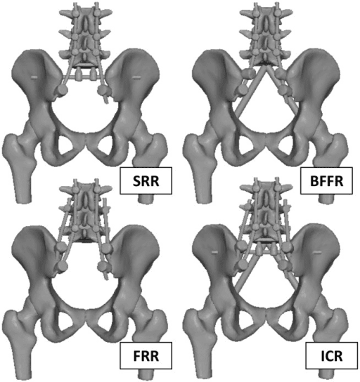
- Total sacrectomy disconnects spine from pelvis: reconstruct with lumbopelvic screw-rod fixation plus anterior column strut (fibula or cage) in a triangulated construct
- Unresectable or high sacral chordoma: definitive carbon-ion or high-dose proton radiotherapy is an established alternative
- “Unilateral root sacrifice at any sacral level generally preserves bladder and bowel through contralateral compensation
- “Bilateral S2-S5 loss produces a flaccid, insensate bladder, incontinent bowel and sexual dysfunction — mandatory pre-operative counselling
- “Silastic sheet placed anterior to the sacrum at the first (anterior) stage protects rectum and vessels during posterior osteotomies
In sacral chordoma, an intralesional or contaminated margin roughly doubles local recurrence versus wide/R0 resection, and local recurrence is the strongest predictor of disease-specific death. Never debulk a resectable chordoma.
Posterior midline, transtumoural, image-guided core biopsy with the tract tattooed or marked for en bloc excision. Transrectal biopsy contaminates the rectum and may convert a curable tumour into one requiring abdominoperineal resection — or an incurable one.
Consent must state expected bladder, bowel and sexual outcome for the planned root sacrifice. Trading margin for function is an oncologic error; trading function without consent is a medicolegal one.
Massive haemorrhage (frequently several litres) is the leading intraoperative threat. Anterior-stage ligation of internal iliac and median sacral vessels, cell salvage, staged surgery and a prepared transfusion protocol are mandatory for high resections.
Oncologic Principles and Tumour Biology
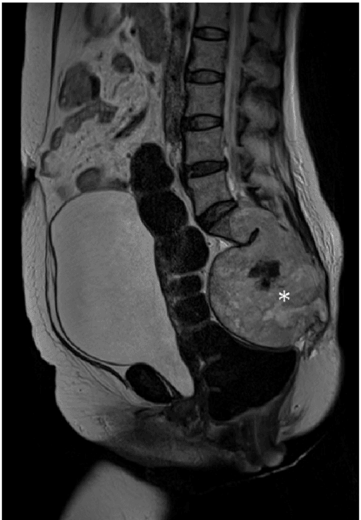
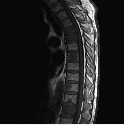
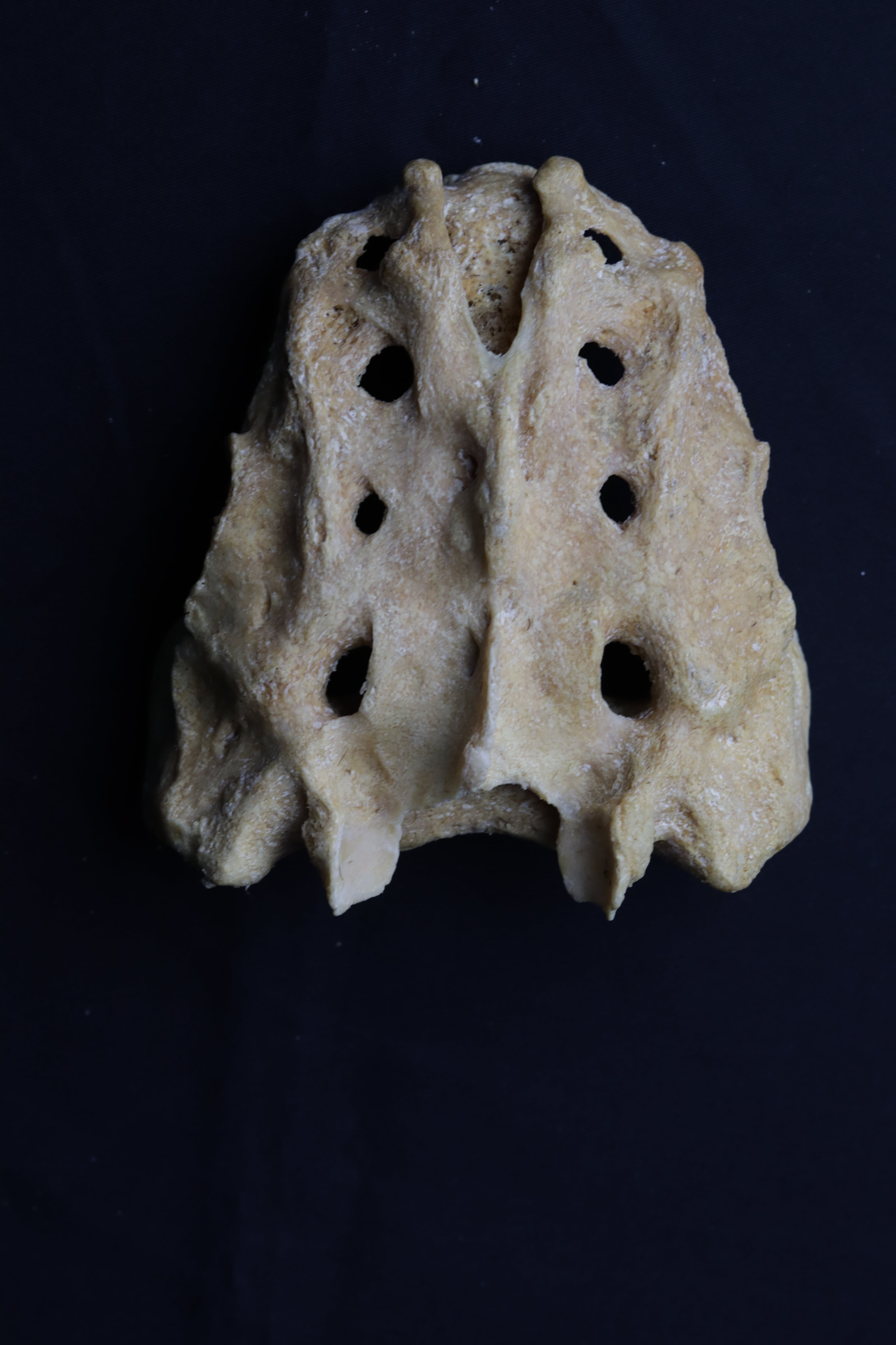
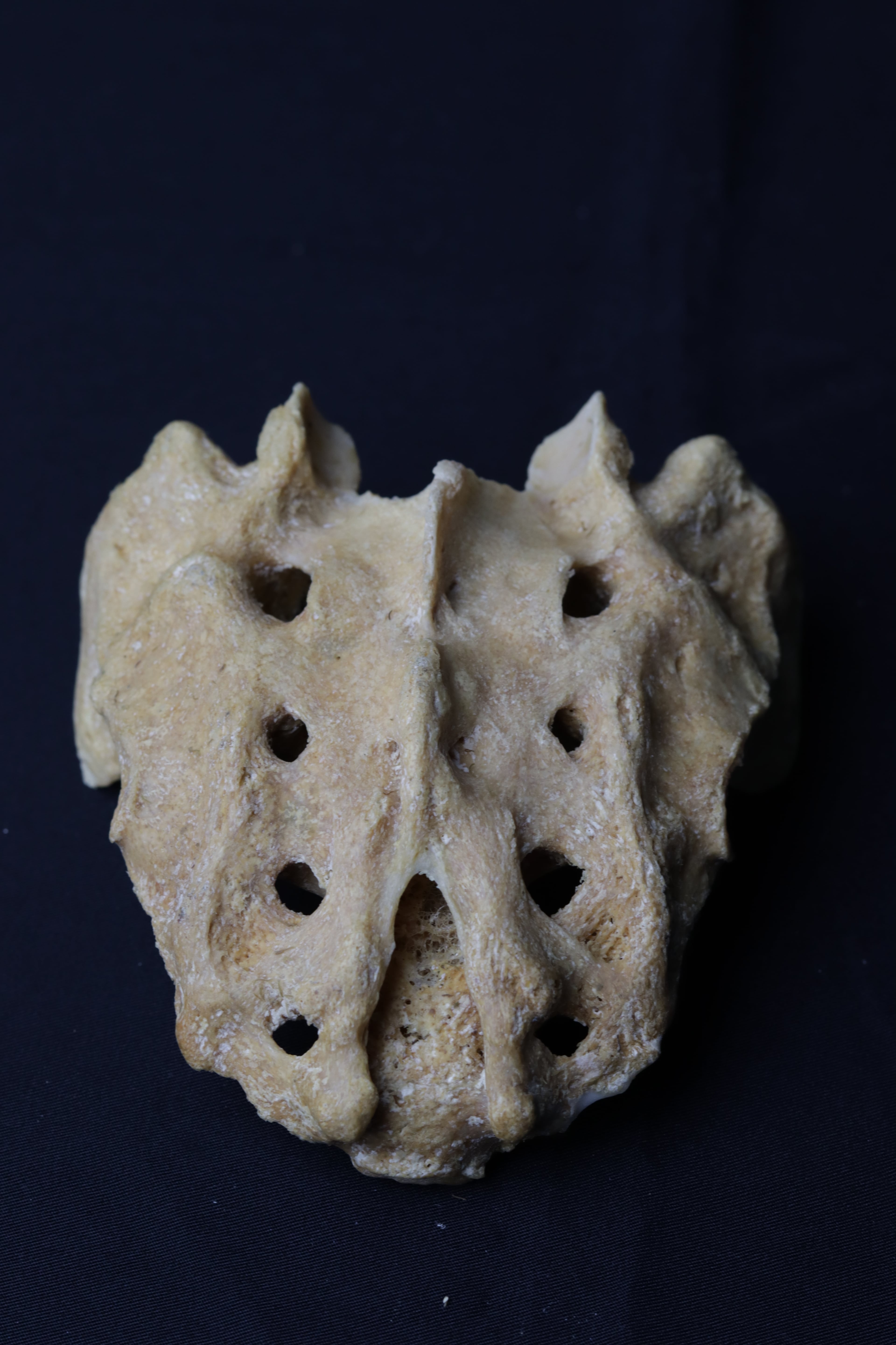
Primary sacral tumours amenable to sacrectomy are dominated by three diagnoses:
- Chordoma — the commonest primary malignant sacral tumour; arises from notochordal remnants in the midline, typically in patients in the sixth and seventh decades. Slow-growing, locally destructive, radioresistant and chemoresistant at conventional doses. Physaliphorous cells, brachyury-positive on immunohistochemistry.
- Chondrosarcoma — eccentric rather than midline; also radio- and chemoresistant, so surgical margin is again the decisive variable.
- Giant cell tumour (GCT) — younger patients (third and fourth decades), eccentric, lytic and expansile. Benign but locally aggressive; denosumab and serial embolisation have shifted many sacral GCTs away from sacrectomy toward intralesional surgery or non-operative control, because sacrificing continence for a benign tumour is rarely justified.
Core principle: for chordoma and chondrosarcoma the operation is planned backwards from the margin. The bony osteotomy level, the roots sacrificed, the amount of gluteus maximus and pelvic floor taken, and whether rectum must be resected are all dictated by achieving a wide margin — never by the desire to preserve function.
Biopsy
- CT-guided core needle biopsy through a posterior midline transtumoural tract, placed in the line of the future incision.
- The tract is considered contaminated and is excised en bloc with the specimen.
- Never transrectal, transvaginal or transperitoneal — contamination of these routes commits the patient to visceral resection or renders the tumour effectively unresectable with margin.
- Presacral masses discovered on rectal examination should NOT be biopsied through the rectum by non-oncology teams; refer intact to a sarcoma centre.
Anatomy and Neurological Consequences of Root Sacrifice
- 1Step 1 — Margin first
Define the oncological level required for a negative margin; the tumour dictates the osteotomy, not the desired function
Function is counselled around the margin, never traded for it
- 2Step 2 — Unilateral or bilateral?
Unilateral S1 to S5 sacrifice (hemisacrectomy) leaves the contralateral pelvic splanchnic (S2 to S4) and pudendal supply intact
Bladder, bowel and sexual function usually preserved by contralateral compensation
- 3Step 3 — Bilateral, but S3 and above preserved
Bilateral S4 to S5 division only
Normal or near-normal bladder, bowel and sexual function; minimal deficit
- 4Step 4 — Bilateral S2 preserved with one S3 retained
S3 sacrificed on one side only — the best compromise for mid sacrectomy
Majority retain socially acceptable, largely preserved voiding, evacuation and sexual function
- 5Step 5 — Bilateral S3-S5 sacrificed, both S2 roots intact
Both S3 roots divided with S2 preserved. This is the step most often under-warned, because 'S2 preserved' sounds reassuring
In Todd's 53-patient series only 25 per cent had normal bladder function and 40 per cent normal bowel function - so roughly three-quarters were abnormal despite bilateral S2 preservation. Keeping S2 without an S3 does not protect continence
- 6Step 6 — Bilateral S2 to S5 sacrificed
Complete loss of bilateral pelvic splanchnic and pudendal supply - every patient in Todd's series was abnormal
Flaccid, areflexic, insensate bladder; incontinent, insensate bowel; erectile and ejaculatory failure with loss of perineal sensation
- 7Step 7 — Extension to S1 and/or L5
Total sacrectomy with high extension adds somatic motor loss to the sacral deficit
Foot drop, plantarflexion weakness and gait deficit superimposed on complete sphincter and sexual loss
The sacral roots exit their foramina and merge to form the sacral plexus on pelvic surface of piriformis. The pelvic splanchnic nerves (S2-S4) carry parasympathetic supply to bladder, rectum and erectile tissue; the pudendal nerve (S2-S4) supplies the external sphincters and perineal sensation.
- Bladder
- Normal or near-normal
- Bowel
- Normal or near-normal
- Sexual function / motor
- Preserved
- Overall pattern
- Minimal deficit
- Bladder
- Usually preserved
- Bowel
- Usually preserved
- Sexual function / motor
- Usually preserved; possible ipsilateral motor/sensory loss
- Overall pattern
- Contralateral roots compensate
- Bladder
- Normal in 60 per cent (Todd)
- Bowel
- Normal in 67 per cent (Todd)
- Sexual function / motor
- Largely preserved
- Overall pattern
- Best compromise for mid sacrectomy - the retained S3 is what buys this, not the S2
- Bladder
- Normal in only 25 PER CENT - three-quarters are abnormal
- Bowel
- Normal in only 40 per cent
- Sexual function / motor
- Variable dysfunction
- Overall pattern
- Not a minor deficit. Preserving S2 alone does NOT protect continence; consent accordingly
- Bladder
- Normal in 69 per cent
- Bowel
- Normal in 100 per cent
- Sexual function / motor
- Preserved
- Overall pattern
- S3 is the root that matters
- Bladder
- Flaccid, areflexic, insensate bladder — intermittent self-catheterisation
- Bowel
- Incontinent, insensate bowel — regimented bowel programme or colostomy
- Sexual function / motor
- Erectile and ejaculatory failure; loss of perineal sensation
- Overall pattern
- Complete sacral deficit
- Bladder
- As above
- Bowel
- As above
- Sexual function / motor
- Foot drop, plantarflexion weakness, gait deficit added
- Overall pattern
- Motor deficit superimposed on sphincter loss
Preserve both S2 roots and at least one S3 root and the majority of patients retain socially acceptable bladder and bowel function. Bilateral S2 sacrifice is the functional watershed: below it, continence is lost. Unilateral sacrifice at any level is generally well compensated.
Classification by Level and Approach Selection
- Osteotomy
- Below S3 — distal S3 body or the S3-S4 junction; both S3 roots or lower sacrificed as the margin dictates
- Approach
- Posterior-only, prone, single stage; rectum mobilised bluntly off the anterior sacrum from below through the retrorectal plane after dividing the anococcygeal ligament
- Root sacrifice and function
- Little or no sphincter deficit if S3 is preserved bilaterally or sacrificed on one side only
- Reconstruction and cover
- None — sacroiliac joints intact, pelvic ring uninvolved; primary closure usually sufficient
- Osteotomy
- Through the S2-S3 region; thecal sac ligated below the roots to be preserved
- Approach
- Usually posterior-only in experienced hands; combined approach if there is a large presacral component, prior contamination or previous radiotherapy
- Root sacrifice and function
- Counsel for partial bladder and bowel dysfunction if bilateral S3 is taken
- Reconstruction and cover
- Usually not required — pelvic ring remains in continuity through the upper sacroiliac joints; stress fracture of the residual sacrum can still occur
- Osteotomy
- S1 involvement mandates total sacrectomy — the lumbar spine is disconnected from the pelvis
- Approach
- Staged combined anterior-posterior: typically laparotomy day 1, posterior stage during the same admission
- Root sacrifice and function
- Complete bilateral sacral root sacrifice from S1 or S2 downward with full sphincter deficit; consider planned colostomy
- Reconstruction and cover
- Mandatory spinopelvic reconstruction plus planned soft-tissue flap cover; highest blood loss, longest operating time and greatest complication burden — sarcoma-centre surgery with colorectal, vascular, plastic and spinal collaboration
- Osteotomy
- Anterior tumour extent modest, rectal plane free, vascular control achievable from behind
- Approach
- Posterior-only feasible — generally low and mid lesions
- Root sacrifice and function
- Root sacrifice limited to the planned margin
- Reconstruction and cover
- No anterior stage needed
- Osteotomy
- Bulky presacral disease, rectal involvement, re-resection after contamination or post-radiotherapy tissue
- Approach
- Combined staged anterior-posterior surgery
- Root sacrifice and function
- En bloc rectal resection with colostomy where the rectum is involved
- Reconstruction and cover
- Reconstruction and flap cover planned from the outset
Approach selection summary: posterior-only is feasible when the anterior tumour extent is modest, the rectal plane is free and vascular control from behind is realistic (generally low and mid lesions). Combined staged anterior-posterior surgery is chosen for high/total sacrectomy, bulky presacral disease, rectal involvement (en bloc rectal resection with colostomy), re-resection after contamination, or post-radiotherapy tissue.
Decision Thresholds in Management
- Sacral GCT: first-line is usually denosumab with or without serial embolisation and, if surgery is needed, nerve-sparing intralesional excision; sacrectomy with sphincter sacrifice for a benign tumour only in exceptional, refractory, destructive disease.
- Chordoma/chondrosarcoma, resectable with acceptable morbidity: en bloc wide resection is the standard of care.
- Chordoma requiring bilateral S2 sacrifice or S1 body resection: discuss in a multidisciplinary sarcoma meeting; options are total sacrectomy accepting complete deficit versus definitive carbon-ion or high-dose proton radiotherapy, which achieves durable local control in a meaningful proportion of patients and is favoured for surgically unfit patients, patients declining sphincter loss, and some high sacral tumours.
- Adjuvant radiotherapy: high-dose photon/proton therapy is commonly added for close margins or after any contamination; pre-operative radiotherapy protocols exist in some centres but raise wound risk.
- Recurrent chordoma: re-resection is rarely curative once the field is contaminated; particle therapy and systemic options (brachyury-targeted trials, imatinib historically with modest activity) are considered.
Operative Technique
Staged total sacrectomy: anterior then posterior
Midline laparotomy (laparoscopic or robotic in selected centres). Mobilise sigmoid colon and rectum and develop the presacral (retrorectal) plane sharply off the tumour pseudocapsule. If the rectum is adherent or involved, resect it en bloc with the specimen and form a colostomy. Identify and protect both ureters.
Ligate the median sacral artery. For high resections ligate or temporarily control both internal iliac arteries and the lateral and median sacral branches, while preserving the superior gluteal arteries to protect gluteal flap options. Ligate internal iliac veins with care: presacral venous plexus injury bleeds torrentially.
Anterior discectomy or partial corpectomy at the planned proximal level (L5-S1 for total sacrectomy); score or start the anterior osteotomies and lateral sacroiliac joint cuts so the posterior surgeon works to a defined plane.
Place a silastic sheet (or mesh/spacer) anterior to the sacrum, separating rectum and iliac vessels from the posterior osteotomy plane — the single most protective manoeuvre for the second stage. Consider harvesting a pedicled vertical rectus abdominis myocutaneous (VRAM) flap and tucking it into the pelvis for retrieval posteriorly. Close and stage two after physiological recovery.
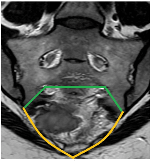
Prone on a four-poster frame, hips slightly flexed; entire back, buttocks and perineum prepared; anus sutured or excluded if being resected. Fluoroscopy or navigation for the osteotomy level, neuromonitoring of preserved roots, ultrasonic bone cutter useful. Midline incision excising the biopsy tract en bloc as an ellipse of skin; detach gluteus maximus origin taking a cuff on the tumour side, divide sacrotuberous and sacrospinous ligaments, release piriformis, protect the sciatic nerves.
Laminectomy above the tumour; ligate and divide the thecal sac below the last root to be preserved, using a watertight double ligature or oversew to prevent CSF leak. Divide roots destined for sacrifice sharply after ligation.
Complete the posterior osteotomies through the pre-scored planes (transverse sacral or bilateral sacroiliac/iliac cuts); the silastic sheet confirms the safe anterior plane. Divide the residual pelvic floor and deliver the specimen en bloc, then orient and margin-ink for the pathologist. At risk: presacral venous plexus, internal iliac vessels, rectum, sciatic and preserved sacral roots, superior gluteal bundles. Pitfalls include entering tumour through unrecognised extraosseous extension and an osteotomy too low leaving an R1 margin at S1.
Posterior tension band of pedicle screws L3-L5 (at least two lumbar levels) to bilateral iliac screws, often double iliac screws each side — the Galveston concept as modular screw-rod lumbopelvic fixation. Anterior column support with vascularised or non-vascularised fibula struts or titanium mesh cages from the L5 endplate to the ilium and acetabular pillars, triangulated so load is shared rather than cantilevered. Cross-links add torsional stability and abundant graft aims for lumbo-iliac fusion, since all-metal constructs eventually fatigue. Rod fracture and screw loosening are common in long-term survivors but often tolerable or revisable once partial fusion occurs; frail patients may be offered deliberate 'soft' reconstruction, accepting trunk shortening.
Deliver the pedicled VRAM through the pelvis as the workhorse for total sacrectomy and any irradiated field — well-vascularised muscle obliterates dead space and covers hardware. Gluteal advancement or V-Y flaps suit smaller low or mid defects when superior gluteal pedicles were preserved; free latissimus is the reserve. Primary closure alone after radiotherapy or total sacrectomy invites the 25 to 40 percent wound complication rate — plan the flap, do not react to breakdown. Drains, avoid direct pressure lying, graded mobilisation per construct, with catheterisation and a bowel regimen from the outset.
supine, midline laparotomy (or laparoscopic/robotic in selected centres).
- Mobilise sigmoid colon and rectum; develop the presacral (retrorectal) plane sharply off the tumour pseudocapsule — if rectum is adherent or involved, resect en bloc with the specimen and form a colostomy.
- Vascular control: ligate the median sacral artery and, for high resections, ligate or temporarily control both internal iliac arteries (and lateral/median sacral branches); preserve superior gluteal arteries where possible to protect gluteal flap options. Ligate internal iliac veins with care — presacral venous plexus injury bleeds torrentially.
- Anterior discectomy or partial corpectomy at the planned proximal level (L5-S1 for total sacrectomy); score or start the anterior osteotomies and lateral SI joint cuts.
- Place a silastic sheet (or mesh/spacer) anterior to the sacrum, separating rectum and iliac vessels from the posterior osteotomy plane — the single most protective manoeuvre for the second stage.
- Consider harvesting a pedicled vertical rectus abdominis myocutaneous (VRAM) flap, tucked into the pelvis for retrieval at the posterior stage.
- Close; stage two after physiological recovery.
Complications: Rates, Prevention, Management
- Mechanism
- Presacral venous plexus is valveless and diffuse — cannot be suture-ligated; torn during blunt rectal mobilisation
- Typical burden
- Blood loss frequently several litres in total sacrectomy
- Prevention
- Anterior-stage vascular ligation, staged surgery, silastic sheet, hypotensive anaesthesia, correct retrorectal fascial plane, activated transfusion protocol
- If it happens
- Packing, thumbtack occlusion, muscle-fragment welding, rapid completion of resection (bleeding often stops once the specimen is out), interventional radiology
- Mechanism
- Large dead space, thin posterior flaps, irradiated tissue bed
- Typical burden
- 25 to 40 percent, higher after radiotherapy
- Prevention
- VRAM or gluteal flap cover, dead-space obliteration, drains, offloading, nutritional optimisation
- If it happens
- Debridement, negative-pressure therapy, secondary flap
- Mechanism
- Rectal proximity and communication with the wound bed
- Typical burden
- Common, frequently linked to wound failure
- Prevention
- Flap cover, consider diverting colostomy in total sacrectomy, antibiotic protocol
- If it happens
- Debridement, retain hardware early and remove after fusion, prolonged antibiotics
- Mechanism
- Sacral root sacrifice — deficit is planned; unplanned injury to preserved roots also occurs
- Typical burden
- Bladder, bowel and sexual dysfunction proportional to level of root division
- Prevention
- Neuromonitoring, sharp division of intended roots only, preserve roots where oncologically safe
- If it happens
- Intermittent catheterisation, bowel programme, sexual health input, ankle-foot orthosis for L5/S1 loss
- Mechanism
- Thecal sac ligation at the transection level
- Typical burden
- Follows every high transection if closure is imperfect
- Prevention
- Double-ligature or watertight oversew of the sac, fibrin sealant, layered closure
- If it happens
- Lumbar drain; re-exploration and direct repair if persistent
- Mechanism
- Loss of spinopelvic continuity loads the construct until a fusion mass forms
- Typical burden
- Substantial in long-term total sacrectomy survivors
- Prevention
- Triangulated anterior strut plus lumbopelvic rods, generous biological fusion mass
- If it happens
- Observe if fused and asymptomatic; revise if painful instability
- Typical burden
- Blood loss frequently several litres in total sacrectomy
- Prevention
- Anterior vascular ligation, staged surgery, silastic sheet, hypotensive anaesthesia, transfusion protocol
- Management
- Packing, rapid completion of resection (bleeding often stops when specimen is out), interventional radiology
- Typical burden
- 25-40 percent, higher after radiotherapy
- Prevention
- VRAM or gluteal flap, dead-space obliteration, drains, offloading, nutrition
- Management
- Debridement, negative-pressure therapy, secondary flap
- Typical burden
- Common, frequently linked to wound failure and rectal proximity
- Prevention
- Flap cover, consider diverting colostomy in total sacrectomy, antibiotic protocol
- Management
- Debridement, hardware retention early / removal after fusion, prolonged antibiotics
- Typical burden
- Follows thecal sac ligation
- Prevention
- Double-ligature or watertight oversew of sac, fibrin sealant, layered closure
- Management
- Lumbar drain, re-exploration and repair if persistent
- Typical burden
- Substantial in long-term total sacrectomy survivors
- Prevention
- Triangulated anterior strut plus lumbopelvic rods, biological fusion mass
- Management
- Observe if fused and asymptomatic; revise if painful instability
- Typical burden
- Deficit is planned by root sacrifice; unplanned injury to preserved roots also occurs
- Prevention
- Neuromonitoring, sharp division only of intended roots
- Management
- Intermittent catheterisation, bowel programme, sexual health input, ankle-foot orthosis for L5/S1 loss
Injury to the presacral venous plexus during blunt rectal mobilisation causes bleeding that is diffuse, valveless and cannot be suture-ligated. Control with packing, thumbtack occlusion or muscle-fragment welding — and prevent it by staying in the correct retrorectal fascial plane and by anterior-stage vascular control.
BLOCKOrder of the posterior stage
Hook:You BLOCK the tumour's escape — every step protects the en bloc margin.
ONE S3Root sacrifice outcomes — count the S3s, not the S2s
Hook:One S3 is the watershed, not two S2s. Todd's own conclusion: unilateral resection, or preservation of at least one S3 root, preserves function in the majority.
Guidelines, Registries & Global Practice
- Epidemiology: chordoma incidence is roughly one per million per year worldwide; sacrococcygeal disease accounts for about half of chordomas, with male predominance and peak in the sixth to seventh decades. Sacral chondrosarcoma and GCT are rarer at this site.
- Society guidance: ESMO/EURACAN and Chordoma Global Consensus Group guidance emphasise referral of presacral masses to sarcoma centres before biopsy, trocar/core biopsy through a resectable posterior tract, en bloc resection with wide margins as standard for chordoma, and high-dose particle therapy (proton or carbon-ion) as definitive treatment where resection is not feasible or declined. NCCN bone tumour guidance mirrors these principles. BOA/BSG sarcoma pathways in the UK mandate management within designated sarcoma networks.
- Registries and multicentre series: there is no arthroplasty-style implant registry for sacrectomy; evidence rests on multicentre collaborations — AOSpine Knowledge Forum Tumour primary spinal tumour cohorts, Rizzoli Institute and Memorial Sloan Kettering chordoma series, and Japanese and Italian carbon-ion cohorts (NIRS/QST Chiba, CNAO Pavia).
- Practice variation by resource setting: carbon-ion facilities exist in only a handful of countries (Japan, Germany, Italy, Austria, China), so surgical resection remains the only curative option in most of the world; in low-resource settings, staged surgery, flap availability and intensive-care capacity constrain total sacrectomy, and non-operative palliation or referral abroad is common for S1-level disease. Denosumab access similarly determines whether sacral GCT is managed medically or surgically.
Controversies & Areas of Uncertainty
- Surgery versus particle therapy for high sacral chordoma: no randomised comparison exists and none is likely, but the field is no longer evidence-free — Yolcu's propensity-matched analysis found no significant difference in overall survival between carbon-ion therapy and en bloc resection (median 68.1 versus 58.6 months, hazard ratio 0.71, p equals 0.53) with markedly less peripheral motor neuropathy after carbon ion (odds ratio 0.13). Read it with its own caveat: the surgical arm was only 54 patients and that confidence interval spans a large benefit either way. Note also that carbon-ion series report ambulation, not continence — Imai's 97 per cent refers to walking, and his paper contains no sphincter data. Long-term insufficiency fracture, late nerve toxicity and the difficulty of salvage after radiotherapy failure remain genuine concerns.
- Rigid spinopelvic reconstruction versus no reconstruction after total sacrectomy: rigid constructs restore load transfer but carry high hardware failure and infection rates; some series report acceptable ambulation without reconstruction.
- Extent of nerve sacrifice for margin: whether a planned marginal (rather than wide) plane over preserved S2/S3 roots is oncologically acceptable in selected chordomas is debated.
- Cell salvage and tumour surgery: traditionally avoided; leucocyte-depletion filters and irradiation of salvaged blood are used in some centres.
- Denosumab in sacral GCT: optimal duration, rebound after cessation, and whether it should replace surgery entirely in the sacrum remain unresolved.
- Prophylactic colostomy in total sacrectomy: reduces perineal wound soiling but adds morbidity; practice varies widely.
MCQ Practice Points
A: Achievement of a wide (R0) surgical margin at the index resection. Contaminated/intralesional resection roughly doubles local recurrence, and local recurrence drives disease-specific mortality.
A: Bilateral S2 preservation with at least one S3 root — the majority retain useful bladder and bowel function. Bilateral S2 sacrifice produces a flaccid bladder and incontinent bowel.
A: CT-guided core biopsy through a posterior midline transtumoural tract placed in the line of the planned incision, excised en bloc at resection. Transrectal biopsy is absolutely contraindicated.
A: Total sacrectomy (S1 resection) disconnecting the lumbar spine from the pelvis. Reconstruct with lumbopelvic screw-rod fixation (lumbar pedicle plus iliac screws) triangulated with a fibular or cage anterior strut.
A: Definitive alternative to surgery for unresectable or high sacral chordoma, or where the patient declines sphincter sacrifice — Japanese cohorts show durable local control with sphincter preservation in most patients.
A: Placed anterior to the sacrum after rectal mobilisation and vascular ligation, it protects the rectum and iliac vessels and defines the safe anterior plane for the posterior-stage osteotomies.
Exam Viva Scenarios
Practise clinical reasoning and management decisions out loud
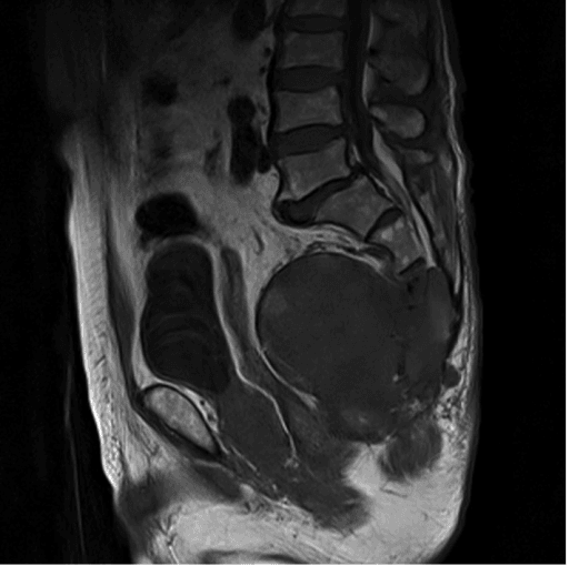
“A 62-year-old man has a firm midline presacral mass palpable on rectal examination and low sacral pain. MRI shows a destructive S3-S5 lesion with a lobulated, T2-hyperintense presacral component. The referring team asks whether to arrange a transrectal biopsy.”
“A 55-year-old woman requires sacrectomy through the S2-S3 junction for chordoma; the plan preserves both S2 roots and one S3 root. She asks exactly what will happen to her bladder, bowel and sexual function.”
“A 48-year-old man has a chordoma involving the S1 body with a large presacral mass abutting but not invading the rectum. He is fit and wishes to pursue curative treatment. Outline your operative strategy.”
“A 26-year-old woman has a large lytic eccentric S1-S2 lesion; biopsy confirms giant cell tumour. A colleague proposes total sacrectomy. What is your view?”
Oncology
- Chordoma: midline, notochordal, brachyury-positive, radioresistant — wide en bloc resection is curative treatment
- R0 margin halves local recurrence versus contaminated resection; recurrence drives mortality
- Biopsy: posterior midline transtumoural core, tract excised en bloc; NEVER transrectal
- Sacral GCT: denosumab and embolisation first; avoid sphincter sacrifice for benign disease
Levels and neurology
- Low (below S3): posterior-only, continence usually preserved
- Mid (S2-S3): posterior-only usually feasible; keep bilateral S2 plus one S3 for continence in the majority
- High/total (S1): staged anterior-posterior, complete sphincter deficit, reconstruction required
- Unilateral root sacrifice at any level: contralateral compensation preserves function
- Bilateral S2-S5 loss: flaccid bladder, incontinent bowel, sexual dysfunction; add L5/S1 loss = motor deficit
Technique
- Anterior stage: rectal mobilisation, ligate internal iliacs and median sacral, anterior L5-S1 discectomy, silastic sheet, harvest VRAM
- Posterior stage: excise biopsy tract, ligate thecal sac below preserved roots, osteotomies onto silastic sheet, en bloc delivery
- Reconstruction: L3-L5 pedicle screws to double iliac screws, triangulated fibula/cage strut, cross-links, fusion graft
- VRAM flap for total sacrectomy and irradiated fields
Complications and follow-up
- Massive haemorrhage — mitigated by anterior vascular control and staging
- Wound breakdown 25-40 percent; infection, CSF leak, hardware failure in long-term survivors
- Carbon-ion/proton therapy: definitive alternative for unresectable/high sacral chordoma
- Surveillance: MRI of the resection bed plus chest imaging at regular intervals for at least ten years — chordoma recurs late
Evidence Base
Operative Management of Sacral Chordoma
- 52 patients operated for sacrococcygeal chordoma at one institution between 1980 and 2001, followed a mean of 7.8 years
- A wide margin was achieved in only 21 of 52 - fewer than half
- ALL patients with a wide margin survived, significantly better than marginal or intralesional excision (p equals 0.0001)
- Local recurrence occurred in 23 patients (44 per cent); recurrence-free survival was 59 per cent at 5 years and 46 per cent at 10 years; overall survival 74, 52 and 47 per cent at 5, 10 and 15 years
- 17 of the 21 wide margins came from a COMBINED ANTEROPOSTERIOR approach and only 4 from a posterior-only approach
Bowel and Bladder Function After Major Sacral Resection
- 53 patients undergoing major sacral resection at one institution over 10 years, analysed retrospectively for bowel and bladder outcome by the roots divided
- UNILATERAL sacrectomy with contralateral roots preserved: normal bowel in 87 per cent, normal bladder in 89 per cent
- Bilateral S4-S5 division with BOTH S3 roots preserved: normal bowel in 100 per cent, normal bladder in 69 per cent
- Asymmetric resection preserving AT LEAST ONE S3 root: normal bowel in 67 per cent, normal bladder in 60 per cent
- Bilateral S3-S5 resection - that is, both S2 roots preserved but no S3: normal bowel in only 40 per cent and NORMAL BLADDER IN ONLY 25 PER CENT
- Bilateral S2-S5 sacrifice: ALL patients had abnormal bowel and bladder function
- The authors' stated conclusion: unilateral resection, OR preservation of at least one S3 root in a bilateral resection, preserves function in the majority
Carbon Ion Radiation Therapy for Unresectable Sacral Chordoma: An Analysis of 188 Cases
- 188 patients with UNRESECTABLE primary sacral chordoma treated at a single Japanese institute between 1996 and 2013, median age 66, median follow-up 62 months
- Proximal invasion reached above S2 in 137 of the 188 - this is a high-level, surgically difficult population
- Five-year local control 77.2 per cent, overall survival 81.1 per cent, disease-free survival 50.3 per cent
- 41 patients had a local recurrence; sex, tumour volume, level of proximal invasion and dose were all UNRELATED to local control
- Toxicity: grade 3 peripheral nerve in 6 patients and grade 4 skin in 2
- AMBULATION was retained in 97 per cent - note that the paper reports ambulation, NOT sphincter function, which it does not measure
Comparison of Oncologic Outcomes and Treatment-Related Toxicity of Carbon Ion Radiotherapy and En Bloc Resection for Sacral Chordoma
- 911 patients in total: 188 treated with carbon ion radiotherapy, 54 with en bloc resection, and 669 from the National Cancer Database as a survival comparator
- After PROPENSITY SCORE MATCHING there was NO significant difference in overall survival between carbon ion therapy and en bloc resection - median 68.1 versus 58.6 months (hazard ratio 0.71, p equals 0.53)
- The carbon ion cohort had substantially LESS peripheral motor neuropathy (odds ratio 0.13, 95 per cent CI 0.04 to 0.40, p less than 0.001)
- Against matched database cohorts, carbon ion therapy gave better survival than margin-positive surgery without adjuvant radiotherapy (64.7 versus 60.6 months, p equals 0.03) and far better than primary photon radiotherapy alone (64.9 versus 31.8 months, p less than 0.001)
- The authors position carbon ion therapy for older patients with high performance status in whom surgery is not preferred, and after margin-positive resection