The Fifth Ligament of the Knee
- The popliteus tendon originates INSIDE the joint capsule, from the popliteal sulcus on the lateral femoral condyle, anterior and distal to the fibular collateral ligament attachment.
- The femoral attachment of the popliteus tendon averages 18.5 mm from the femoral attachment of the fibular collateral ligament.
- The popliteus tendon, the popliteofibular ligament and the fibular collateral ligament are the three primary static restraints of the posterolateral corner.
- Popliteus unlocks the screw-home mechanism by internally rotating the tibia (or externally rotating the femur on a fixed tibia) at the start of flexion.
- The tibial nerve branch to popliteus winds around the muscle's lower border to enter its deep (anterior) surface — a recurrent innervation.
- “Popliteus is the only muscle in the body whose tendon of origin is intracapsular but extrasynovial, running through the popliteal hiatus.
- “The dial test at 30 degrees only implicates the posterolateral corner; positive at both 30 and 90 degrees implicates the PLC and the PCL.
- “Popliteus tendon impingement after total knee arthroplasty presents as lateral pain and a painful snap, often from an oversized femoral component or a retained posterolateral osteophyte.
- “An unaddressed posterolateral corner injury is a leading cause of failure of an otherwise well-performed cruciate reconstruction.
Overview
Popliteus is a flat, triangular muscle forming the floor of the lower popliteal fossa. It is anatomically peculiar in a way that matters clinically: it is the only muscle whose tendon of origin lies inside the knee joint. The tendon arises from the lateral femoral condyle, deep to the fibular collateral ligament, runs intracapsular but extrasynovial through the popliteal hiatus between the lateral meniscus and the capsule, and only then emerges to become a muscle belly on the posteromedial tibia.
This gives popliteus a dual identity that every question about it exploits:
- As a muscle, it is the unlocker of the knee — the muscle that initiates flexion from full extension by reversing the screw-home mechanism.
- As a ligament, the popliteus tendon and its extension the popliteofibular ligament (PFL) are among the primary static restraints of the posterolateral corner (PLC), resisting tibial external rotation, varus and posterior translation. LaPrade's group have called it the fifth ligament of the knee, and that phrase is worth having ready.
This is the single biomechanical concept an examiner will always push on for popliteus.
- Screw-home (terminal rotation) in extension: during the last 20-30 degrees of extension, the tibia externally rotates roughly 10-15 degrees relative to the femur. Three factors drive it:
- The medial femoral condyle has a longer articular surface than the lateral, so the tibia must rotate to complete its excursion.
- The anterior cruciate ligament tightens and guides the tibia into external rotation.
- The lateral pull of the quadriceps through the patellar tendon, and the shape of the lateral tibial plateau, complete the movement.
- Why it exists: in the screwed-home position the knee is a rigid, close-packed, locked column with the collaterals and cruciates taut, allowing standing with minimal quadriceps activity. It is an energy-saving mechanism.
- The problem: to flex, the knee must first be unlocked.
- Popliteus is the unlocker. With the foot off the ground (open chain), it internally rotates the tibia on the femur. With the foot planted (closed chain), it externally rotates the femur on the fixed tibia. Either way, the effect is the same: the screw-home is reversed and flexion can begin.
- The single most examinable statement: "Popliteus initiates flexion of the fully extended knee by unlocking the screw-home mechanism."
- Corollary in gait: the closed-chain function is the physiologically important one. Popliteus fires at heel strike and in early stance, when the knee must unlock and begin flexing while the foot is fixed.
3 Layers, 3 PrimariesThe Posterolateral Corner in Layers
Hook:Seebacher described the three layers; LaPrade defined the three primary structures. Reconstruct all three or the corner is not reconstructed.

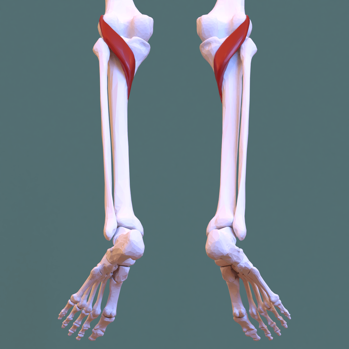
Attachments, Innervation and Relations
Origin — Intra-Articular
- Site: the popliteal sulcus (groove) on the lateral aspect of the lateral femoral condyle, at the anterior fifth of the sulcus.
- Relationship to the fibular collateral ligament (FCL): the popliteus tendon attaches anterior and distal to the FCL. In quantitative cadaveric mapping, the popliteus femoral attachment averages 18.5 mm from the FCL femoral attachment, and the FCL itself attaches approximately 1.4 mm proximal and 3.1 mm posterior to the lateral epicondyle.
- These two numbers are the anatomical foundation of every anatomical posterolateral corner reconstruction. Reproducing the 18.5 mm separation is what distinguishes an anatomical reconstruction from a non-anatomical sling.
- Character: the tendon is intracapsular but extrasynovial, invested by a synovial reflection that keeps it outside the joint fluid space while it lies inside the capsule.
Course — the Popliteal Hiatus
- From its femoral attachment the tendon runs posteriorly, medially and distally, passing deep to the fibular collateral ligament.
- It traverses the popliteal hiatus, the gap in the coronary (meniscotibial) attachment of the lateral meniscus through which the tendon reaches the posterior compartment.
- The popliteomeniscal fascicles — usually described as anteroinferior, posterosuperior and posteroinferior — bridge the hiatus, tethering the tendon to the lateral meniscus and forming the borders of the hiatus.
- What a lesion at the hiatus is worth as a sign. Stäubli's arthroscopic series found popliteus-system lesions in 95 per cent of acute and 85.7 per cent of chronic ACL-deficient knees — but also in 18.7 per cent of 107 knees with intact cruciates. Nearly one in five. So a frayed or detached fascicle seen at the hiatus is a common finding, not automatically the patient's problem, and it earns treatment only when the history, the dial test and the lateral meniscal behaviour agree with it.
The Popliteofibular Ligament
- Arises from the musculotendinous junction of popliteus and runs inferolaterally to attach to the posteromedial downslope of the fibular styloid process.
- Usually has anterior and posterior divisions, the posterior division being the larger and stronger.
- Functionally: it converts popliteus from a purely femorotibial structure into a femoro-fibular one, and it is a primary restraint to tibial external rotation. It is the structure most consistently disrupted in a PLC injury.
Muscle Belly and Insertion
- The muscle belly is triangular, arising from the tendon and expanding medially and distally.
- Insertion: the posterior surface of the tibia, above the soleal line, over a triangular area on the posteromedial proximal tibia.
- Additional attachments described in cadaveric work include slips to the posterior horn of the lateral meniscus, the posterior capsule, and in a substantial proportion of knees to the fibula and the posterior cruciate ligament region.
Related Structures
- Popliteal sesamoid (cyamella): a small sesamoid bone in the popliteus tendon, present in a minority of knees, occasionally mistaken for a loose body on radiographs.
- Arcuate ligament and fabellofibular ligament: variable posterolateral capsular thickenings; the fabellofibular ligament runs from the fabella to the fibular styloid and is present when a fabella is present.
- The nerve runs along the posteromedial border of the biceps femoris tendon, crosses the fibular neck roughly 2-4 cm distal to the tip of the fibular head, and then divides into superficial and deep branches as it passes through peroneus longus.
- In every posterolateral corner reconstruction, the nerve is identified and protected before any drilling, and formally neurolysed if it is scarred. A drill exiting the posteromedial fibular head passes within millimetres of it.
- Direct the fibular tunnel from the fibular styloid posteromedially to the anterolateral fibular head, with the nerve retracted and visualised.
- Beware the anterior tibial artery as it passes through the interosseous membrane just distal to the tibiofibular joint when drilling the tibial tunnel.
- Nerve palsy is present at the time of injury in a substantial proportion of PLC injuries — document it before surgery, or you will be blamed for it afterwards.
Action and Biomechanics
Actions
- Unlocking the knee: internal rotation of the tibia on the femur (open chain) or external rotation of the femur on the fixed tibia (closed chain), reversing the screw-home mechanism to initiate flexion.
- Weak knee flexion — a minor contribution compared with the hamstrings and gastrocnemius.
- Retraction of the lateral meniscus during flexion, through the popliteomeniscal fascicles, preventing entrapment of the posterior horn between the femoral condyle and the tibial plateau.
- Static restraint (as a ligament complex): with the popliteofibular ligament and the FCL, it resists tibial external rotation, varus angulation and posterior tibial translation.
Restraint Hierarchy — the Table Examiners Want
- Primary restraint
- Fibular collateral ligament
- Secondary restraint
- Popliteus complex, cruciates
- Clinical test
- Varus stress at 30 degrees
- Primary restraint
- FCL plus cruciates and posterolateral capsule
- Secondary restraint
- Popliteus complex
- Clinical test
- Varus stress in full extension — laxity here means a cruciate is also out
- Primary restraint
- Popliteus tendon and popliteofibular ligament
- Secondary restraint
- FCL, posterolateral capsule
- Clinical test
- Dial test at 30 degrees
- Primary restraint
- Posterior cruciate ligament with the PLC
- Secondary restraint
- Popliteus complex
- Clinical test
- Dial test at 90 degrees
- Primary restraint
- Posterior cruciate ligament
- Secondary restraint
- Popliteus complex and posterolateral structures
- Clinical test
- Posterior drawer
- Primary restraint
- Popliteomeniscal fascicles
- Secondary restraint
- Coronary ligament
- Clinical test
- Arthroscopic probing for a hypermobile lateral meniscus
The Dial Test — Interpretation
- Performed prone (or supine) with the knees flexed to 30 degrees, then repeated at 90 degrees, comparing the thigh-foot angle side to side. A difference of more than 10-15 degrees is positive.
- Positive at 30 degrees only: an isolated posterolateral corner injury.
- Positive at both 30 and 90 degrees: a combined posterolateral corner and posterior cruciate ligament injury.
- Positive at 90 degrees only: unusual, and should raise suspicion of an isolated PCL injury or a testing error.
- Why the difference: the PLC is the primary restraint to external rotation near extension; at 90 degrees the PCL becomes the dominant restraint, so external rotation laxity at 90 degrees requires the PCL to be involved.
Other Posterolateral Tests
- How to perform
- Prone, knees flexed 30 then 90 degrees; externally rotate both feet and compare thigh-foot angles
- Positive finding
- More than 10-15 degrees side-to-side difference
- What it means
- PLC injury at 30; combined PLC and PCL at 30 and 90
- False positives
- Physiological hyperlaxity; a rotated pelvis; medial-sided injury allowing external rotation
- How to perform
- Supine, lift both great toes and compare hyperextension and varus recurvatum
- Positive finding
- Recurvatum, varus and external rotation on the affected side
- What it means
- Combined PLC and cruciate injury, usually severe
- False positives
- Generalised ligamentous laxity
- How to perform
- Knee flexed 80-90 degrees, foot externally rotated 15 degrees, posterior force applied
- Positive finding
- Posterior rotation of the lateral tibial plateau
- What it means
- Posterolateral rotatory instability
- False positives
- PCL injury producing global posterior sag
- How to perform
- From flexion, extend the knee with a valgus force and the tibia externally rotated
- Positive finding
- A clunk as the laterally subluxed plateau reduces near 20-30 degrees
- What it means
- Posterolateral rotatory instability
- False positives
- Positive in up to a third of normal knees under anaesthesia — always compare sides
- How to perform
- Varus force applied in full extension and at 30 degrees
- Positive finding
- Laxity at 30 alone = FCL; laxity at 0 as well = FCL plus cruciate
- What it means
- Distinguishes isolated FCL from a combined injury
- False positives
- Bony varus alignment producing apparent laxity
- How to perform
- Weight-bearing full-length radiograph, mechanical axis measured
- Positive finding
- Varus mechanical axis passing through or medial to the medial compartment
- What it means
- Varus thrust that will overload any PLC reconstruction
- False positives
- Compensated stance masking the true alignment
The Failure Cascade
- Loss of the PLC allows the lateral compartment to open and the tibia to externally rotate. The cruciates become the primary restraint to motions they were designed only to resist secondarily.
- Consequence: the graft force in an ACL or PCL reconstruction rises substantially in a PLC-deficient knee, and this is the recognised mechanism by which an unaddressed PLC injury causes cruciate graft failure.
- Varus alignment compounds it. A varus mechanical axis puts a chronic tensile load on the reconstructed lateral structures. In a chronic PLC injury with a varus thrust, a corrective osteotomy must be considered before or with the ligament reconstruction, or the reconstruction will stretch out.
Surface Anatomy and Examination
Palpation
- The popliteus muscle belly is not directly palpable — it lies deep to the neurovascular bundle and the gastrocnemius heads in the popliteal fossa.
- The popliteus tendon can sometimes be appreciated as a tender cord in the posterolateral sulcus, just posterior to the fibular collateral ligament, with the knee flexed and in the figure-of-four position.
- The figure-of-four position (hip flexed, abducted and externally rotated with the knee flexed 90 degrees) is the key examination position for the posterolateral corner. It tensions the FCL, which becomes palpable as a distinct cord running from the lateral epicondyle to the fibular head, and it opens the lateral compartment for palpation and arthroscopic access.
- Landmarks: the lateral epicondyle, the fibular head and styloid, the Gerdy tubercle anteriorly, and the biceps femoris tendon posteriorly, with the common peroneal nerve rolling under the finger just posteromedial to it.
Examination Sequence for the Posterolateral Corner
- Gait: look for a varus thrust — lateral opening of the knee during stance. This is the single most important observation, because it changes management from soft tissue reconstruction to osteotomy first.
- Alignment: standing, and on a full-length weight-bearing radiograph.
- Neurological: document common peroneal nerve function before touching the patient surgically. A foot drop present at injury is common in PLC injuries.
- Varus stress at 0 and 30 degrees.
- Dial test at 30 and 90 degrees.
- Posterolateral drawer, reverse pivot shift, external rotation recurvatum.
- Cruciate testing: Lachman, pivot shift, posterior drawer, quadriceps active test.
- Vascular assessment, particularly in the acute multiligament knee.
Imaging
- Radiographs: an arcuate sign — a small avulsion fracture of the fibular styloid — is pathognomonic of a PLC injury and is easily missed. Also look for a Segond fracture (lateral tibial rim avulsion, associated with ACL injury) and a lateral capsular sign.
- Varus stress radiographs: the objective measure. Side-to-side lateral compartment gapping of more than about 2.7 mm at 20 degrees of flexion suggests an isolated FCL injury; more than about 4 mm suggests a complete posterolateral corner injury.
- MRI: thin-slice coronal oblique sequences along the plane of the FCL and popliteus tendon best demonstrate the corner. Report the FCL, the popliteus tendon, the popliteofibular ligament, the biceps insertion, the peroneal nerve and the presence of a fibular styloid avulsion.
- Full-length standing alignment radiographs in every chronic case.
Complications
Neurological
- Common peroneal nerve injury — the defining complication of posterolateral corner surgery. Mechanisms are direct injury, drill or retractor trauma at the fibular neck, and traction. A substantial proportion of PLC injuries have a pre-existing palsy, which must be documented before surgery.
- Tibial nerve branch to popliteus injury during a posterior approach to the tibia, from stripping the muscle off the tibia anteriorly. Functionally minor but a source of posterolateral pain.
- Sural nerve injury from posteromedial or posterolateral incisions.
Vascular
- Popliteal artery injury — the catastrophe. It lies directly on popliteus, only millimetres behind the posterior capsule. Prevention is knee flexion for all posterior work, and never drilling from anterior to posterior with the knee extended.
- Anterior tibial artery injury during tibial tunnel drilling near the interosseous membrane.
- Inferior lateral genicular artery bleeding during lateral compartment work.
Reconstruction-Related
- Graft failure and recurrent laxity — most commonly from an unaddressed varus alignment, from non-anatomical femoral tunnel placement, or from missing the corner injury altogether.
- Tunnel convergence in a combined ACL, PCL and PLC reconstruction; the femoral tunnels for the FCL, popliteus and ACL can collide. Plan divergent trajectories and consider a staged reconstruction if the tunnel geometry is impossible.
- Fibular head fracture through the transfibular tunnel, particularly in a small fibular head or with an over-large drill.
- Stiffness and arthrofibrosis after multiligament reconstruction, particularly with prolonged immobilisation.
- Over-constraint from over-tensioning the popliteus limb, producing a knee that cannot externally rotate and is painful in the figure-of-four position.
Arthroplasty-Related
- Popliteus tendon impingement and snapping — from component oversizing, retained osteophytes or cement.
- Iatrogenic popliteus division during lateral release or posterior osteophyte removal, producing flexion instability if the tendon was contributing to the lateral flexion space.
- Flexion instability from an inappropriate popliteus release in valgus correction.
Preventing Each
- Mechanism
- Drilling or retracting at the fibular neck without identifying the nerve
- Prevention
- Identify and protect the nerve first, every time; neurolyse if scarred; document pre-existing palsy
- Mechanism
- Posterior instrumentation or drilling with the knee extended
- Prevention
- Flex to at least 90 degrees for all posterior work
- Mechanism
- Unaddressed varus alignment or varus thrust
- Prevention
- Full-length standing radiographs; osteotomy before or with the reconstruction
- Mechanism
- Guessing the femoral attachment points
- Prevention
- Reference the lateral epicondyle; reproduce the 18.5 mm FCL to popliteus separation
- Mechanism
- Over-large transfibular tunnel in a small fibula
- Prevention
- Match tunnel diameter to the fibular head; consider a fibular-sparing technique
- Mechanism
- Releasing popliteus to correct an extension-space problem
- Prevention
- Match the release to the tight space; reassess gaps after each step
- Mechanism
- Oversized femoral component, retained posterolateral osteophyte, prominent cement
- Prevention
- Accurate sizing; remove posterolateral osteophytes under vision; control cement
Clinical Relevance
Mechanism and Epidemiology
- Mechanism: a varus force to a hyperextended knee, a posterolaterally directed blow to the anteromedial tibia, a twisting injury, or a knee dislocation.
- Rarely isolated: the great majority of PLC injuries occur with a cruciate injury — most commonly the PCL, and frequently both.
- Common peroneal nerve injury is present at the time of injury in a substantial minority, and its presence should raise suspicion of a severe corner injury or a spontaneously reduced knee dislocation.
Classification
- Hughston grading (by lateral gapping): grade I, 0-5 mm; grade II, 6-10 mm; grade III, more than 10 mm.
- Fanelli classification:
- Type A: increased external rotation only — injury to the popliteofibular ligament and popliteus tendon.
- Type B: increased external rotation plus mild varus opening — the above plus attenuation of the FCL.
- Type C: increased external rotation plus marked varus opening — disruption of the popliteus tendon, popliteofibular ligament, FCL and lateral capsule.
- The clinically useful version: an isolated FCL injury behaves differently from a complete corner injury, and only the latter reliably causes rotatory instability.
Management Principles
- Grade I and II isolated injuries: non-operative, in extension for 3-4 weeks with protected weight-bearing and progressive rehabilitation.
- Grade III injuries: operative. This is the point candidates get wrong — a complete PLC injury treated non-operatively is a recognised cause of chronic instability and cruciate graft failure.
- Acute (within 2-3 weeks): repair of avulsions with augmentation, or primary reconstruction. The published outcomes consistently favour reconstruction over isolated repair, because repaired attenuated tissue stretches out.
- Chronic: anatomical reconstruction, preceded or accompanied by a corrective osteotomy if there is a varus mechanical axis or a varus thrust.
- Combined injuries: address the corner at the same sitting as the cruciate. Reconstructing the cruciate alone in a PLC-deficient knee predictably fails.
Reconstruction Techniques
- What it reconstructs
- FCL, popliteus tendon and popliteofibular ligament, on two separate femoral tunnels 18.5 mm apart
- Advantages
- Anatomical; restores all three primary restraints separately
- Limitations
- Technically demanding; two femoral tunnels and a tibial tunnel; longer graft requirement
- What it reconstructs
- FCL and popliteofibular ligament via a figure-of-eight graft through a transfibular tunnel
- Advantages
- Simpler, fewer tunnels, widely used
- Limitations
- Does not reconstruct the popliteus tendon; non-anatomical femoral position in some descriptions
- What it reconstructs
- FCL and PFL with anatomical femoral fixation
- Advantages
- Balance of simplicity and anatomical femoral placement
- Limitations
- Still does not reproduce the popliteus tendon
- What it reconstructs
- Avulsed structures reattached
- Advantages
- Preserves native tissue in acute avulsions
- Limitations
- High failure rate if used alone for midsubstance or attenuated tissue
- What it reconstructs
- Non-anatomical lateral tenodesis
- Advantages
- Simple
- Limitations
- Largely superseded; does not restore rotational control
- What it reconstructs
- Corrects varus and increases the sagittal slope effect
- Advantages
- Essential in chronic varus; may alone relieve symptoms in some patients
- Limitations
- Does not restore rotational stability by itself
Rehabilitation
- Non-weight-bearing or protected weight-bearing in extension for 6 weeks.
- No active hamstring contraction early in combined PCL and PLC reconstruction, because the hamstrings pull the tibia posteriorly and load the graft.
- Avoid tibial external rotation loading for at least 3-4 months.
- Return to pivoting sport at 9-12 months, and only with restored strength and no varus thrust.
The classic cause of cruciate graft failure.
- A PLC injury raises graft force in an ACL or PCL reconstruction and stretches it out.
- Look for the arcuate sign (fibular styloid avulsion) on plain radiographs, do a dial test at 30 and 90 degrees, and get varus stress radiographs.
- In a chronic case, obtain full-length standing alignment films. A varus thrust means osteotomy first.
Creates flexion instability in a total knee.
- Popliteus is a flexion-space structure. The iliotibial band and posterolateral capsule are extension-space structures.
- Release only what matches the tight space, and reassess the gaps after every step.
- Many surgeons preserve the popliteus tendon entirely during valgus correction and pie-crust the iliotibial band instead.
Surgical Relevance
Structures at Risk with Distances
- Location relative to a landmark
- Posteromedial to the biceps femoris tendon; crosses the fibular neck 2-4 cm distal to the fibular head tip
- How to protect it
- Identify and protect before any fibular drilling; neurolyse if scarred
- Location relative to a landmark
- Immediately posterior to the popliteus muscle belly, a few millimetres to 1 cm behind the posterior capsule
- How to protect it
- Flex the knee to at least 90 degrees for all posterior work; never drill posteriorly with the knee extended
- Location relative to a landmark
- Passes through the interosseous membrane just distal to the proximal tibiofibular joint
- How to protect it
- Aim the tibial tunnel obliquely and avoid over-penetration anteriorly
- Location relative to a landmark
- Deep to the fibular collateral ligament, along the lateral meniscus
- How to protect it
- Anticipate bleeding during lateral meniscal and PLC surgery
- Location relative to a landmark
- Immediately adjacent to the popliteal hiatus
- How to protect it
- Do not damage the meniscal attachment when exposing the popliteus tendon
- Location relative to a landmark
- 1.4 mm proximal and 3.1 mm posterior to the lateral epicondyle
- How to protect it
- Reference point for anatomical reconstruction tunnels
- Location relative to a landmark
- Anterior fifth of the popliteal sulcus, averaging 18.5 mm from the FCL attachment
- How to protect it
- The second femoral tunnel in an anatomical reconstruction
The Posterolateral Approach to the Knee
- Position: supine with a bump under the ipsilateral hip, knee flexed 70-90 degrees; a figure-of-four position for the lateral compartment work.
- Incision: a curvilinear lateral incision from just posterior to the lateral epicondyle, curving distally toward the Gerdy tubercle, or a hockey-stick incision between the iliotibial band and the biceps.
- First step, always: identify the common peroneal nerve on the posteromedial border of the biceps femoris tendon and follow it distally past the fibular neck. Formal neurolysis is routine in chronic cases.
- Intervals:
- The iliotibial band is split or reflected anteriorly to expose the FCL femoral attachment and the lateral epicondyle.
- The interval between the iliotibial band and the biceps femoris gives access to the posterolateral capsule and the popliteus tendon.
- The FCL and popliteus tendon femoral attachments are found by dissecting through the iliotibial band split onto the lateral epicondyle and then identifying the two attachments, separated by roughly 18.5 mm.
- Uses: posterolateral corner reconstruction, fibular head fracture fixation, lateral meniscal root repair, peroneal nerve decompression, and access to posterolateral tibial plateau fragments.
Anatomical Posterolateral Corner Reconstruction — the Steps
- Expose and protect the common peroneal nerve.
- Create the fibular tunnel from the fibular styloid posteromedially (the popliteofibular ligament attachment) to the anterolateral fibular head (the FCL attachment), roughly 7 mm in diameter, with the nerve under direct vision.
- Create the tibial tunnel from the flat spot on the anterolateral proximal tibia, distal and medial to the Gerdy tubercle, exiting at the popliteal sulcus of the posterior tibia, just proximal and medial to the fibular styloid.
- Identify the two femoral attachments: the FCL (1.4 mm proximal and 3.1 mm posterior to the lateral epicondyle) and the popliteus tendon (18.5 mm anterior and distal to it, in the popliteal sulcus). Confirm the separation with a ruler — the number is the point.
- Drill two femoral tunnels at those points and pass a graft (typically split Achilles allograft or two separate grafts).
- Pass the FCL limb through the fibular tunnel and the popliteus limb through the tibial tunnel.
- Tension and fix: the FCL graft at 20-30 degrees of flexion with a slight valgus force and the tibia in neutral rotation; the popliteus graft at 60 degrees with the tibia in neutral rotation.
- Confirm correction of the dial test and varus stress intra-operatively.
The Osteotomy Question
- A varus mechanical axis or a varus thrust in a chronic PLC injury mandates consideration of a proximal tibial opening-wedge osteotomy, either as a first stage or concurrently.
- Reconstructing soft tissue into a varus limb is a recognised recipe for failure: the graft is loaded in tension with every step.
- A proportion of patients become sufficiently asymptomatic after the osteotomy alone that the ligament reconstruction becomes unnecessary — a point worth making, because it changes the counselling.
- Slope considerations: an opening-wedge osteotomy done anteromedially increases posterior tibial slope, which is favourable in a PCL-deficient knee and unfavourable in an ACL-deficient one. Plan the wedge geometry deliberately, not by default.
Guidelines, Registries & Global Practice
Anatomical Variation Across Populations
- The core posterolateral anatomy is highly consistent. The fibular collateral ligament, the popliteus tendon and the popliteofibular ligament are present in essentially all knees across European, North American and Asian cadaveric series, and the quantitative attachment relationships reproduce well.
- The variable structures are the capsular thickenings. The arcuate ligament and the fabellofibular ligament are inconsistently present, and their prevalence varies both between series and between populations, largely tracking the prevalence of the fabella itself, which is reported as more common in some East Asian populations than in European ones. This variability is why classifications based on the arcuate complex have been superseded by descriptions based on the three primary structures.
- The cyamella (popliteal sesamoid) is present in a minority of knees and is more commonly reported in some populations; it matters chiefly as a radiographic mimic of a loose body and as an occasional cause of snapping.
- Discoid lateral meniscus prevalence is substantially higher in East Asian populations, and a discoid meniscus alters the anatomy of the popliteal hiatus and the popliteomeniscal fascicles, increasing the relevance of lateral meniscal hypermobility in those populations.
Differences in Described Technique
- Position on the posterolateral corner
- Reconstruct all three primary structures — FCL, popliteus tendon and popliteofibular ligament — using two femoral tunnels 18.5 mm apart, a transfibular tunnel and a tibial tunnel.
- Position on the posterolateral corner
- A figure-of-eight graft through a transfibular tunnel reconstructing the FCL and popliteofibular ligament; simpler, widely used, does not reconstruct the popliteus tendon.
- Position on the posterolateral corner
- Classification-driven approach with simultaneous reconstruction of cruciates and corner in the multiligament knee.
- Position on the posterolateral corner
- Recognises that grade III posterolateral corner injuries warrant surgical treatment and that concomitant cruciate injury must be addressed.
- Position on the posterolateral corner
- Emphasises early specialist referral for the multiligament knee and for suspected knee dislocation, with mandatory vascular assessment.
- Position on the posterolateral corner
- Supports anatomical reconstruction over repair for grade III injuries and correction of malalignment before or with soft tissue reconstruction.
Practice Signals
- Across published series and systematic reviews, the consistent findings are that reconstruction outperforms repair for grade III posterolateral corner injuries, that failure to address concomitant varus alignment predicts failure, and that untreated posterolateral corner injury is a leading contributor to cruciate graft failure. These three statements are safe to make in any examination.
- National ligament registries capture cruciate reconstruction well but capture posterolateral corner surgery poorly, because the volume is low and coding is inconsistent. Registry data nonetheless identify concomitant collateral and rotatory instability as a risk factor for revision.
- Popliteus impingement after total knee arthroplasty is not captured in any registry and is almost certainly under-reported; it belongs in the differential of the painful total knee alongside infection, loosening, instability and malrotation.
High- and Limited-Resource Practice
- Well-resourced settings: MRI with coronal oblique posterolateral sequences, varus stress radiography, allograft availability for anatomical multi-tunnel reconstruction, and navigation for osteotomy planning.
- Limited-resource settings: the diagnosis is entirely clinical and radiographic — the dial test at 30 and 90 degrees, varus stress at 0 and 30 degrees, a plain radiograph looking for an arcuate sign, and full-length standing alignment films identify almost every posterolateral corner injury without an MRI. A fibular-based Larson reconstruction using autograft hamstring requires only a drill, a tunnel and fixation, and is a reasonable, reproducible operation where allograft and multi-tunnel instrumentation are unavailable. The universal, cost-free principle remains: identify and protect the common peroneal nerve first, and correct varus alignment before or with the soft tissue reconstruction.
MCQ Practice Points
Q: Where does the popliteus tendon originate? A: The anterior fifth of the popliteal sulcus on the lateral aspect of the lateral femoral condyle, anterior and distal to the fibular collateral ligament attachment — inside the capsule but outside the synovium.
Q: How far is the popliteus femoral attachment from the fibular collateral ligament attachment? A: An average of 18.5 mm. Reproducing this separation is what makes a posterolateral reconstruction anatomical.
Q: What is the principal action of popliteus? A: It unlocks the screw-home mechanism — internally rotating the tibia on the femur, or externally rotating the femur on the fixed tibia, to initiate flexion from full extension.
Q: How does the nerve to popliteus reach the muscle? A: The tibial nerve branch (L4-S1) descends over the posterior surface, winds around the lower border and enters the deep (anterior) surface — a recurrent course reflecting limb bud rotation.
Q: What does a dial test positive at 30 degrees but negative at 90 degrees mean? A: An isolated posterolateral corner injury. Positive at both 30 and 90 degrees implicates the PCL as well.
Q: What is the arcuate sign? A: An avulsion fracture of the fibular styloid on a plain radiograph — pathognomonic of a posterolateral corner injury and easily overlooked.
Q: Where does the popliteofibular ligament attach? A: From the musculotendinous junction of popliteus to the posteromedial downslope of the fibular styloid. It is a primary restraint to tibial external rotation.
Q: What lateral gapping on varus stress radiographs indicates a complete posterolateral corner injury? A: More than about 4 mm side-to-side difference. Around 2.7 mm suggests an isolated fibular collateral ligament injury.
Q: In a valgus total knee, is popliteus a flexion or an extension space structure? A: Flexion. Releasing it for extension tightness creates flexion instability. The iliotibial band and posterolateral capsule are the extension-space structures.
Q: A patient has lateral mechanical symptoms and a normal MRI. What should you probe at arthroscopy? A: The lateral meniscus for hypermobility from disrupted popliteomeniscal fascicles at the popliteal hiatus.
Exam Viva Scenarios
Practise clinical reasoning and management decisions out loud
“A 27-year-old rugby player is examined three weeks after a knee injury. The dial test shows 20 degrees more external rotation on the injured side at 30 degrees, and the same at 90 degrees. Varus stress opens at 30 degrees and also at 0 degrees. What is your diagnosis and how do you manage it?”
“A 68-year-old woman is 14 months after a total knee arthroplasty. She has lateral knee pain and a painful snap she can reproduce at about 40 degrees of flexion. Radiographs show well-fixed, well-aligned components. Inflammatory markers are normal. What is your approach?”
“Describe the screw-home mechanism and explain the role of popliteus. Then tell me why the muscle's tendon of origin is such an anatomical oddity.”
Core Anatomy
- Origin: popliteal sulcus, lateral femoral condyle — intracapsular, extrasynovial
- 18.5 mm from the FCL femoral attachment, anterior and distal to it
- Insertion: posterior tibia above the soleal line
- Nerve: tibial nerve L4-S1, recurrent, entering the deep surface
Biomechanics
- Unlocks the screw-home mechanism to initiate flexion
- Primary restraint to tibial external rotation with the popliteofibular ligament
- Retracts the lateral meniscus posterior horn during flexion
- Secondary restraint to varus and posterior translation
Examination
- Dial 30 only = PLC; dial 30 and 90 = PLC plus PCL
- Varus at 30 = FCL; varus at 0 = FCL plus cruciate
- Arcuate sign = fibular styloid avulsion, pathognomonic
- Varus stress radiographs: about 2.7 mm FCL, more than 4 mm complete corner
Surgery
- Identify the peroneal nerve first, always
- FCL femoral point: 1.4 mm proximal, 3.1 mm posterior to the lateral epicondyle
- Correct varus alignment before or with soft tissue reconstruction
- Popliteus is a flexion-space structure in TKA balancing
- Popliteus impingement after TKA: snap at 30-45 degrees, arthroscopic release
Evidence Base
The Posterolateral Attachments of the Knee: A Qualitative and Quantitative Morphologic Analysis
- Dissections and measurements on 10 non-paired fresh-frozen cadaveric knees
- The fibular collateral ligament attached on average 1.4 mm proximal and 3.1 mm posterior to the lateral epicondyle
- The popliteus tendon attached at the most proximal and anterior fifth of the popliteal sulcus, always anterior to the fibular collateral ligament
- The average distance between the femoral attachments of the popliteus tendon and the fibular collateral ligament was 18.5 mm
- The popliteofibular ligament had anterior and posterior divisions in all cases, attaching 2.8 mm and 1.6 mm distal to the tip of the fibular styloid
Analysis of the Static Function of the Popliteus Tendon and Evaluation of an Anatomic Reconstruction: The Fifth Ligament of the Knee
- Eleven non-paired cadaveric knees tested intact, with the popliteus tendon sectioned, and after anatomic reconstruction with an autogenous semitendinosus graft
- Sectioning the popliteus tendon significantly increased external rotation, and produced small but significant increases in internal rotation, varus angulation and anterior translation
- Reconstruction significantly reduced external rotation compared with the sectioned state at 20, 30, 60 and 90 degrees of flexion
- Reconstruction did not restore internal rotation, varus angulation or anterior translation, and external rotation remained significantly reduced relative to intact at 30, 60 and 90 degrees
- The authors concluded the popliteus tendon functions essentially as the fifth major ligament of the knee
The Posterolateral Aspect of the Knee: Anatomy and Surgical Approach
- Thirty cadaveric knees dissected to define the posterolateral structures, and the resulting surgical approach used in 71 consecutive patients operated on for posterolateral knee injury
- The approach used three fascial incisions and one lateral midcapsular incision to provide surgical access
- Structures individually identified included the layers of the iliotibial tract, the long and short heads of biceps femoris, the fibular collateral ligament, the mid-third lateral capsular ligament, the fabellofibular and posterior arcuate ligaments, the popliteus muscle complex, the lateral coronary ligament and the posterior capsule
- The authors concluded that the approach should allow proper assessment of the individual injured structures before repair
The Popliteus Tendon and Its Fascicles at the Popliteal Hiatus: Gross Anatomy and Functional Arthroscopic Evaluation With and Without Anterior Cruciate Ligament Deficiency
- Gross dissection of the popliteus muscle, tendon and fascicles and their relation to the lateral meniscus in 14 adult cadaver knees
- Videoarthroscopy through an anterolateral portal assessed the popliteus tendon and the fascicles forming the popliteal hiatus in vivo
- Structural lesions of the popliteus system were present in 18.7 per cent of 107 cruciate-intact control knees
- Lesions were present in 95 per cent of 40 acute anterior cruciate ligament disruptions and 85.7 per cent of 28 chronic ACL-deficient knees
- Arthroscopic evaluation of the popliteus tendon and its fascicles was a valuable adjunct in assessing secondary posterolateral restraints
Arthroscopic Treatment of Popliteus Tendon Dysfunction Following Total Knee Arthroplasty
- After total knee arthroplasty the popliteus tendon may cause a snap as it rolls over a retained lateral femoral condylar osteophyte
- It may alternatively subluxate over the posterior condyle of the femoral component
- Where the condition is painful and fails to respond to conservative treatment, arthroscopic release of the popliteus tendon has been beneficial
- Indexed by PubMed as a CASE REPORT - no cohort size, follow-up interval or failure rate is given anywhere in the paper
Popliteus Tendon Dysfunction Following Total Knee Arthroplasty
- The popliteus tendon is a potential source of internal derangement after total knee arthroplasty
- It can subluxate anteriorly and posteriorly over a retained lateral femoral condylar osteophyte
- It can also subluxate over the overhanging edge of the metallic posterior femoral condyle
- Surgical release of the tendon from its femoral insertion relieves the problem
- Indexed by PubMed as a CASE REPORT - the abstract contains no patient numbers, no follow-up and no outcome measure