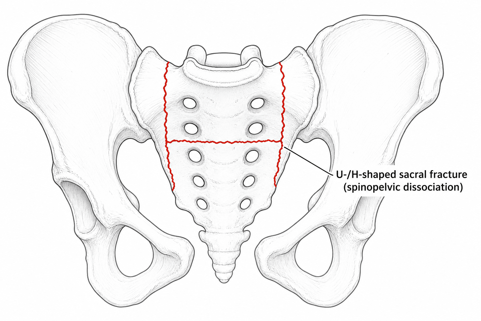
U-shaped Sacral Fracture / Lumbosacral Dissociation
- Traumatic spinopelvic (lumbosacral) dissociation is a TRANSVERSE sacral fracture combined with BILATERAL vertical (longitudinal) sacral fractures, which together DISCONNECT the upper-central sacrum and the spine above it from the rest of the sacrum/pelvis - producing the 'U-shaped' (also H-, Y-, lambda- or T-shaped) sacral fracture.
- It is a RARE, HIGH-ENERGY axial-loading injury - classically a fall from height ('jumper's fracture') or high-speed collision - and is HIGHLY UNSTABLE, with a HIGH INCIDENCE of NEUROLOGICAL injury (cauda equina / sacral nerve roots: bladder, bowel, sexual and saddle dysfunction).
- It is EASILY MISSED and diagnosis is often DELAYED because patients usually have severe associated (poly)trauma and the TRANSVERSE component is hard to see on the AP pelvis - look for it on the LATERAL sacral view (sacral kyphosis/step) and confirm with CT; if missed it leads to progressive deformity and chronic pain.
- The TRANSVERSE component is graded by the ROY-CAMILLE classification (Type 1 flexion/kyphosis, Type 2 flexion + posterior translation, Type 3 extension + anterior translation, Type 4 comminuted), and the vertical components are usually DENIS ZONE 3 (central) - the zone with the highest neurological risk.
- Imaging requires plain pelvic radiographs (AP, INLET, OUTLET), a LATERAL SACRAL radiograph, and a CT with reconstructions to define the pattern; MRI assesses the lumbosacral roots/cauda equina.
- Because the lumbopelvic junction is unstable, treatment is usually OPERATIVE: REALIGNMENT and LUMBOPELVIC FIXATION (spinopelvic fixation - e.g. L4/L5 pedicle to iliac or S2-alar-iliac screws, with or without iliosacral screws = TRIANGULAR OSTEOSYNTHESIS) to restore stability and allow EARLY MOBILISATION, with SACRAL DECOMPRESSION of compromised nerve roots when there is neurological deficit or canal compromise.
- “Spinopelvic dissociation = transverse + bilateral vertical sacral fractures (U/H shape) separating spine from pelvis - high-energy, highly unstable, high neuro-injury rate.
- “It's easily missed: look for the transverse component on the LATERAL sacral view (sacral kyphosis) and CT; suspect it in a jumper/polytrauma patient with sacral neurology.
- “Treat with lumbopelvic (triangular) fixation +/- sacral decompression; classify the transverse part by Roy-Camille (zone 3 Denis = highest neuro risk).
The patient is usually a polytrauma victim, and the transverse sacral component is hard to see on the AP pelvis. Look at the LATERAL sacrum (kyphosis/step), get a CT, and actively suspect it after a fall from height or with unexplained sacral neurology.
The vertical fractures are usually Denis zone 3 (central canal/transforaminal), so cauda equina and sacral-root injury is common - assess bladder, bowel, perianal sensation and sexual function in every suspected case.
What It Is & Why It Happens
In spinopelvic dissociation, bilateral vertical sacral fractures (one through each ala/foraminal region) are joined by a transverse fracture of the sacral body, so the upper-central sacrum - and the entire spine above it - is mechanically dissociated from the rest of the sacrum and the pelvis. The configuration of the transverse and vertical lines gives the characteristic shapes - 'U', 'H', 'Y', 'lambda' or 'T'. The mechanism is high-energy axial loading, classically a fall from a height (the 'jumper's fracture', often with calcaneal/tibial-plateau/spinal burst fractures) or a high-speed collision, so it is typically part of severe polytrauma.
Classification
| 0 | 1 |
|---|---|
| Type 1 | Simple flexion - kyphotic ANGULATION of the upper sacral fragment (no translation) |
| Type 2 | Flexion with POSTERIOR translation of the upper fragment |
| Type 3 | Extension with ANTERIOR translation of the upper fragment |
| Type 4 (Strange-Vognsen & Lebech) | Comminuted/segmental fracture of the S1 body |
The vertical components of the sacral fracture are graded by the Denis zones - Zone 1 (lateral to the foramina/ala), Zone 2 (transforaminal) and Zone 3 (medial to the foramina / central canal). Spinopelvic dissociation involves the central sacrum (Zone 3) and the transverse canal, which is why the neurological-injury rate is high (cauda equina and sacral roots controlling bladder, bowel and sexual function). The Roy-Camille type (especially the degree of translation/kyphosis) correlates with the risk and the need for decompression/reduction.
Assessment
- Pelvic radiographs: AP, inlet and outlet views
- LATERAL sacral radiograph - key to seeing the transverse component / sacral kyphosis
- CT with reconstructions - essential to define the full pattern (the diagnosis is often made here)
- MRI to evaluate the lumbosacral roots / cauda equina
- High-energy mechanism (fall from height, MVC) - treat as polytrauma (ATLS)
- Examine sacral neurology: bladder/bowel function, perianal sensation, anal tone, sexual function, lower-limb roots
- Inspect the soft tissues (degloving/Morel-Lavallée); assess associated spinal/limb injuries
Management
Because the lumbopelvic junction is grossly unstable, treatment is usually surgical, with three aims:
- Realignment / reduction of the displaced/kyphotic sacrum.
- Stable fixation of the lumbopelvic junction - LUMBOPELVIC (spinopelvic) FIXATION, connecting the lower lumbar spine (L4/L5 pedicle screws) to the pelvis (iliac or S2-alar-iliac (S2AI) screws), often combined with iliosacral screws as 'triangular osteosynthesis'. This provides robust, load-sharing fixation that resists the high shear at the lumbosacral junction and allows EARLY MOBILISATION of the polytrauma patient.
- Sacral decompression (e.g. sacral laminectomy / foraminal decompression) when there is a neurological deficit or canal/root compromise. Modern series using lumbopelvic fixation with S2AI screws report good reduction and function, reliable union and neurological recovery with low complication rates. (Minimally displaced, neurologically intact fractures may occasionally be managed non-operatively, but the unstable, displaced or neurologically involved patterns need fixation.)
Evidence & Key Studies
Traumatic spinopelvic dissociation or U-shaped sacral fracture: a review of the literature
- Spinopelvic dissociation is a rare high-energy injury - a transverse sacral fracture with bilateral sacral fracture-dislocations - with a high incidence of neurological complications.
- It is easily missed/delayed because patients commonly have severe associated injuries; if untreated it leads to progressive deformity and chronic pain. Imaging needs AP/inlet/outlet pelvis, a lateral sacral radiograph and CT.
- Early realignment and fixation of the unstable lumbopelvic junction (lumbopelvic fixation / triangular osteosynthesis) with decompression of compromised roots provides the best environment for early mobilisation.
Lumbopelvic fixation with S2 alar-iliac screws for U-shaped sacral fractures
- In 14 patients with U-shaped sacral fractures treated by lumbopelvic fixation with S2 alar-iliac (S2AI) screws, 92% had excellent/good reduction and 100% excellent/good pelvic function, with all fractures healed and neurological recovery in all.
- Complications were minimal (one superficial infection); no implant failure, loss of reduction or deep infection occurred.
- S2AI-screw lumbopelvic fixation gives firm fixation with low complication rates and supports early rehabilitation.
According to PubMed, the injury definition, the missed-diagnosis pitfall, the imaging requirements and the lumbopelvic-fixation/triangular-osteosynthesis principle come from the cited Yi & Hak review, and the S2AI lumbopelvic-fixation outcomes from the cited Luo series. The Roy-Camille classification of the transverse component and the Denis zones are standard, well-established sacral-fracture teaching. (See also our Sacral Fracture and Pelvic Ring Injury material.)
Clinical Decision Scenarios
Practise clinical reasoning and management decisions out loud
“A patient who fell from a height has sacral pain and difficulty voiding. What injury must you consider, what is the fracture pattern, and how would you image and assess it?”
“CT confirms a U-shaped sacral fracture with kyphotic displacement and a cauda equina deficit. How would you classify and manage it?”
Mnemonics & Memory Aids
U-SHAPE
Hook:U-SHAPE: the spine separates from the pelvis - high-energy, high-neuro, see it on the lateral/CT, fix it lumbopelvic.
ROY
Hook:Roy-Camille: 1 angulation, 2 posterior, 3 anterior translation, 4 comminuted.
What it is
- Transverse + bilateral vertical sacral fractures = U/H/Y/lambda shape
- Spine dissociated from pelvis - highly unstable
- High-energy (fall from height/jumper's fracture); usually polytrauma
Classify
- Transverse component: Roy-Camille 1 (flexion/kyphosis), 2 (flexion+posterior), 3 (extension+anterior), 4 (comminuted)
- Vertical components: Denis zone 3 (central) - highest neuro risk
- High rate of cauda equina/sacral-root injury
Diagnose
- Easily missed - AP/inlet/outlet + LATERAL sacrum (kyphosis) + CT (+/- MRI)
- Full sacral neuro exam (bladder/bowel/perianal/sexual)
- Screen for associated spinal/limb injuries
Treat
- Realign + lumbopelvic (spinopelvic) fixation - L4/L5 to iliac/S2AI screws +/- iliosacral (triangular osteosynthesis)
- Sacral decompression for neurological deficit/canal compromise
- Robust fixation -> early mobilisation; counsel re incomplete neuro recovery