The Dorsiflexor That Controls Heel Strike
- Origin: lateral condyle and upper half to two-thirds of the lateral tibial surface plus the interosseous membrane and deep fascia.
- Insertion: medial and plantar aspect of the medial cuneiform and the base of the first metatarsal - it spans the medial column.
- Innervation: deep peroneal (deep fibular) nerve, roots L4 and L5, entering the proximal third of the muscle.
- It is the only muscle that both dorsiflexes and inverts, so isolated loss produces drop foot without fixed varus at rest.
- It is the most superficial and most medial muscle of the anterior compartment - the palpable landmark for compartment assessment.
- “Steppage gait means excessive hip and knee flexion in swing to clear a foot that cannot dorsiflex.
- “Tibialis anterior rupture in the patient over 60 is painless, slowly progressive and routinely mislabelled as an L5 radiculopathy or a peroneal palsy.
- “SPLATT transfers the lateral half of the tendon to the cuboid or lateral cuneiform to convert a deforming invertor into a balanced dorsiflexor.
- “Delta P (diastolic minus compartment pressure) of 30 mmHg or less is the accepted decompression threshold, not an absolute pressure.
Overview
Tibialis anterior is the largest and most superficial muscle of the anterior (extensor) compartment of the leg. It is the workhorse of swing-phase foot clearance and the eccentric brake that lowers the forefoot to the ground in the first 10 per cent of stance. Because it inserts on the medial column at the medial cuneiform and first metatarsal base, it is simultaneously a dorsiflexor of the ankle and a supinator of the foot, and this dual vector explains almost all of its clinical behaviour: when the muscle fails the foot drops, and when it is spastic or unopposed the foot goes into varus.
Surgically it is the muscle you meet first in every anterior approach to the tibia and ankle, the muscle whose tendon is thick enough to be split and transferred without sacrificing dorsiflexion, and the muscle whose belly is a poor local flap because of its segmental vascular pattern.
Examiners like to push on when the muscle works rather than what it does:
- Swing phase (concentric): tibialis anterior shortens to lift the foot into dorsiflexion so the toes clear the floor. Failure produces a steppage (high-stepping) gait, in which the patient exaggerates hip and knee flexion to hoist the whole limb.
- Loading response (eccentric): immediately after heel strike the muscle lengthens under load to control the plantarflexion moment created by ground reaction force acting posterior to the ankle. Failure produces foot slap - the audible slap of the forefoot hitting the floor.
- Mid-stance (silent): the muscle is essentially electrically quiet once the foot is flat; the soleus takes over as the controller of tibial advance.
The clinical consequence: a patient can have a completely ruptured tibialis anterior tendon and still walk, because the extensor digitorum longus, extensor hallucis longus and peroneus tertius provide accessory dorsiflexion. They cannot, however, heel-walk, and eccentric control is lost first.
TEDSAnterior Compartment Contents
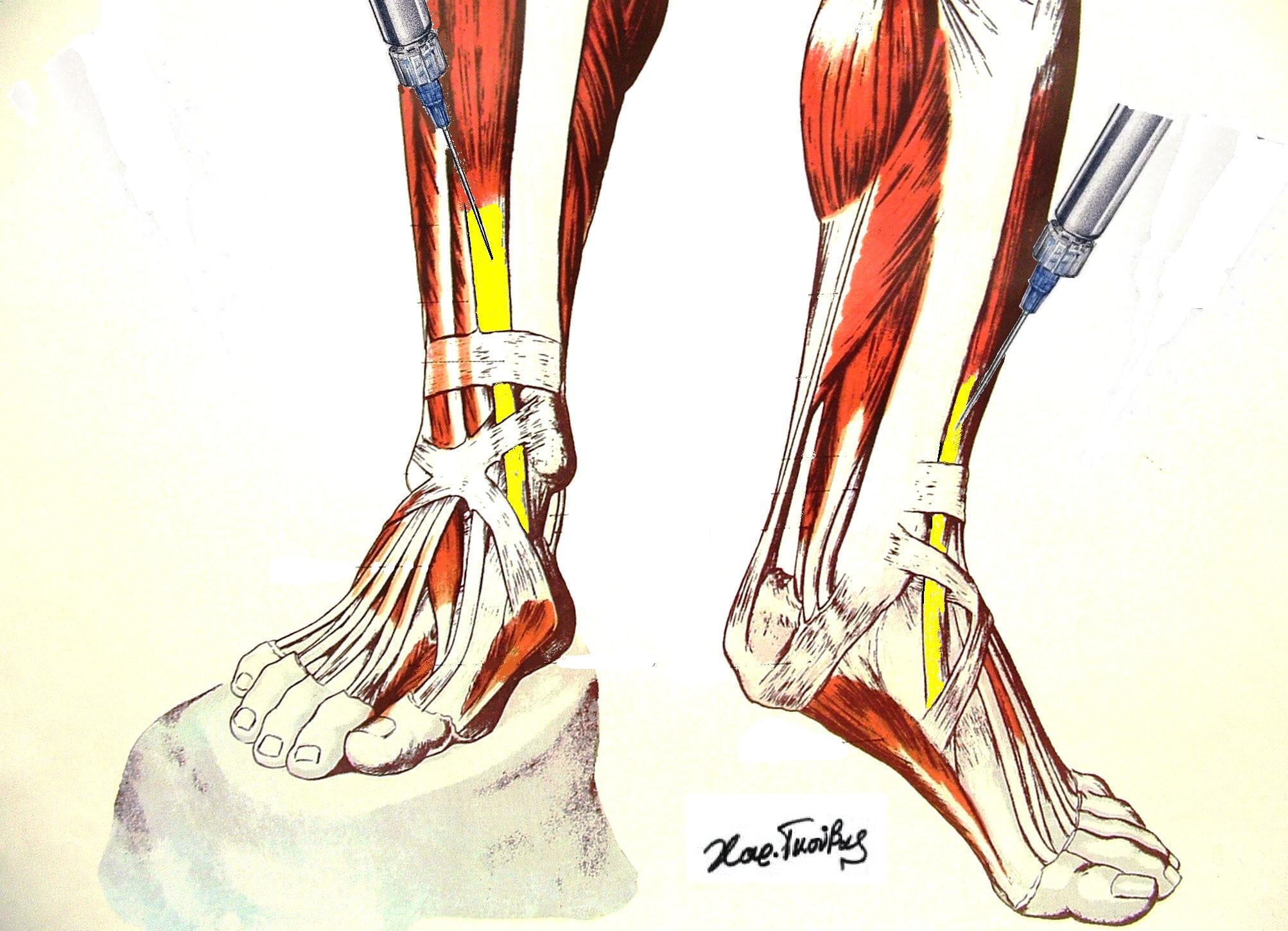
Hook:Plus one artery and one nerve: anterior tibial artery and deep peroneal nerve. Medial to lateral at the ankle - TA, EHL, artery and nerve, EDL, peroneus tertius.
Attachments, Innervation and Relations
Origin
- Lateral condyle of the tibia and the upper half to upper two-thirds of the lateral surface of the tibial shaft.
- Interosseous membrane (adjacent portion).
- Deep fascia of the leg and the anterior intermuscular septum shared with extensor digitorum longus.
- Fibres are unipennate proximally and bipennate distally, converging on a tendon that begins around the junction of the middle and distal thirds of the leg - considerably more proximal than most trainees expect, which is why a distal anterior tibial exposure encounters tendon rather than muscle.
Tendon course
- Passes deep to the superior extensor retinaculum and then through the medial (superomedial) limb of the inferior extensor retinaculum, which forms a discrete synovial-lined tunnel for it.
- The tendon is the most medial structure at the front of the ankle, running obliquely from the anterior ankle downward and medially across the talonavicular joint.
Insertion
- Medial and plantar surface of the medial cuneiform (the dominant and more constant slip).
- Base of the first metatarsal, medial and plantar aspect.
- The relative split between the two is variable; a common description is roughly equal division. A purely cuneiform or purely metatarsal insertion each occur in a minority of feet, and this variability matters when planning where a transfer will be re-anchored.
- There is a consistent fibrous expansion into the plantar fascia and the first tarsometatarsal capsule, coupling the tendon to medial-column stability.
The deep peroneal nerve is tethered where it pierces the anterior intermuscular septum and where it lies on the interosseous membrane. In anterior compartment syndrome the first sensory sign is numbness in the first dorsal web space, and it precedes motor loss. Never wait for foot drop to make the diagnosis of an anterior compartment syndrome - by then the muscle is already infarcting.
Action and Biomechanics
Actions by plane
- Sagittal: ankle dorsiflexion. Tibialis anterior contributes the majority of dorsiflexion torque; extensor digitorum longus, extensor hallucis longus and peroneus tertius supply the remainder.
- Frontal: inversion (supination) of the foot at the subtalar and transverse tarsal joints, because the tendon passes medial to the subtalar joint axis.
- Transverse: mild adduction of the forefoot through its medial-column insertion.
- First ray: dorsiflexion and elevation of the first metatarsal - the direct antagonist of peroneus longus, which plantarflexes the first ray. This antagonist pair is the axis around which cavovarus deformity turns.
Moment arms and force
- The dorsiflexion moment arm at the ankle is around 3.5 to 4 cm, the largest of the dorsiflexors, which is why the muscle dominates the group.
- In the classic cadaveric analysis of relative strength and excursion about the foot and ankle (Silver, de la Garza and Rang, 1985), tibialis anterior contributes only a small percentage of total lower-leg muscle strength, yet it is stronger than all other dorsiflexors combined. That study found the plantarflexors of the ankle to be six times as strong as the dorsiflexors, which is why an equinus contracture always beats a dorsiflexor and why a dorsiflexion transfer must be accompanied by lengthening of a tight gastrocsoleus.
- Excursion is approximately 3 cm. Any tendon transfer must respect excursion matching: a donor with 3 cm of excursion cannot reproduce the function of a recipient needing 5 cm.
Length-tension and transfer principles
- Tibialis anterior is a phasic swing-phase muscle. Transferring it into a stance-phase role requires either phase conversion (unreliable in spasticity) or a design that exploits its natural firing, which is exactly what a split transfer does.
- One grade of power is lost on transferring any tendon; a donor must therefore be at least MRC grade 4, and preferably grade 5, before transfer.
- A transfer must be straight-line, deep to the retinaculum, and tensioned in the corrected position with a supple, passively correctable joint.
What happens when it fails
- Gait consequence
- Drop foot with steppage gait and foot slap
- Compensation
- Hip and knee hyperflexion in swing
- Discriminating sign
- Eversion also lost, sensory loss in first web space and dorsum
- Gait consequence
- Drop foot, eversion preserved
- Compensation
- Peroneals evert but cannot dorsiflex
- Discriminating sign
- Numbness confined to the first web space
- Gait consequence
- Mild drop foot, foot slap, pseudo-drop
- Compensation
- EDL and EHL recruit - visible clawing of toes
- Discriminating sign
- Absent tendon cord on active dorsiflexion, normal sensation
- Gait consequence
- Weak dorsiflexion AND weak EHL AND weak hip abduction
- Compensation
- Trendelenburg may coexist
- Discriminating sign
- EHL weakness with dermatomal L5 sensory change
- Gait consequence
- Equinovarus, forefoot supination in swing
- Compensation
- Weightbearing on lateral border
- Discriminating sign
- Dynamic varus that corrects passively
When the tibialis anterior tendon is ruptured but the deep peroneal nerve is intact, the patient recruits extensor digitorum longus and extensor hallucis longus as substitute dorsiflexors. Because these muscles insert on the toes, the recruitment produces visible hyperextension of the toes during attempted dorsiflexion - the toes claw upward while the ankle barely moves. Combine this with a palpably absent tibialis anterior cord in the anteromedial ankle and normal sensation, and you have made the diagnosis at the bedside without imaging.
Surface Anatomy and Examination
Palpation
- Ask the patient to dorsiflex and invert against resistance. The tendon stands out as the most medial and most prominent cord at the front of the ankle, running from the anterior ankle to the medial cuneiform.
- Trace it distally to the medial cuneiform to confirm continuity. In rupture the cord vanishes distal to the retinaculum, and a soft-tissue fullness or pseudotumour may be palpable just proximal to the ankle where the retracted stump sits.
- The muscle belly is palpable immediately lateral to the anterior tibial crest in the upper two-thirds of the leg. Firmness here is the clinical sign of a tense anterior compartment.
Isolation and grading
- Isolate: the ankle is dorsiflexed with the foot inverted to exclude the toe extensors and peroneus tertius. Dorsiflexion in eversion recruits extensor digitorum longus and peroneus tertius preferentially.
- MRC grading: grade 5 is full resistance in dorsiflexion-inversion; grade 3 is dorsiflexion against gravity only, which is the threshold below which foot clearance fails.
- Heel walking is the single best functional screen. A patient who cannot walk on their heels for 10 metres has meaningful dorsiflexor weakness whatever the bench test suggests.
Named clinical tests and signs
- How to perform
- Ankle dorsiflexed with the foot inverted against the examiner hand
- Positive finding
- Weakness or an absent palpable cord
- What it means
- Tibialis anterior weakness or rupture
- False positives
- Pain inhibition after trauma; a tight gastrocsoleus blocking motion
- How to perform
- Walk 10 metres on the heels
- Positive finding
- Cannot maintain forefoot clearance
- What it means
- Functional dorsiflexor insufficiency
- False positives
- Balance disorder, hallux rigidus pain, ankle arthritis
- How to perform
- Inspect the anteromedial ankle during active dorsiflexion
- Positive finding
- Cord absent, pseudotumour proximal to the retinaculum, toes claw
- What it means
- Tendon rupture rather than nerve palsy
- False positives
- Obesity or oedema obscuring the cord - always compare sides
- How to perform
- Passive dorsiflexion with the knee extended, then flexed to 90 degrees, hindfoot held neutral
- Positive finding
- Dorsiflexion improves with the knee flexed
- What it means
- Isolated gastrocnemius contracture - recession rather than TAL
- False positives
- Allowing midfoot dorsiflexion to substitute if the hindfoot is not locked in neutral
- How to perform
- Stand with a block under the lateral border, first ray allowed to drop
- Positive finding
- Hindfoot varus corrects
- What it means
- Flexible hindfoot driven by a plantarflexed first ray
- False positives
- Rigid hindfoot with a subtalar coalition will not correct
- How to perform
- Watch swing phase from the side
- Positive finding
- Exaggerated hip and knee flexion, then foot slap at loading
- What it means
- Dorsiflexor failure of any cause
- False positives
- Circumduction from a stiff knee, vaulting from limb-length inequality
False positives and traps
- Pain inhibition after ankle trauma mimics weakness. Re-test after analgesia or a block.
- A tight gastrocsoleus limits dorsiflexion passively and can be misread as dorsiflexor weakness. Always test dorsiflexion with the knee flexed.
- Extensor substitution can produce apparently useful dorsiflexion with a completely ruptured tendon; look at the toes.
- A partial tear may retain a palpable cord but with a painful nodule and loss of endurance rather than loss of power.
Complications
- Mechanism
- Deep dissection in the proximal anterior compartment; retraction in the anterior ankle approach
- Prevention
- Stay subperiosteal on the lateral tibia; retract the bundle laterally with EHL, not medially with TA
- Management
- Observe; explore if a sharp injury is suspected; AFO and tendon transfer if permanent
- Mechanism
- Transverse or oblique anterior ankle incision
- Prevention
- Longitudinal incisions; identify the nerve with the foot inverted and the fourth toe plantarflexed before incising
- Management
- Desensitisation; neuroma excision and burial in muscle if refractory
- Mechanism
- Thin subcutaneous cover with no muscle beneath; a tight retinacular repair
- Prevention
- Full-thickness flaps, no undermining, careful tension-free retinacular closure
- Management
- Local wound care, negative pressure dressing, flap cover if the tendon is exposed
- Mechanism
- Routing superficial to the extensor retinaculum
- Prevention
- Always pass the transfer deep to the retinaculum
- Management
- Revision routing; a bowstrung tendon looks poor and loses effective excursion
- Mechanism
- Transferring too much tendon or too far laterally; a whole transfer in a spastic foot
- Prevention
- Split rather than whole transfer in spasticity; recipient at cuboid or lateral cuneiform
- Management
- Orthotic management; revision or medialising calcaneal osteotomy if fixed
- Mechanism
- Untreated tibialis posterior overactivity, untreated equinus, or fixed bony deformity
- Prevention
- Full dynamic assessment before surgery; Silfverskiold test in every case
- Management
- Address the missed component: posterior tibial lengthening, gastrocnemius recession, calcaneal osteotomy
- Mechanism
- Early aggressive dorsiflexion loading; poor tissue quality in a degenerate tendon
- Prevention
- Protect in dorsiflexion for 4 to 6 weeks, then graduated loading over 3 months
- Management
- Revision with graft augmentation
- Mechanism
- Missed compartment syndrome with muscle infarction and fibrosis
- Prevention
- Timely fasciotomy on delta P criteria
- Management
- Tendon lengthening, transfer of a functioning muscle, or arthrodesis in salvage
Donor morbidity of tibialis anterior transfer
- Whole tendon transfer to the lateral cuneiform does not create a drop foot because the muscle still dorsiflexes; the risk is a change in the frontal-plane vector producing pronation if the recipient site is placed too laterally.
- Split transfer preserves the medial insertion, so frontal-plane balance is retained by design; the principal risk is inadequate correction if too little tendon is transferred.
- Neither transfer causes meaningful loss of dorsiflexion power in a normally innervated muscle, which is precisely why this tendon is such a favoured donor.
Clinical Relevance
Foot drop
- Common peroneal nerve palsy at the fibular neck is the commonest cause: compression from plaster, positioning, a thin patient habitually crossing the legs, a proximal fibular fracture, a lateral knee dislocation, or a peroneal intraneural ganglion arising from the superior tibiofibular joint.
- Deep peroneal branch alone is affected in anterior compartment syndrome and in anterior tarsal tunnel syndrome (entrapment beneath the inferior extensor retinaculum).
- L5 radiculopathy is the great mimic; the discriminators are weakness of extensor hallucis longus and of hip abduction (gluteus medius is L5), and a positive straight leg raise.
- Sciatic nerve injury after hip surgery preferentially affects the peroneal division because of its lateral position, larger and fewer fascicles, and less protective connective tissue.
Anterior compartment syndrome
- The anterior compartment is the most commonly affected compartment in tibial diaphyseal fracture, the commonest overall cause of acute compartment syndrome of the leg.
- Clinical diagnosis: pain out of proportion, pain on passive plantarflexion and passive toe flexion (stretching the compartment contents), a tense compartment, and first web space paraesthesia. Pulses and capillary refill remain normal until very late and must never be used to exclude the diagnosis.
- Pressure thresholds: the accepted trigger for fasciotomy is a delta P (diastolic blood pressure minus compartment pressure) of 30 mmHg or less, measured within 5 cm of the fracture. Absolute thresholds of 30 mmHg or 45 mmHg used alone over-diagnose the condition.
- Chronic exertional compartment syndrome of the anterior compartment presents with reproducible exercise-related tightness and transient drop foot. Diagnostic pressures classically used are a resting pressure of 15 mmHg or more, a one-minute post-exercise pressure of 30 mmHg or more, or a five-minute post-exercise pressure of 20 mmHg or more.
In an unconscious, intubated or regionally blocked patient the clinical signs are unavailable. Continuous compartment pressure monitoring is the only reliable surveillance, and the decision remains a delta P of 30 mmHg or less sustained, not a single reading. A missed anterior compartment syndrome produces a fixed equinovarus foot with a fibrotic, contracted tibialis anterior and an insensate first web space - a devastating and entirely preventable outcome.
Tibialis anterior tendon rupture
- The classically missed diagnosis. The typical patient is over 45 and often over 60, with a spontaneous or trivially traumatic rupture in the watershed zone 1 to 3 cm proximal to the insertion, beneath or just distal to the inferior extensor retinaculum.
- Risk factors: diabetes, gout, inflammatory arthropathy, prior local corticosteroid injection, fluoroquinolone exposure, and pre-existing tendinosis.
- Presentation: painless or mildly painful anterior ankle swelling that settles, followed by a slowly progressive foot slap. Many patients present months to years later. Because it does not hurt, it is repeatedly attributed to a nerve problem.
- Acute traumatic rupture occurs with forced plantarflexion against a contracting muscle, or with a laceration at the anterior ankle where the tendon is subcutaneous.
- Imaging: ultrasound is rapid and dynamic and shows the discontinuity and the retracted stump. MRI defines the gap and the quality of the stump, and sagittal images best display retraction. A stump caught at the superior extensor retinaculum limits how far it can be pulled down at surgery.
- Degenerative ruptures in the older patient are typically painless after the first fortnight.
- The patient reports only that the foot slaps or catches on carpet.
- There is no event in the history to anchor the diagnosis.
- Foot slap plus mild dorsiflexion weakness reads as drop foot.
- Referral goes to neurology or to spine for a presumed L5 root.
- Fix: look for the absent cord, the pseudotumour of the retracted stump, the clawing toes, and normal first web space sensation.
Tibialis anterior tendinopathy and insertional disease
- Insertional tendinopathy causes anteromedial midfoot pain aggravated by uphill walking and by resisted dorsiflexion-inversion, often with a dorsal midfoot osteophyte or first tarsometatarsal degenerative change.
- Stenosing tenosynovitis beneath the inferior extensor retinaculum produces a painful creaking cord and is a recognised precursor of rupture. Corticosteroid injection into the sheath is best avoided given the rupture risk in an already degenerate tendon.
Spastic and neuromuscular deformity
- In cerebral palsy, spastic tibialis anterior (often with a spastic tibialis posterior and a tight gastrocsoleus) produces equinovarus. The characteristic gait shows swing-phase supination and weightbearing on the lateral border of the foot, with callus over the fifth metatarsal base.
- In Charcot-Marie-Tooth disease the pattern is the opposite: tibialis anterior and peroneus brevis weaken early while peroneus longus and tibialis posterior remain strong, so the first ray plantarflexes and the hindfoot goes into varus - the classic cavovarus foot.
- In relapsed clubfoot after Ponseti treatment, dynamic supination in swing driven by a relatively overactive tibialis anterior is the commonest relapse pattern in the walking child.
Surgical Relevance
Approaches that use or respect the muscle
- Interval
- Between tibialis anterior and the tibial crest
- Tibialis anterior role
- Muscle elevated off the lateral tibial surface subperiosteally
- Structure at risk
- Deep peroneal nerve if dissection goes deep in the proximal third
- Interval
- Between tibialis anterior and extensor hallucis longus
- Tibialis anterior role
- TA retracted medially, its sheath preserved
- Structure at risk
- Deep peroneal nerve and anterior tibial artery lie in the interval - retract them laterally with EHL
- Interval
- Medial to tibialis anterior
- Tibialis anterior role
- TA retracted laterally
- Structure at risk
- Saphenous nerve and vein anterior to the medial malleolus; medial branch of the superficial peroneal nerve
- Interval
- Anterolateral incision about 2 cm lateral to the tibial crest
- Tibialis anterior role
- TA is the medial muscle of the anterior compartment
- Structure at risk
- Superficial peroneal nerve in the lateral compartment - the incision must be centred over the intermuscular septum
- Interval
- Deep to the inferior extensor retinaculum
- Tibialis anterior role
- TA tendon is the medial boundary of the tunnel
- Structure at risk
- Deep peroneal nerve and its terminal branches
Distances that matter
- The common peroneal nerve lies approximately 2 to 4 cm distal to the tip of the fibular head where it winds around the fibular neck; the deep peroneal branch is formed within peroneus longus at that level.
- The anterior tibial artery enters the anterior compartment through the interosseous membrane roughly at the level of the tibial tuberosity, some 4 to 5 cm distal to the knee joint line.
- The medial branch of the superficial peroneal nerve becomes subcutaneous 10 to 12 cm proximal to the tip of the lateral malleolus and crosses the anterior ankle superficial to the retinaculum. Any anterior ankle incision should be planned with this in mind, and the branch is best identified with the fourth toe plantarflexed and the ankle inverted before the skin is cut.
- The inferior extensor retinaculum occupies roughly the distal 2 to 4 cm of the anterior ankle. In tibialis anterior repair a portion may need release, and it should be repaired to prevent bowstringing.
Tibialis anterior tendon repair and reconstruction
- Acute rupture, good tissue: direct end-to-end repair with a locking core suture (Krackow or Bunnell), the ankle held in dorsiflexion.
- Chronic rupture with a gap of less than 3 cm: direct repair may still be possible after proximal mobilisation of the retracted stump, releasing adhesions at the superior extensor retinaculum, and holding the ankle in dorsiflexion. The tendon shortens and the muscle contracts over time, so early referral matters.
- Gap of 3 cm or more: interposition graft (semitendinosus or gracilis autograft, an extensor hallucis longus turn-down, or allograft), or tendon transfer using extensor hallucis longus re-routed into the medial cuneiform. The distal EHL stump is then tenodesed to extensor digitorum longus to preserve hallux extension.
- Insertion site: whether repairing or reconstructing, re-attach to the medial cuneiform with an interference screw or suture anchor rather than to the first metatarsal base alone; the cuneiform gives stronger, more central and more forgiving fixation and reproduces the native supination vector.
- Low-demand elderly patient: non-operative management with an ankle-foot orthosis is entirely legitimate. Repair gives better strength and function in the literature, but the difference in patient-perceived outcome in the low-demand patient is small.
Split anterior tibial tendon transfer (SPLATT)
This is the transfer examiners return to again and again.
- Indication: dynamic, flexible equinovarus in a walking child with cerebral palsy or in an adult after stroke or head injury, where the tibialis anterior is the dominant deforming force in swing. The deformity must be passively correctable - a fixed varus needs bony correction.
- Prerequisite assessment: confirm with dynamic examination or gait analysis that the varus is tibialis anterior driven rather than tibialis posterior driven. If tibialis posterior is the deforming muscle (varus present through stance, continuous EMG activity), the operation is a split posterior tibial tendon transfer or a posterior tibial lengthening instead.
- Principle: the tendon is split longitudinally; the lateral half is transferred laterally to the cuboid, the lateral cuneiform, or into the peroneus brevis, while the medial half remains attached. The muscle is thereby converted from a pure invertor into a balanced dorsiflexor with a neutral frontal-plane vector, preserving its natural swing-phase firing without needing phase conversion.
- Technique points: three incisions - one over the insertion, one at the musculotendinous junction, one over the lateral recipient site. Split the tendon along its axis from the insertion proximally. Pass the lateral limb deep to the extensor retinaculum to avoid bowstringing. Tension with the ankle in neutral dorsiflexion and the hindfoot neutral, then secure into a drill hole in the cuboid or with an interference screw.
- Always assess and address equinus at the same sitting. A Silfverskiold-positive gastrocnemius contracture is treated with a gastrocnemius recession; a combined contracture needs a tendo-Achilles lengthening. Transferring a dorsiflexor into a fixed equinus foot guarantees failure.
- Adjuncts: consider concurrent tibialis posterior intramuscular lengthening, toe flexor releases for clawing, and in older children a calcaneal osteotomy for fixed hindfoot varus.
Whole tendon transfer for relapsed clubfoot
- After Ponseti treatment, dynamic supination in the walking child (typically aged around 2.5 to 5 years, once the lateral cuneiform has ossified) is managed by transferring the whole tibialis anterior tendon to the lateral cuneiform.
- Prerequisite: the deformity must be dynamic and the foot must be passively correctable - if it is not, repeat Ponseti casting first, then transfer.
- Technique: detach the tendon from the medial cuneiform and first metatarsal base with a whipstitch, tunnel it subcutaneously and deep to the retinaculum to the lateral cuneiform, confirm the recipient bone radiologically (the lateral cuneiform, not the cuboid - transferring too laterally risks a valgus, pronated foot), and fix through a drill hole over a button or with an interference screw with the foot in neutral.
- Why it works: it removes the deforming supination force entirely while retaining dorsiflexion power in the midline, and it does not sacrifice a muscle.
- Whole tibialis anterior transfer to the lateral cuneiform is the operation for the relapsed clubfoot with dynamic supination in a child with normal neurology. The muscle has normal phasic control, so moving the whole vector laterally is safe.
- Split transfer (SPLATT) is the operation for spastic equinovarus, where the muscle fires abnormally and unpredictably. Splitting balances the vector regardless of when the muscle fires, so it is intrinsically forgiving of abnormal phasic activity.
- Getting these the wrong way round - a whole transfer in a spastic child - risks over-correction into planovalgus, which is far harder to manage than the original varus.
The muscle as a donor and as a flap
- Tendon donor: the tibialis anterior tendon is thick (roughly 4 to 6 mm), long and strong, and its split halves each remain functional. It is a superb transfer donor.
- Muscle flap: it is a poor flap for the reasons given above (segmental type IV supply) and its loss would create a drop foot. It is never used for tibial soft-tissue cover.
- Graft donor: a hemitendon strip is occasionally harvested for local reconstruction, but this is uncommon compared with peroneus brevis, semitendinosus or allograft.
Guidelines, Registries & Global Practice
Anatomical variation across populations
- The relative distribution of the insertion between the medial cuneiform and the first metatarsal base varies considerably between cadaveric series and between populations. The medial cuneiform slip is the more constant, and reports of a dominant first metatarsal insertion range widely. Practically, the surgeon should expose the insertion and see it rather than assume a fixed pattern.
- An accessory tibialis anterior slip inserting into the talonavicular capsule, navicular or first metatarsal is described in a minority of feet and can present as an anteromedial mass.
- Absence of tibialis anterior is exceedingly rare, in contrast with peroneus tertius, which is commonly absent.
Side-by-side guidance
- Position relevant to tibialis anterior
- Emphasises subperiosteal elevation of tibialis anterior in anterolateral tibial plating, and full-length four-compartment release in fasciotomy with the anterolateral incision centred over the intermuscular septum.
- Position relevant to tibialis anterior
- Fasciotomy wounds and open tibial fractures managed jointly by orthopaedic and plastic surgical teams; middle-third tibial cover by soleus flap, distal third by free tissue transfer.
- Position relevant to tibialis anterior
- Compartment syndrome guidance supports continuous pressure monitoring in the unreliable or obtunded patient and a differential pressure threshold rather than an absolute value.
- Position relevant to tibialis anterior
- Tibialis anterior transfer to the lateral cuneiform is the accepted management of dynamic supination relapse in the walking child, performed only once the deformity has been rendered passively correctable.
- Position relevant to tibialis anterior
- Instrumented gait analysis with dynamic EMG is recommended before tendon transfer in spastic equinovarus to identify the deforming muscle; where unavailable, careful clinical phasic assessment is required.
Resource-dependent practice
- Well-resourced settings: instrumented gait analysis with dynamic EMG before tendon transfer in cerebral palsy; ultrasound and MRI readily available for suspected tendon rupture; continuous compartment monitoring where indicated.
- Limited-resource settings: clinical phasic assessment substitutes for gait analysis, and careful observation of when in the gait cycle the deformity appears is a legitimate basis for choosing between anterior and posterior split transfers. Compartment syndrome is diagnosed clinically with a low threshold for fasciotomy, which is the safer error. Ankle-foot orthoses remain the mainstay of drop foot management worldwide.
- Tibialis anterior tendon rupture is under-diagnosed globally; the diagnosis is clinical and costs nothing, so awareness rather than technology is the limiting factor.
Registry and outcome signals
- There is no dedicated registry for tendon transfer around the foot and ankle. The evidence base for SPLATT and for tibialis anterior transfer in clubfoot relapse rests on institutional cohort series with medium-term follow-up, consistently reporting good correction in appropriately selected flexible deformity and failure where equinus or fixed bony deformity was not addressed.
- National open fracture and major trauma audits consistently link delayed fasciotomy to poor limb outcomes, reinforcing early decompression as the standard of care.
Related pages: Anterior Tibial Tendon Rupture is the dedicated page for the rupture summarised here - a painless drop foot in an older patient that is routinely mistaken for a nerve lesion. Deep Peroneal Nerve Anatomy and Common Peroneal Nerve Anatomy supply the discriminator that localises a foot drop: a deep peroneal lesion spares eversion because peroneus longus and brevis are supplied by the superficial branch, covered in Superficial Peroneal Nerve Anatomy. Lumbar Radiculopathy is the other cause of a weak dorsiflexor and the reason the L4 and L5 contributions matter at the bedside, while Charcot-Marie-Tooth Disease is the hereditary cause that presents with a cavovarus foot rather than a simple drop. Leg Compartments Anatomy and Compartment Syndrome of the Leg carry the anterior compartment and the delta-P threshold discussed above. For the transfers, Spastic Equinovarus Foot is the SPLATT indication and Clubfoot the setting for whole transfer in dynamic supination relapse, with Cerebral Palsy Gait and SEMLS giving the gait-analysis context. Gastrocnemius Equinus Contracture is the Silfverskiold problem that must be excluded before any dorsiflexion transfer.
MCQ Practice Points
Q: What are the root values of tibialis anterior? A: L4 and L5, via the deep peroneal (deep fibular) nerve, with L4 predominant. L4 is tested by dorsiflexion-inversion; L5 by great toe extension.
Q: Where does tibialis anterior insert? A: The medial and plantar aspect of the medial cuneiform and the base of the first metatarsal. The medial cuneiform slip is the more constant and is the preferred re-attachment site in reconstruction.
Q: What is the medial-to-lateral order of structures at the anterior ankle? A: Tibialis anterior, extensor hallucis longus, anterior tibial artery and deep peroneal nerve, extensor digitorum longus, peroneus tertius. Extensor hallucis longus crosses superficial to the bundle from lateral to medial in the distal leg.
Q: What compartment pressure criterion triggers fasciotomy? A: A delta P of 30 mmHg or less, where delta P equals diastolic blood pressure minus compartment pressure, measured within 5 cm of the fracture. Absolute thresholds alone over-diagnose.
Q: What is the earliest sensory sign of an anterior compartment syndrome? A: Paraesthesia in the first dorsal web space - the autonomous sensory zone of the deep peroneal nerve.
Q: Where is the lateral half of the tendon transferred in a SPLATT? A: The cuboid, the lateral cuneiform, or into the peroneus brevis tendon, always passed deep to the extensor retinaculum.
Q: Where is the whole tibialis anterior transferred for dynamic supination after Ponseti treatment? A: The lateral cuneiform, once it has ossified. Transferring to the cuboid risks over-correction into pronation.
Q: Why is tibialis anterior never used as a local muscle flap? A: It is Mathes and Nahai type IV, with segmental pedicles along its length, and its loss would create a drop foot. Soleus (type II) covers the middle third of the tibia and medial gastrocnemius (type II) the proximal third.
Q: Which muscle is the direct antagonist of tibialis anterior at the first ray? A: Peroneus longus, which plantarflexes the first ray. Weak tibialis anterior with strong peroneus longus plantarflexes the first ray and drives the cavovarus foot of Charcot-Marie-Tooth disease.
Q: What sign suggests tendon rupture rather than nerve palsy in a foot drop? A: Compensatory clawing of the toes on attempted dorsiflexion, from extensor digitorum longus and extensor hallucis longus recruitment, with normal sensation and an absent tibialis anterior cord.
Exam Viva Scenarios
Practise clinical reasoning and management decisions out loud
“A 68-year-old woman with type 2 diabetes is referred by neurology with a six-month history of her right foot catching on the carpet. An MRI of the lumbar spine is reported as degenerative change at L4-5. She has no pain and no back pain. Ankle dorsiflexion is MRC grade 4 minus, eversion is grade 5, great toe extension is grade 5, and sensation is intact throughout. What is the diagnosis and how do you confirm it?”
“A 9-year-old boy with spastic hemiplegic cerebral palsy, GMFCS level II, walks on the lateral border of his right foot with a callus over the fifth metatarsal base. The hindfoot varus corrects fully on passive examination. Ankle dorsiflexion is minus 5 degrees with the knee extended and plus 10 degrees with the knee flexed. Describe your assessment and surgical plan.”
“A 24-year-old man is 8 hours after intramedullary nailing of a closed tibial shaft fracture. He needs escalating opioid analgesia. His foot is warm with a palpable dorsalis pedis pulse. He reports tingling between his first and second toes. Walk me through your management.”
Anatomy
- Origin: lateral tibial condyle, upper two-thirds lateral tibia, interosseous membrane
- Insertion: medial cuneiform (medial and plantar) plus first metatarsal base
- Nerve: deep peroneal, L4-L5, motor entry in the proximal third
- Artery: anterior tibial, segmental - Mathes and Nahai type IV
- Most medial and superficial muscle of the anterior compartment
Function
- Dorsiflexion (moment arm about 3.5-4 cm) plus inversion
- Concentric in swing, eccentric at loading response, silent in mid-stance
- Antagonist of peroneus longus at the first ray
- Excursion about 3 cm - matters for transfer planning
Examination
- Dorsiflex with inversion to isolate; heel walking is the functional screen
- Steppage gait in swing, foot slap at loading response
- Absent cord plus toe clawing plus normal sensation equals rupture
- Silfverskiold test before any dorsiflexion transfer
Pathology
- Foot drop: peroneal palsy vs deep peroneal palsy vs L5 vs tendon rupture
- Anterior compartment syndrome: first web space numbness, delta P 30 mmHg
- Tendon rupture over 60, painless, watershed 1-3 cm from insertion
- Spastic equinovarus and CMT cavovarus both involve TA imbalance
Surgery
- SPLATT: lateral half to cuboid or lateral cuneiform for spastic varus
- Whole transfer to lateral cuneiform for clubfoot relapse
- Always route a transfer deep to the extensor retinaculum
- Chronic rupture: hamstring graft or EHL transfer to the medial cuneiform
- Never use as a local muscle flap - soleus covers the middle third of the tibia
Evidence Base
The Myth of Muscle Balance - Relative Strengths and Excursions of Normal Muscles About the Foot and Ankle
- Muscle fibre lengths and muscle weights below the knee were measured in the lower limbs of five cadavers to derive the relative strength and excursion of each muscle
- The plantarflexors of the ankle were found to be SIX times as strong as the dorsiflexors
- The authors discarded the concept of muscle balance in tendon transfer surgery and proposed that task appropriateness should be the guide
- Because muscle fibre length and excursion are constantly related, contracture is accompanied by decreased excursion - and tendon lengthening improves the deformity but does NOT improve the decreased active range of movement
The Split Anterior Tibial Tendon Transfer for Spastic Varus Hindfoot
- Described splitting the tibialis anterior and transferring the lateral half laterally in spastic equinovarus of childhood
- Converts a deforming supinator into a balanced dorsiflexor without requiring phase conversion
- Suitable only for flexible, passively correctable deformity
- Concomitant equinus must be addressed at the same operation
Split Posterior Tibial Tendon Transfer in Spastic Equinovarus
- 37 operations in 31 hemiplegic, quadriplegic and diplegic children, followed for a mean of 8 years
- The posterior half of the split posterior tibial tendon was transferred laterally into peroneus brevis, supplementing heel-cord lengthening
- 30 excellent, 4 good and 3 poor results, which did not deteriorate with time
- 32 of the 34 children with good or excellent results had functioning foot dorsiflexors
- Appropriate when the tibialis posterior rather than the tibialis anterior is the dominant deforming force
Anterior Tibial Tendon Transfer in Residual Dynamic Clubfoot Deformity
- 55 patients with 71 feet with residual dynamic clubfoot deformity treated by anterior tibial tendon transfer
- 42 full transfers and 29 split transfers, at a mean age of 6 years at operation
- Clinical appearance improved in both groups by Garceau criteria, with improved dorsiflexion and eversion
- Addresses the residual dynamic forefoot adduction and supination component, not fixed deformity