The Largest Quadriceps Head and the Door to the Femur
- Largest of the four quadriceps heads, contributing roughly 35-40 percent of total quadriceps cross-sectional area and the greatest extension force.
- Origin is a continuous line: the intertrochanteric line, the greater trochanter, the gluteal tuberosity, the lateral lip of the linea aspera and the lateral intermuscular septum.
- Innervated by the femoral nerve, posterior division; root values L2, L3, L4, the branch descending on the deep surface alongside the descending branch of the lateral circumflex femoral artery.
- The lateral approach to the femur elevates the vastus lateralis anteriorly off the lateral intermuscular septum, dividing the perforating branches of the profunda femoris that pierce that septum.
- The vastus lateralis obliquus (distal oblique fibres) inserts into the lateral patella and the deep lateral retinaculum, generating the lateral patellar tracking vector.
- “Bleeding in the lateral approach to the femur is almost always a perforating branch of the profunda femoris retracting into the lateral intermuscular septum — control it at the septum, not in the muscle.
- “The descending branch of the lateral circumflex femoral artery runs in the interval between rectus femoris and vastus lateralis: it is the axial pedicle of the anterolateral thigh flap and of vastus lateralis free functional transfer.
- “The mid-third of the vastus lateralis is the recommended intramuscular injection site in infants precisely because no major named nerve or vessel lies in that zone.
- “Vastus lateralis and vastus medialis obliquus fibre angles are asymmetric — roughly 30-35 degrees laterally versus 50-55 degrees for the VMO — the anatomical basis of the lateral patellar vector.
Overview
The vastus lateralis is the largest muscle of the quadriceps and the largest single muscle of the anterior thigh. It is a broad, flat, multipennate sheet lying on the anterolateral aspect of the femur, wrapping from the lateral intermuscular septum posteriorly around to the rectus femoris anteriorly. Its posterior boundary — the lateral intermuscular septum — is the single most important surgical fact about it, because that septum is both the plane of access to the femoral shaft and the conduit through which the perforating branches of the profunda femoris reach the muscle.
Functionally the muscle is a monoarticular knee extensor: unlike the rectus femoris it does not cross the hip, so its length and force output are unaffected by hip position. That is why the vasti are irrelevant to swing-phase stiff-knee gait but central to stance-phase knee stability and to extensor lag after injury or arthroplasty.
The lateral intermuscular septum runs from the lateral lip of the linea aspera to the iliotibial tract and fascia lata, separating the anterior compartment (the vasti) from the posterior compartment (the hamstrings).
- The vastus lateralis arises in part from the anterior surface of the septum, so the muscle is genuinely adherent to it: you cannot simply push the muscle forward.
- The perforating branches of the profunda femoris artery cross from the posterior compartment to the anterior compartment by piercing the septum. Typically three to four perforators do so along the length of the femur.
- Therefore elevating the vastus lateralis anteriorly off the septum inevitably divides these perforators. They are short, thin-walled, under arterial pressure and — critically — they retract into the septum and into the posterior compartment when cut. Chasing a retracted perforator into muscle is a recipe for a large haematoma.
- The technical answer is a controlled sequence: incise the fascia lata, sweep the vastus lateralis forward under direct vision, and as each perforator is encountered clip or ligate it at the septum before dividing it. Diathermy alone on a retracted vessel is unreliable.
- The same septum is the deep boundary in anterior thigh compartment syndrome and the structure released in a lateral thigh decompression.
I-G-G-L-SVastus Lateralis Origin — One Continuous Line
Hook:One continuous aponeurotic line from the intertrochanteric region down the lateral femur.
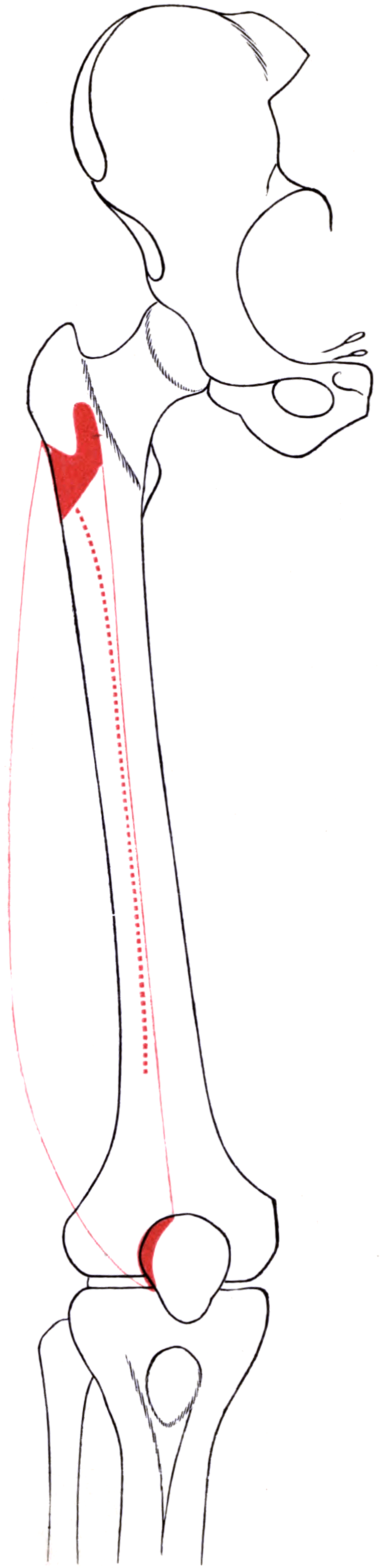
Attachments, Innervation and Relations
Origin — a continuous aponeurotic line
- Upper part of the intertrochanteric line (lateral portion).
- Anterior and inferior borders of the greater trochanter.
- Lateral lip of the gluteal tuberosity.
- Upper half of the lateral lip of the linea aspera.
- Anterior surface of the lateral intermuscular septum — the fibres here interdigitate with the septum, which is why the muscle must be dissected off rather than pushed forward.
- A tendinous aponeurosis covers the superficial surface of the proximal muscle; many fibres arise from its deep surface, making the muscle multipennate.
Insertion
- Fibres converge into a flat aponeurotic tendon on the deep surface of the muscle, which becomes the lateral portion of the quadriceps tendon, inserting on the lateral aspect of the patellar base and the proximal lateral patellar border.
- Vastus lateralis obliquus (VLO): the distal oblique fibres, orientated roughly 30-35 degrees to the femoral shaft, insert into the lateral patellar border and the deep lateral retinaculum. These are the fibres that generate the lateral tracking vector.
- An expansion continues distally into the lateral retinaculum, blends with the iliotibial tract, and reaches the lateral tibial condyle and the lateral capsule.
Named landmarks and measurements
- Relationship
- Ridge on the anterolateral greater trochanter marking the proximal origin
- Practical use
- Reference for the proximal end of a lateral incision and for plate positioning
- Relationship
- Posterior boundary; source of origin fibres; conduit for perforators
- Practical use
- The plane of the lateral approach; perforators clipped here
- Relationship
- Origin along the upper half of the shaft
- Practical use
- Defines how far posteriorly the muscle must be released
- Relationship
- No major named nerve or vessel within it
- Practical use
- Recommended intramuscular injection site in infants
- Relationship
- VLO insertion and retinacular blend
- Practical use
- Target of lateral lengthening or release for tilt
The commonest avoidable bleed in femoral surgery.
- Perforators cross the lateral intermuscular septum and are short.
- When divided they retract into the septum and the posterior compartment.
- Consequence: a large occult posterior thigh haematoma; occasionally postoperative anterior thigh compartment syndrome.
- Answer: clip or tie at the septum under direct vision before dividing. Never chase a retracted vessel blindly toward the sciatic nerve.
It has a lateral patellar vector.
- The vastus lateralis obliquus fibres insert into the lateral patella and the deep lateral retinaculum.
- Their fibre angle (roughly 30-35 degrees) is much shallower than the VMO's (50-55 degrees).
- Consequence: the resultant quadriceps vector is laterally biased; loss of VMO function or a tight lateral retinaculum exaggerates it, producing tilt, maltracking and a J-sign.
Action and Biomechanics
Actions by plane
- Action
- Extension (dominant contributor)
- Note
- Monoarticular — force output independent of hip position
- Action
- Laterally directed vector via the VLO and lateral retinaculum
- Note
- Normally balanced by the VMO; imbalance produces tilt and maltracking
- Action
- Eccentric control of knee flexion at initial contact and loading response
- Note
- The principal anti-buckling mechanism of the knee
- Action
- Compressive load along the shaft; partial tension band effect on the lateral cortex
- Note
- Relevant to lateral plate mechanics and to atypical femoral fracture location
Force contribution and architecture
- Cross-sectional area: roughly 35-40 percent of the quadriceps; the single largest contributor to knee extension torque.
- Architecture: multipennate with relatively short fibres and a large aponeurotic surface. High force, limited excursion — the classic design of a stance-phase stabiliser.
- Moment arm: shares the patellar tendon lever with the other heads. Its distinctive mechanical signature is its lateral line of pull, which combines with the Q-angle to generate the laterally directed patellar force.
Length-tension
- Because the muscle is monoarticular, its length depends only on knee angle. Peak force generation occurs in mid-range flexion; in terminal extension the quadriceps is at a mechanical disadvantage, which is why the last 15-20 degrees of extension demands disproportionate quadriceps force — and why extension lag is the first deficit to appear and the last to recover.
Synergists and antagonists
- Synergists: vastus medialis, vastus intermedius, rectus femoris. The tensor fascia lata and iliotibial tract act as passive lateral stabilisers pulling in the same direction.
- Antagonists: hamstrings and gastrocnemius for knee flexion; for the patellar vector, the vastus medialis obliquus and the medial patellofemoral ligament oppose the lateral pull.
What happens when it fails
- Isolated weakness or denervation: loss of the largest extension contributor produces extensor lag, difficulty with stair descent and giving way. A quadriceps-avoidance gait pattern may develop.
- Fibrosis or contracture (post-traumatic, post-compartment syndrome, post-injection): loss of knee flexion, an extension contracture, and in severe cases habitual or fixed lateral patellar dislocation — the classic sequel of a scarred, shortened vastus lateralis tethering the patella laterally.
- Tight lateral structures including a shortened VLO and retinaculum: lateral patellar tilt, excessive lateral facet loading, and the excessive lateral pressure syndrome.
An extensor lag is the difference between full passive extension and achievable active extension. It is a quadriceps power or continuity problem, and the vasti — especially vastus lateralis as the largest — dominate it.
- The terminal 15-20 degrees of extension requires the greatest quadriceps force, because the mechanical advantage of the extensor mechanism is least favourable there.
- Any process reducing quadriceps force (denervation, disuse atrophy, effusion-mediated arthrogenic inhibition, pain, fibrosis) shows up first as terminal extension lag.
- Distinguish from a fixed flexion deformity: if passive extension is also limited, the problem is capsular or bony, not muscular. Always measure both.
- After a lateral approach to the distal femur or a quadriceps-splitting exposure, extensor lag is common early and usually recovers; a lag persisting beyond three months should prompt assessment for a structural extensor mechanism deficit or denervation.
Surface Anatomy and Examination
Palpation and positioning
- Belly: supine with the knee extended, ask for isometric quadriceps contraction. The vastus lateralis is the large convex mass on the anterolateral thigh, lateral to the rectus femoris ridge, extending from just below the greater trochanter to the superolateral patella.
- Greater trochanter and vastus ridge: palpate the trochanter; the vastus ridge is the transverse ridge on its anterolateral aspect marking the proximal origin. It is the reference point for the proximal end of a lateral incision.
- Lateral intermuscular septum: its line approximates the posterior border of the palpable vastus mass, running from the posterior aspect of the greater trochanter to the lateral femoral epicondyle. Deep palpation between the vastus and the hamstrings finds the septal plane.
- Distal insertion and VLO: palpable at the superolateral patellar border; tenderness here in an adolescent with anterior knee pain is often retinacular.
- Intramuscular injection site: the middle third of the muscle on the anterolateral thigh — safe because no major named nerve or vessel lies in that zone.
Named tests and their interpretation
- How to perform
- Seated, hip at 90 degrees, resist extension; palpate the lateral belly
- Positive finding
- Weakness or reduced belly firmness compared with the other side
- What it means
- Biases the vasti because the rectus femoris is at active insufficiency
- False positives
- Pain inhibition, patient effort, poor stabilisation of the pelvis
- How to perform
- Measure passive terminal extension, then active terminal extension, with a goniometer
- Positive finding
- Active extension falls short of passive extension
- What it means
- Quadriceps power or continuity deficit, not a capsular problem
- False positives
- Fixed flexion deformity mistaken for lag; tense effusion causing arthrogenic inhibition
- How to perform
- Supine, lift the extended leg actively
- Positive finding
- Inability to lift the extended limb
- What it means
- Extensor mechanism discontinuity or profound quadriceps weakness
- False positives
- Pain, hip flexor pathology, poor comprehension of the instruction
- How to perform
- Knee extended, quadriceps relaxed; attempt to elevate the lateral patellar border to horizontal
- Positive finding
- Lateral border cannot be brought to neutral
- What it means
- Tight lateral retinaculum and vastus lateralis obliquus
- False positives
- Performing it with the quadriceps contracting; generalised hypomobility
- How to perform
- Knee extended and relaxed; translate the patella medially, expressed in quadrants
- Positive finding
- Less than one quadrant of medial glide
- What it means
- Tight lateral structures
- False positives
- Guarding, effusion, examiner inconsistency in quadrant estimation
- How to perform
- Observe the patella during active extension from flexion
- Positive finding
- Patella deviates laterally near terminal extension
- What it means
- Maltracking, often with lateral tightness or trochlear dysplasia
- False positives
- Voluntary quadriceps substitution; observation from the wrong angle
- How to perform
- Side-lying, hip extended and adducted with the knee flexed
- Positive finding
- Thigh fails to adduct below horizontal
- What it means
- Iliotibial band tightness — relevant because the tract blends with the lateral retinaculum
- False positives
- Pelvic rotation not controlled; hip abductor spasm
- How to perform
- Measure at a fixed distance above the superior pole of the patella, both sides
- Positive finding
- Difference between limbs
- What it means
- Objective surrogate for quadriceps atrophy, which is hard to see through the fascia lata
- False positives
- Oedema, differing measurement level, obesity
Pitfalls in examination
- Selective vastus lateralis wasting is hard to see because the muscle lies deep to the fascia lata and iliotibial tract. Measure mid-thigh circumference at a fixed distance above the patella and compare sides.
- Compartment assessment: anterior thigh compartment syndrome presents with a tense, tender anterolateral thigh, pain out of proportion, pain on passive knee flexion (which stretches the vasti) and pain on active extension. Absent pulses are a late and unreliable sign. A high index of suspicion after femoral fracture, prolonged pressure or a large perforator bleed is essential.
- What Schwartz's 17 patients add to that. Tense swelling of the thigh was present in ALL of them, which makes it the most consistent sign — pain is not. Most of the cohort could not give a reliable history because they were obtunded or anaesthetised, so compartment pressure measurement was the primary diagnostic tool, not an adjunct. Two thirds had no femoral fracture at all: eleven of the 21 syndromes followed blunt thigh trauma, prolonged compression by body weight, or vascular injury, and five followed femoral intramedullary nailing — the operation is a cause. Predisposing factors were systemic hypotension, external compression, coagulopathy and vascular injury. Fasciotomy is not a free rescue: six of the nine survivors developed infection at the fasciotomy site.
- Post-injection quadriceps contracture in children presents as loss of knee flexion, an extension contracture and sometimes lateral patellar subluxation; the history of repeated thigh injections may have to be sought explicitly.
- Grading: MRC grading applies to the quadriceps as a group. Use positional change (seated versus hip-extended) to infer whether the deficit is vastus-dominant or rectus-dominant.
Complications
- Mechanism
- Perforating branches of profunda femoris divided without control; they retract into the septum
- Avoidance / management
- Clip or ligate at the septum before dividing; do not chase retracted vessels posteriorly
- Mechanism
- Retracted perforator bleeding into the posterior compartment
- Avoidance / management
- Meticulous haemostasis, consider a drain, monitor haemoglobin and thigh girth
- Mechanism
- Postoperative bleeding, fracture, external compression, reperfusion
- Avoidance / management
- High suspicion; pain on passive knee flexion; urgent lateral fasciotomy releasing fascia lata and septum
- Mechanism
- Muscle splitting, denervation of the dominant branch, extensive elevation, flap harvest
- Avoidance / management
- Elevate rather than split; avoid deep proximal dissection in the anterior interval; early rehabilitation
- Mechanism
- Intramuscular scar after splitting, distal plating, prolonged immobilisation
- Avoidance / management
- Minimal intramuscular dissection, early controlled flexion, quadricepsplasty for established contracture
- Mechanism
- Blind dissection or clamping posterior to the lateral intermuscular septum chasing a bleeding vessel
- Avoidance / management
- Never work blindly beyond the septum; pack and gain vision
- Mechanism
- Muscle trauma, hip surgery, head or spinal cord injury
- Avoidance / management
- Gentle handling, haemostasis, prophylaxis in high-risk patients; excise only when mature
- Mechanism
- Direct blow with intramuscular haematoma
- Avoidance / management
- Ice and compression in flexion, gentle range of motion, avoid deep massage, delay excision until mature
- Mechanism
- Vastus lateralis harvest reduces peak extension torque
- Avoidance / management
- Counsel and rehabilitate; avoid where the quadriceps is already deficient
- Mechanism
- Over-aggressive lateral release including VLO fibres
- Avoidance / management
- Prefer lengthening; never release for instability alone
- Mechanism
- Superior lateral genicular artery divided with a medial arthrotomy and fat pad excision
- Avoidance / management
- Preserve the superior lateral genicular vessels where possible
- Mechanism
- Repeated intramuscular injection causing fibrosis
- Avoidance / management
- Rotate sites, inject the mid-third, use the smallest effective volume
Denervation pattern
- The nerve to vastus lateralis enters the deep anteromedial surface with segmental branches. A distal split therefore denervates only a limited territory, whereas division of the dominant proximal branch — or harvest of the muscle as a flap — denervates the whole muscle.
- Clinical picture of complete denervation: visible flattening of the anterolateral thigh, disproportionate loss of extension torque, and on MRI selective fatty atrophy of vastus lateralis with preserved rectus femoris, vastus medialis and vastus intermedius.
Clinical Relevance
The lateral approach to the femur
This is the workhorse exposure of the femoral shaft and the exposure that defines vastus lateralis surgical anatomy.
Positioning and incision
- Supine (or lateral) with the limb free; a radiolucent table if fluoroscopy is required.
- Incision: longitudinal, along a line from the greater trochanter to the lateral femoral epicondyle, over the required length of shaft. For distal femoral work the incision curves anteriorly toward the lateral patellar border.
Layers
- Skin and subcutaneous fat.
- Fascia lata and iliotibial tract — incised in line with its fibres.
- Vastus lateralis — two options:
- Elevation (preferred for the shaft): develop the plane at the posterior border of the muscle where it arises from the lateral intermuscular septum, and sweep the muscle anteriorly off the septum and the femur. This preserves the innervation (which enters anteromedially) and gives a clean subvastus plane onto the lateral femoral cortex.
- Splitting: divide the muscle longitudinally in line with its fibres. Quicker, but denervates the fibres lateral to the split, bleeds from within the muscle, and creates intramuscular scar. Reserved for limited exposures.
- Periosteum — incise only over the plate footprint.
Controlling the perforators — the key step
- As the muscle is swept forward, perforating branches of the profunda femoris are encountered crossing the septum; typically three to four along the shaft.
- Technique: work in a disciplined proximal-to-distal (or distal-to-proximal) sequence, keep a retractor on the septum, and clip, tie or seal each perforator at the septum before dividing it.
- If a perforator retracts, do not chase it into the posterior compartment — the sciatic nerve is there. Pack, extend the exposure along the septum, and identify the stump under direct vision.
Extensions
- Proximally: to the greater trochanter and the vastus ridge, converting to a lateral approach to the proximal femur — as used for cephalomedullary nails, dynamic hip screws and lateral plating of intertrochanteric and subtrochanteric fractures.
- Distally: to the lateral femoral condyle and a lateral parapatellar arthrotomy, as used for distal femoral fracture plating and lateral condylar exposure. The superior lateral genicular artery is encountered here.
Swashbuckler and mini-swashbuckler
- A modified anterolateral exposure of the distal femur that elevates the vastus lateralis anteriorly and gives wide articular access through a lateral incision, reducing the extensor mechanism insult compared with a quadriceps-splitting or turndown exposure.
A divided perforating branch of the profunda femoris retracts into the lateral intermuscular septum and into the posterior compartment, where it is invisible and unreachable through the anterior wound. The consequences are a large occult haematoma, a transfusion requirement, an increased infection risk and, rarely, postoperative anterior thigh compartment syndrome. Control each perforator at the septum, under vision, before it is divided.
Surgical Relevance
Approach-by-approach summary
- How vastus lateralis is handled
- Elevated anteriorly off the lateral intermuscular septum (preferred) or split in line with fibres
- Structures at risk with location
- 3-4 profunda femoris perforators at the septum; sciatic nerve if the septum is breached
- How vastus lateralis is handled
- Split or elevated distal to the vastus ridge on the greater trochanter
- Structures at risk with location
- First perforator within the proximal third, roughly 8-12 cm distal to the vastus ridge; ascending branch of LCFA proximally
- How vastus lateralis is handled
- Elevated anteriorly from the septum, preserving the extensor mechanism
- Structures at risk with location
- Superior lateral genicular artery at the lateral condyle; distal perforators
- How vastus lateralis is handled
- Retinacular incision continuous with the distal vastus lateralis expansion
- Structures at risk with location
- Superior lateral genicular artery; lateral meniscus and popliteus if extended posteriorly
- How vastus lateralis is handled
- Dissected to identify musculocutaneous perforators; muscle usually preserved
- Structures at risk with location
- Descending branch of LCFA (must be preserved); nerve to vastus lateralis in the same interval
- How vastus lateralis is handled
- Harvested on the descending or transverse branch of the LCFA
- Structures at risk with location
- Quadriceps power loss; the dominant motor branch is divided at harvest
- How vastus lateralis is handled
- Fascia lata incised over the muscle; septum released for the posterior compartment
- Structures at risk with location
- Perforators at the septum; superficial nerves
- How vastus lateralis is handled
- Distal VLO fibres and retinaculum divided or lengthened
- Structures at risk with location
- Superior lateral genicular artery (patellar vascularity); medial instability if over-released
Distances and landmarks worth quoting
- The vastus ridge on the anterolateral greater trochanter marks the proximal origin and is the reference for the proximal end of a lateral incision.
- The line from the greater trochanter to the lateral femoral epicondyle approximates both the skin incision and the underlying septal plane.
- Perforator zones: expect one in the proximal third (roughly 8-12 cm distal to the vastus ridge), one in the middle third and one or two in the distal third. Treat each transition as a moment to look for a vessel rather than to advance faster.
- ALT flap perforator: near the midpoint of the ASIS-to-superolateral-patella line; pedicle length commonly 8-12 cm, diameter about 2-2.5 mm.
- Superior lateral genicular artery: encountered at the lateral femoral condyle just proximal to the joint line during distal exposures and lateral release. It is a major contributor to the peripatellar anastomotic ring, hence the concern about patellar vascularity when lateral release is combined with a medial arthrotomy and fat pad excision.
- Intramuscular injection safe zone: the middle third of the muscle on the anterolateral thigh.
Both are legitimate; know the trade-off.
- Elevation off the septum preserves the segmental innervation entering the deep anteromedial surface, keeps the muscle as a vascularised soft-tissue envelope over the plate, and gives a bloodless subvastus plane onto bone — but requires deliberate control of every perforator.
- Splitting in line with fibres is faster and gives direct access at a single level, but denervates the fibres lateral to the split, bleeds from within the muscle, and creates intramuscular scar that tethers the extensor mechanism.
- Default: elevate for any exposure of significant length (shaft plating, tumour resection, revision surgery); split only for a limited window, for example a percutaneous plate insertion point or a single screw site.
Guidelines, Registries & Global Practice
Variation and prevalence
- The perforator pattern of the descending branch of the lateral circumflex femoral artery varies: the majority are musculocutaneous (traversing vastus lateralis) but a minority are septocutaneous, and in a small proportion of individuals the descending branch is diminutive or is replaced by a dominant oblique branch. Some centres use preoperative Doppler or CT angiography for this reason; most surgeons rely on intraoperative perforator identification.
- The number and position of profunda femoris perforators is generally described as three to four; treat the count as a prompt to inspect each third of the femur rather than as an anatomical guarantee.
- The vastus lateralis obliquus is described as a discrete distal oblique segment in many but not all anatomical studies. The functional point — a shallow lateral vector inserting on the proximolateral patella and the deep retinaculum — is consistent regardless of whether it is named as a separate entity.
Differences in described technique
- Emphasis relating to vastus lateralis
- Describes the lateral approach to the femur with explicit instruction to elevate the vastus lateralis anteriorly off the lateral intermuscular septum and to ligate perforating vessels as they are encountered; warns against blind haemostasis posterior to the septum.
- Emphasis relating to vastus lateralis
- Wider adoption of minimally invasive and percutaneous lateral plating with limited muscle windows, reducing but not eliminating perforator encounters; emphasis on submuscular tunnelling over the lateral cortex.
- Emphasis relating to vastus lateralis
- Emphasises early recognition of thigh compartment syndrome after femoral trauma and surgery, and structured physiotherapy-led quadriceps rehabilitation to address extensor lag.
- Emphasis relating to vastus lateralis
- Standardises the anterolateral thigh flap as a workhorse free flap, with marking along the ASIS-to-patella line and explicit preservation of the descending branch of the LCFA in trauma patients.
- Emphasis relating to vastus lateralis
- Restricts isolated lateral release to demonstrable lateral tightness with a positive tilt test, favours lengthening over release, and warns explicitly against release as a treatment for instability.
Registry and outcome signals
- National trauma and arthroplasty registries do not record vastus lateralis handling directly, but they do capture transfusion, deep infection and reoperation after femoral fracture surgery — outcomes to which uncontrolled perforator bleeding and the resulting haematoma contribute.
- Patellofemoral cohort and registry data consistently show poor outcomes for isolated lateral release performed for instability, which has shifted international practice decisively toward MPFL reconstruction with or without bony correction, and toward lengthening rather than release when tilt must be addressed.
- Free flap outcome series report anterolateral thigh flap survival comparable to other workhorse flaps, with donor-site morbidity limited mainly to sensory change and, where muscle is harvested, a measurable extension torque deficit.
High- versus limited-resource practice
- Well-resourced settings: percutaneous or minimally invasive lateral plating under fluoroscopy, vessel-sealing devices for perforator control, preoperative perforator mapping for flap planning, and compartment pressure monitoring.
- Limited-resource settings: the open lateral approach with careful elevation and suture ligation of perforators remains entirely reliable and is arguably safer than percutaneous technique without fluoroscopy. Anterior thigh compartment syndrome is diagnosed and treated clinically; a single lateral fasciotomy incision releasing the fascia lata and, where needed, the lateral intermuscular septum decompresses both compartments without special equipment.
- Universal principles: elevate rather than split for long exposures; control perforators at the septum; never dissect blindly beyond it; and do not perform a lateral retinacular release for patellar instability.
MCQ Practice Points
Q: Which is the largest head of the quadriceps and roughly what proportion of its cross-sectional area does it contribute? A: Vastus lateralis, roughly 35-40 percent of total quadriceps cross-sectional area — the greatest single contributor to knee extension torque.
Q: List the origin of the vastus lateralis. A: Intertrochanteric line (upper, lateral part), anterior and inferior borders of the greater trochanter, lateral lip of the gluteal tuberosity, upper half of the lateral lip of the linea aspera, and the anterior surface of the lateral intermuscular septum.
Q: What bleeds when you elevate the vastus lateralis in the lateral approach to the femur? A: Perforating branches of the profunda femoris artery, typically three to four, which pierce the lateral intermuscular septum. Control them at the septum before dividing.
Q: Why must you never clamp blindly posterior to the lateral intermuscular septum? A: The sciatic nerve lies in the posterior compartment immediately behind the septum. Pack, extend the exposure and identify the vessel under vision instead.
Q: Which vessel is the pedicle of both the anterolateral thigh flap and the vastus lateralis muscle flap? A: The descending branch of the lateral circumflex femoral artery, running in the rectus femoris-vastus lateralis interval.
Q: Where is the dominant perforator for an anterolateral thigh flap usually found? A: Near the midpoint of a line from the ASIS to the superolateral corner of the patella, most often a musculocutaneous perforator traversing the vastus lateralis.
Q: How do the vastus lateralis and vastus medialis obliquus fibre angles compare? A: Vastus lateralis roughly 30-35 degrees to the femoral shaft; vastus medialis obliquus roughly 50-55 degrees. The asymmetry underlies the net lateral patellar vector.
Q: Why is the mid-third of the vastus lateralis the recommended intramuscular injection site in infants? A: No major named nerve or vessel lies within that zone, and the muscle is bulky and accessible. Repeated injection at the same site risks fibrosis and an extension contracture.
Q: Why is the vastus lateralis irrelevant to swing-phase stiff-knee gait? A: It is monoarticular — it crosses only the knee and is normally electrically silent in swing. The biarticular rectus femoris is the culprit in stiff-knee gait.
Q: Which passive movement provokes pain in anterior thigh compartment syndrome? A: Passive knee flexion, because it stretches the vasti within the tight anterior compartment. Active knee extension is also painful. Absent pulses are late and unreliable.
Exam Viva Scenarios
Practise clinical reasoning and management decisions out loud
“You are plating a mid-shaft femoral fracture through a lateral approach. As you elevate the vastus lateralis you encounter sudden brisk arterial bleeding from deep in the wound, and the vessel disappears posteriorly. What is it and what do you do?”
“A 71-year-old man has a chronically infected total hip arthroplasty with a discharging sinus over the greater trochanter and a large cavity after debridement. The plastic surgeon asks what local muscle options exist. Talk me through the vastus lateralis flap.”
“A 16-year-old girl has anterior knee pain and no history of frank dislocation. The lateral patellar border cannot be lifted to neutral and medial glide is less than one quadrant. Her trochlea is normal on MRI, the TT-TG distance is 12 mm and there is no patella alta. How do the lateral structures contribute and what would you offer?”
Anatomy
- Largest quadriceps head, 35-40 percent of CSA
- Origin: intertrochanteric line, greater trochanter, gluteal tuberosity, lateral linea aspera, lateral intermuscular septum
- Insert: lateral quadriceps tendon, lateral patella, lateral retinaculum
- Nerve: femoral, posterior division, L2-L4, deep surface, segmental
- Pedicle: descending branch of LCFA
Lateral Approach
- Incision: greater trochanter to lateral epicondyle
- Split fascia lata and iliotibial tract
- Elevate muscle anteriorly off the septum (preferred)
- 3-4 profunda femoris perforators pierce the septum
- Clip or tie AT the septum before dividing
- Never clamp blindly behind the septum — sciatic nerve
Flap
- Pedicled muscle flap: trochanter, ischium, groin
- ALT flap: ASIS to superolateral patella, midpoint perforator
- Pedicle 8-12 cm, diameter 2-2.5 mm
- Free functional transfer possible (nerve and vessel together)
Patellofemoral
- VLO fibre angle 30-35 degrees versus VMO 50-55 degrees
- Contributes to the deep lateral retinaculum
- Positive tilt test equals lateral tightness
- Lengthening preferred over release
- Release treats tilt, NOT instability
- Superior lateral genicular artery at risk
Complications
- Perforator haemorrhage and occult haematoma
- Anterior thigh compartment syndrome (pain on passive knee flexion)
- Extensor lag and quadriceps adhesions
- Myositis ossificans after contusion
- Post-injection contracture in children
Evidence Base
The Vastus Lateralis Muscle: An Anatomical Investigation
- TEN cadaveric lower limbs macrodissected specifically to map architecture, attachments, innervation and internal partitioning
- THE HEADLINE FINDING: vastus lateralis comprises FOUR distinct partitions on the basis of architecture and innervation, and EACH RECEIVES ITS OWN UNIQUE NERVE BRANCH - it is not one muscle with a segmental supply
- Beyond the proximal origin at the base of the greater trochanter and the distal insertion into the superolateral border and base of the patella, THREE ADDITIONAL attachment sites were found: the lateral intermuscular septum, the ILIOTIBIAL BAND, and the RECTUS FEMORIS TENDON
- Mean fascicular length 7 cm; mean fascicular physiological cross-sectional area 1.2 cm squared; mean whole-muscle physiological cross-sectional area 21.6 cm squared
- The authors conclude the gross morphology is more complex than previously described
The Free Thigh Flap: A New Free Flap Concept Based on the Septocutaneous Artery
- Original description of the anterolateral, anteromedial and posterior thigh free flaps
- Established the descending branch of the lateral circumflex femoral artery as the axial vessel of the anterolateral thigh flap
- Perforators reach the skin through or alongside the vastus lateralis
- Demonstrated a long pedicle with large-calibre vessels suitable for microvascular transfer
Characteristics of the Anterolateral Thigh Flap in a Western Population and Its Application in Head and Neck Reconstruction
- 72 CONSECUTIVE anterolateral thigh flaps for head and neck reconstruction, reviewed retrospectively in a Western population
- The number of cutaneous perforators ranged from ONE to THREE - the study's purpose was to show that the 'confusing' reported variation follows predictable patterns
- A simple classification of perforators by location and origin is introduced to guide dissection
- 68 of 72 flaps (94 per cent) were RAISED SUCCESSFULLY - four could not be
- The authors conclude the flap is well suited to head and neck reconstruction in Westerners, countering the perception that Asian anatomical series did not transfer
Quadriceps Function: An Anatomical and Mechanical Study Using Amputated Limbs
- Anatomical and mechanical study of quadriceps function carried out on amputated limbs
- Indexed subject headings cover muscle anatomy and physiology, knee physiology, movement, the femur and the patella
- No abstract is indexed for this 1968 paper, so only the title and subject headings are cited here rather than specific numerical results
- It is conventionally cited as the source for the differing fibre orientations of the individual quadriceps heads and for the disproportionate quadriceps force demanded by terminal knee extension
Lateral Retinacular Release: A Survey of the International Patellofemoral Study Group
- Questionnaire survey of the members of an international group with a specific interest in patellofemoral disorders, with a 60 percent response rate
- Isolated lateral release was rarely performed - an estimated 1 to 5 surgical cases per respondent per year, about 2 per cent of cases performed annually
- Strong consensus that objective evidence is required to justify a lateral release and that isolated release should not be undertaken without it
- Agreement was poor as to which clinical finding constitutes the most appropriate indication
- 74 per cent of respondents believed that lateral release in the setting of arthroscopy or exploration calls for SPECIFIC informed consent
Acute Compartment Syndrome of the Thigh: A Spectrum of Injury
- 21 thigh compartment syndromes in 17 patients, reviewed retrospectively at a major trauma centre
- TEN had an ipsilateral femoral fracture, five of them open - and FIVE followed femoral INTRAMEDULLARY STABILISATION, so the operation itself is a recognised cause
- The other eleven followed blunt thigh trauma, prolonged compression by body weight, or vascular injury - a femoral fracture is NOT required
- Predisposing factors: systemic hypotension, external compression of the thigh, military antishock trousers, coagulopathy, vascular injury and thigh trauma
- About HALF developed a crush syndrome with myoglobinuria, renal failure and multi-organ collapse; EIGHT of the 17 patients (47 per cent) DIED of their multiple injuries
- Of the nine survivors, SIX developed infection at the fasciotomy site, and follow-up showed marked sensory deficit and motor weakness