The Flexor That the Extensor Nerve Supplies
- Origin: proximal two-thirds of the LATERAL SUPRACONDYLAR RIDGE of the humerus and the lateral intermuscular septum; insertion: the lateral distal radius just proximal to the RADIAL STYLOID.
- Innervated by the RADIAL NERVE (C5, C6) proximal to the elbow — it is the first radial-nerve branch below the triceps and is spared in a posterior interosseous nerve lesion.
- It is the most superficial and most anterior of the mobile wad; it forms the LATERAL boundary of the cubital fossa and the lateral wall of the Henry volar approach.
- Longest flexion moment arm of any elbow flexor because of its far-distal insertion, but a small physiological cross-section — a fast, high-excursion, low-force flexor, most efficient with the forearm mid-prone.
- It is the workhorse tendon donor in C6 tetraplegia (brachioradialis to flexor pollicis longus for key pinch) and in high median nerve palsy (brachioradialis to flexor pollicis longus for thumb interphalangeal flexion).
- “The paradox is explained embryologically: brachioradialis develops in the extensor (dorsal) muscle mass and migrates anteriorly, taking its dorsal nerve supply with it.
- “A patient with a posterior interosseous nerve palsy has NORMAL brachioradialis and NORMAL wrist extension in radial deviation — the branch to brachioradialis and to extensor carpi radialis longus arises proximal to the supinator.
- “In C6 tetraplegia the brachioradialis is often the ONLY muscle below the elbow under voluntary control, which is why it is the first donor spent.
- “Brachioradialis is expendable as a transfer because biceps and brachialis provide ample elbow flexion — but harvesting it in a patient who relies on it as a shunt muscle for heavy lifting costs endurance.
Overview
Brachioradialis is the long, strap-like muscle that stands out as a cord on the lateral aspect of the flexed, mid-prone forearm. It lies anterior to the elbow axis and works as a flexor, yet it is supplied by the radial nerve, the nerve of the extensor compartment. That single contradiction generates more exam questions than any other feature of the muscle.
Companion muscle. The other elbow flexor that does not share the biceps's radius insertion is the brachialis — the deep, musculocutaneous-innervated prime mover that lies under the biceps on the anterior humerus. The two are read together: brachialis is the workhorse the brachioradialis is expendable because of, and the brachialis-brachioradialis interval is where the radial nerve is found in the distal arm.
Why the surgeon cares. It is expendable, because biceps and brachialis provide all the elbow flexion power a patient needs, so it can be taken as a tendon transfer donor without functional cost. It has long excursion, more than most forearm donors, which suits it to driving a long-excursion recipient such as the flexor pollicis longus. And it defines two intervals used constantly: the brachialis-brachioradialis interval, where the radial nerve is found in the distal arm, and the brachioradialis-flexor carpi radialis interval, the Henry volar approach to the radius.
The examiner will ask why. Two complementary answers, and you should give both.
The embryological answer. The limb bud musculature divides into a ventral (flexor) mass supplied by the median and ulnar nerves and a dorsal (extensor) mass supplied by the radial nerve. Brachioradialis develops within the dorsal mass on the lateral aspect of the developing limb and then migrates around to lie anterior to the axis of the elbow joint, carrying its nerve supply with it. The same logic explains why the supinator and extensor carpi radialis longus, which also lie on the radial side, are radial-innervated.
The mechanical answer. What defines a muscle as a flexor is not the compartment it grew in but where its line of pull falls relative to the joint axis. The brachioradialis tendon passes well anterior to the flexion-extension axis of the elbow, so its contraction flexes. Because it inserts far from that axis, its flexion moment arm is the longest of any elbow flexor, while its physiological cross-sectional area is small, so the force it generates is modest.
Attachments, Innervation and Relations
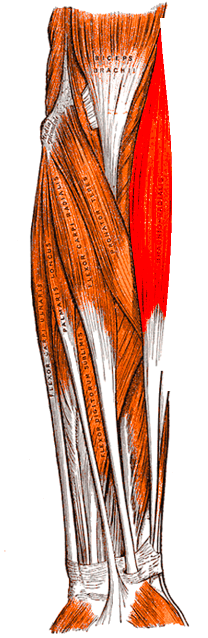
Where it starts. Brachioradialis arises from the proximal two-thirds of the lateral supracondylar ridge of the humerus, from roughly 10 cm to 3 cm proximal to the lateral epicondyle, and from the anterior surface of the lateral intermuscular septum. That is the highest origin of any forearm muscle, and it leaves a substantial part of the muscle belly lying in the arm rather than the forearm — which is what makes the muscle available for elbow cover as a proximally based flap.
Belly and tendon. The belly is fleshy for the proximal half of the forearm and then becomes a long, flat tendon for the distal half. That tendon is long, typically 8 to 12 cm, and a long tendon on a short belly is a liability in transfer: usable excursion has to be won by mobilising the muscle-tendon unit extensively from proximal (see the tendon transfer tab of Surgical Relevance).
Where it ends. The tendon inserts on the lateral surface of the distal radius, at the base of and just proximal to the radial styloid process, about 1 to 2 cm proximal to the styloid tip. It frequently blends with the fascia over the first dorsal compartment.
Action and Biomechanics
Primary action. Elbow flexion, maximally efficient with the forearm in the mid-prone (neutral, thumb-up) position.
Rotation. From full pronation it produces some supination toward neutral, and from full supination some pronation toward neutral. It drives the forearm toward mid-prone from either extreme, and is not a strong rotator in either direction.
The shunt muscle concept. Because the muscle runs nearly parallel to the forearm, only a small component of its force turns the elbow; the larger component runs along the forearm and compresses the joint. Muscles with this geometry are called shunt muscles; those whose line of pull is more perpendicular to the bone, like brachialis, are spurt muscles. Practically, brachioradialis contributes disproportionately during rapid flexion and during carrying a heavy load with the elbow flexed, where resisting distraction of the joint matters.
- Brachialis
- Musculocutaneous (+ radial)
- Biceps brachii
- Musculocutaneous
- Brachioradialis
- RADIAL
- Brachialis
- Ulnar tuberosity / coronoid
- Biceps brachii
- Radial tuberosity
- Brachioradialis
- Distal lateral radius (styloid)
- Brachialis
- Largest
- Biceps brachii
- Large
- Brachioradialis
- Small
- Brachialis
- Moderate, rotation-independent
- Biceps brachii
- Moderate, falls in pronation
- Brachioradialis
- LONGEST
- Brachialis
- Short (pennate)
- Biceps brachii
- Moderate
- Brachioradialis
- Long (about 6 cm) — key for transfer
- Brachialis
- Any (ulnar insertion)
- Biceps brachii
- Supination
- Brachioradialis
- Mid-prone
- Brachialis
- No
- Biceps brachii
- No
- Brachioradialis
- YES
What happens when it fails. Within a radial nerve palsy the loss is compounded by wrist drop and finger drop, and the absence of the visible brachioradialis cord on resisted mid-prone flexion is a useful bedside localiser. In tetraplegia at C6 brachioradialis may be the only voluntarily controlled muscle below the elbow, so its loss to a transfer is deliberate and calculated, and the choice of recipient has to be right first time.
Surface Anatomy and Examination
Palpation. Put the elbow at 90 degrees with the forearm mid-prone (thumb up) and resist flexion: brachioradialis leaps out as a prominent oblique cord running from the lateral supracondylar ridge to the radial side of the forearm. In a slim patient it is the most easily visualised muscle in the upper limb and the first landmark to find on the lateral forearm.
At the wrist. The distal tendon is palpable on the lateral side of the radial artery. Roll the tendon under the finger and the artery can be appreciated immediately medial to it.
- How to perform
- Elbow 90 degrees, thumb-up, resist flexion; observe the lateral cord
- Positive finding
- No visible or palpable cord
- What it means
- Radial nerve lesion proximal to the brachioradialis branch (spiral groove or above)
- False positives
- Obesity or oedema obscures the cord; always compare sides
- How to perform
- Tap the distal radius 3 to 5 cm proximal to the styloid with the forearm mid-prone and relaxed
- Positive finding
- Absent or reduced elbow flexion jerk
- What it means
- C5-C6 root or radial nerve lesion
- False positives
- Poor relaxation; reinforce with jaw clench or Jendrassik manoeuvre
- How to perform
- Same tap; observe the FINGERS
- Positive finding
- Absent brachioradialis contraction with brisk finger flexion instead
- What it means
- Cord lesion at C5-C6 with reflex arc preserved at C8 — a myelopathy sign
- False positives
- None significant; this is a specific sign
- How to perform
- Ask the patient to extend the wrist against gravity
- Positive finding
- Extension present but in RADIAL DEVIATION
- What it means
- Posterior interosseous nerve palsy — ECRL working, ECU not
- False positives
- Wrist tenodesis effect from finger flexion can mimic extension
- How to perform
- Palpate the lateral proximal forearm mass; assess pain on passive wrist and finger flexion
- Positive finding
- Tense wad with disproportionate pain on passive stretch
- What it means
- Compartment syndrome of the mobile wad — a separate compartment requiring its own release
- False positives
- Fracture pain confounds; measure pressures if in doubt
Grading. Grade against the contralateral side. A brachioradialis of grade 4 or better is required for a useful tendon transfer.
Substitution to watch for. Patients supinate or pronate the forearm to recruit biceps or pronator teres. Fix the forearm mid-prone and stabilise the elbow against the trunk.
What fools you. A prominent extensor carpi radialis longus can be mistaken for brachioradialis; brachioradialis is the more anterior and more proximal of the two and its cord appears on mid-prone flexion, whereas extensor carpi radialis longus becomes prominent on resisted wrist extension. After a distal humeral or radial head fracture, pain inhibition makes power testing unreliable — test sensation in the dorsal first web space to assess the radial nerve instead.
Complications
Donor-site morbidity. Loss of elbow flexion after harvest is clinically undetectable in a patient with functioning biceps and brachialis, and reported deficits are limited to endurance in sustained heavy carrying — which is the whole basis of using the muscle as a donor. In C5 tetraplegia, with no biceps, brachioradialis may be the sole elbow flexor and harvesting it is contraindicated, so test biceps and brachialis independently before committing.
A spent pedicle. After a brachioradialis flap the radial recurrent pedicle is committed: a subsequent Henry approach through the same field is more difficult, and the flap option is gone.
Adhesions and bowstringing. The transferred tendon crossing the volar wrist can adhere or bowstring if it is routed too superficially or over a sharp angle. Route it in a smooth subcutaneous tunnel and begin protected active motion early under hand therapy supervision.
Clinical Relevance
Why this muscle localises the lesion. Brachioradialis is the single most useful muscle for localising a radial nerve lesion, because its branch arises above the elbow and above the division into the posterior interosseous and superficial radial nerves. A weak brachioradialis with wrist drop, finger drop and numbness in the dorsal first web places the lesion at or proximal to the spiral groove or lateral intermuscular septum — classically after a humeral shaft fracture, the Holstein-Lewis distal-third spiral pattern tethering the nerve at the septum.
Prognosis. Radial nerve palsy after a closed humeral shaft fracture recovers spontaneously in the large majority of cases, and observation with splinting and serial examination is standard. Brachioradialis, being the most proximal muscle supplied below the injury, is usually the first to recover, and its return is the earliest clinical evidence of reinnervation.
What to document at each review. A Tinel sign advancing distally, and the return of the brachioradialis cord on resisted mid-prone flexion.
Surgical Relevance
The interval. Brachioradialis laterally, supplied by the radial nerve, against flexor carpi radialis medially, supplied by the median — a true internervous plane. The skin incision runs from just lateral to the biceps tendon at the elbow crease to the radial styloid, following the medial border of the brachioradialis.
Sorting the structures. Once the interval is open, the radial artery lies deep and immediately medial to brachioradialis and is retracted medially with the flexor carpi radialis, while the superficial radial nerve lies on the deep surface of brachioradialis and is retracted laterally with the muscle. Getting these two on the correct sides is the whole trick of the approach.
Each third of the radius is exposed by moving a different muscle:
- Proximal third — fully supinate, which brings the supinator insertion anteriorly and moves the posterior interosseous nerve laterally and posteriorly away from the field, then elevate the supinator subperiosteally off the radius from its insertion, protecting the nerve within its substance.
- Middle third — release and reflect the pronator teres insertion from the lateral radius, with the forearm pronated to bring it anteriorly.
- Distal third — elevate the pronator quadratus from its radial origin, with the flexor pollicis longus retracted ulnarly.
The leash. The radial recurrent vessels (leash of Henry) cross the field proximally and are ligated to allow mobilisation. That sacrifices the dominant brachioradialis pedicle, so a brachioradialis flap has to be planned before a Henry approach, not after it.
- Where
- Within supinator, proximal third
- Distance from a landmark
- Crosses the radial shaft a mean of 5.2 cm distal to the radiocapitellar joint in PRONATION, falling to 3.3 cm in SUPINATION (Diliberti, posterolateral approach)
- Protection
- For the VOLAR approach, fully SUPINATE and elevate supinator subperiosteally from its insertion
- Where
- Deep surface of brachioradialis
- Distance from a landmark
- Emerges from beneath the tendon 8 to 9 cm proximal to the radial styloid
- Protection
- Keep it with the brachioradialis and retract LATERALLY
- Where
- Medial to brachioradialis
- Distance from a landmark
- Immediately medial to the tendon throughout the distal two-thirds
- Protection
- Retract MEDIALLY with flexor carpi radialis
- Where
- Crossing the proximal field
- Distance from a landmark
- Just distal to the radial artery origin, at the level of the radial neck
- Protection
- Identify and ligate; do not avulse
Guidelines, Registries & Global Practice
Anatomical Variation
- Brachioradialis is one of the most anatomically constant muscles of the upper limb; complete agenesis is exceedingly rare.
- Accessory or duplicated bellies and an accessory tendon slip to the first dorsal compartment or the abductor pollicis longus are described in a small percentage of dissections; a duplicated tendon can be a useful bonus in transfer surgery but can also cause diagnostic confusion on ultrasound.
- The level at which the superficial radial nerve emerges from beneath the tendon shows meaningful individual variation around its 8 to 9 cm mean, which is why the nerve should be found rather than assumed.
Side-by-Side Guidance on Tendon Transfer Timing
- Position
- Reconstruction is offered once the neurological level is stable, usually at least 12 months after injury, with a documented ICSHT group and a formal assessment of every candidate donor's MRC grade.
- Position
- Early presenters with a reconstructable nerve may be offered nerve transfer, often combined with a pronator teres to extensor carpi radialis brevis transfer as an immediate internal splint; late presenters or those with established muscle atrophy receive tendon transfers.
- Position
- Describes the volar Henry approach with explicit instruction to supinate for the proximal third and elevate supinator subperiosteally, and to identify and protect the superficial radial nerve on the deep surface of brachioradialis.
- Position
- Emphasise that the forearm has three compartments and that the mobile wad requires separate fascial release; a volar decompression alone is inadequate.
Global Practice Differences
- Tetraplegia upper limb reconstruction is markedly under-utilised worldwide relative to the number of eligible patients. Access is concentrated in a small number of specialist centres in Sweden, the United Kingdom, North America, Australia and India; single-stage combined reconstructions are more common in high-volume European centres, whereas staged flexor-then-extensor reconstruction remains standard elsewhere.
- Nerve transfer for radial nerve palsy requires microsurgical infrastructure and early referral. In settings where presentation is typically late, tendon transfer — with brachioradialis as one of the standard donors — remains the appropriate and reliable answer.
- Brachioradialis flap cover for the elbow is a low-technology, single-stage, equipment-light option that performs well in limited-resource settings where free tissue transfer is not available.
Rehabilitation Consensus
- Early active mobilisation after tendon transfer, under supervised hand therapy, is now widely preferred over prolonged immobilisation. It reduces adhesion formation and accelerates motor relearning, and is explicitly built into contemporary single-stage tetraplegia reconstruction protocols.
- Motor re-education for a brachioradialis transfer exploits its synergy with elbow flexion: patients are taught to initiate the new movement by thinking about flexing the elbow, then progressively dissociate the two.
Related pages: Radial Nerve Anatomy supplies this paradoxical flexor above the elbow, which is why an intact brachioradialis with a finger drop localises the lesion distally — see Posterior Interosseous Nerve Anatomy, Posterior Interosseous Syndrome and Radial Tunnel Syndrome for the arcade of Frohse and the drop-versus-pain distinction. Superficial Radial Nerve Anatomy is the nerve lying on this muscle's deep surface and at risk in every distal extension of the approach, and Wartenberg's Syndrome is what happens when it is compressed between brachioradialis and extensor carpi radialis longus in pronation. Brachialis Anatomy is the medial neighbour and the true prime elbow flexor, with the radial nerve running in the interval between the two. Distal Radius Fractures is where brachioradialis becomes a deforming force on the styloid fragment, and Forearm Shaft Fracture in the Adult and Both-Bone Forearm Fractures are the operations approached through its interval. Forearm Compartment Syndrome concerns the mobile wad as a compartment in its own right. Spinal Cord Injury is the setting for the brachioradialis-to-flexor pollicis longus transfer, and Cervical Myelopathy is where the inverted supinator jerk earns its place.
MCQ Practice Points
Q: Which nerve supplies brachioradialis and why is that surprising? A: The radial nerve (C5, C6). It is an elbow flexor supplied by the nerve of the extensor compartment, because it develops in the dorsal muscle mass and migrates anteriorly with its nerve.
Q: Where does brachioradialis arise and insert? A: Proximal two-thirds of the lateral supracondylar ridge of the humerus to the lateral distal radius just proximal to the radial styloid.
Q: A patient has finger drop, normal brachioradialis and normal sensation. Where is the lesion? A: The posterior interosseous nerve, most commonly at the arcade of Frohse. Wrist extension is present but in radial deviation.
Q: What are the three muscles of the mobile wad? A: Brachioradialis, extensor carpi radialis longus and extensor carpi radialis brevis. It is a separate compartment and must be released separately in forearm fasciotomy.
Q: In the volar Henry approach, which side does the radial artery go? A: Medially, with flexor carpi radialis. The superficial radial nerve goes laterally with brachioradialis.
Q: Which transfer restores thumb interphalangeal flexion in high median nerve palsy? A: Brachioradialis to flexor pollicis longus. The donor is radial-innervated, expendable and excursion-matched.
Q: How much excursion does brachioradialis provide, and what is the catch? A: Approximately 6 cm — but only after extensive proximal mobilisation. The muscle has a long tendon and short resting amplitude.
Q: What is the dominant pedicle of the brachioradialis muscle flap? A: The radial recurrent artery, entering the deep surface of the proximal third. It is ligated in a standard Henry approach.
Q: Where does the superficial radial nerve become subcutaneous? A: Emerging from beneath the brachioradialis tendon 8 to 9 cm proximal to the radial styloid, between brachioradialis and extensor carpi radialis longus.
Q: How does brachioradialis deform a distal radius fracture? A: It pulls the radial styloid fragment proximally and into radial deviation, resisting restoration of radial length. Release of its insertion aids reduction.
Exam Viva Scenarios
Practise clinical reasoning and management decisions out loud
“The examiner asks: brachioradialis is an elbow flexor, yet it is supplied by the radial nerve, which is the nerve of the extensor compartment. Explain this, and tell me how you use it clinically.”
“A 24-year-old man is 18 months after a C6 complete spinal cord injury. He has grade 5 biceps and brachialis, grade 4 brachioradialis, grade 4 extensor carpi radialis longus, and nothing else below the elbow. He wants to be able to hold a fork and a card. What do you offer him and why?”
“You are exposing a mid-shaft radius fracture through a volar Henry approach. Talk me through the interval and tell me exactly what you do with the brachioradialis, the radial artery and the superficial radial nerve.”
Anatomy
- Origin: proximal two-thirds of the lateral supracondylar ridge
- Insertion: lateral distal radius just proximal to the styloid
- Most anterior and superficial of the MOBILE WAD
- Lateral boundary of the cubital fossa
Innervation
- RADIAL nerve C5, C6 - the paradox
- Branch arises PROXIMAL to the elbow, above the PIN division
- Spared in PIN palsy - the key localiser
- Mediates the brachioradialis (supinator) reflex, C5-C6
Biomechanics
- Longest flexion moment arm, smallest cross-section
- Optimal in MID-PRONE position
- Shunt muscle - resists joint distraction in fast/loaded flexion
- Excursion about 6 cm AFTER full mobilisation
Surgical Numbers
- Superficial radial nerve emerges 8-9 cm proximal to the radial styloid
- PIN crosses the radius 5.2 cm distal to radiocapitellar joint in PRONATION, 3.3 cm in supination
- Flap covers elbow defects up to 3 cm; 100 per cent anterior/posterior coverage proximally pedicled
- Dominant pedicle: radial recurrent artery, proximal third, deep surface
Transfers
- BR to FPL: key pinch in C6 tetraplegia; thumb IP flexion in high median palsy
- BR to ECRB: active wrist extension when ECRL absent
- Expendable ONLY if biceps and brachialis are working
- Expect loss of one MRC grade after transfer
Evidence Base
Brachioradialis Muscle Flap: Clinical Anatomy and Use in Soft-Tissue Reconstruction of the Elbow
- Fifty-three upper extremities dissected to define the muscular and vascular anatomy of brachioradialis and its arc of rotation
- A consistent major pedicle was found near the elbow, arising in descending order of frequency from the radial recurrent, radial and brachial arteries
- A variable number of minor pedicles were found along the length of the muscle
- Proximally pedicled, the distal muscle covered both the anterior and posterior elbow in 100 per cent of specimens; distally pedicled, it covered the anterior elbow in 100 per cent and the posterior elbow in 91 per cent
- Arc of rotation encompassed the distal half of the arm and the proximal two-thirds of the forearm, reliably covering defects up to 3 cm, with no loss of upper extremity function and no major vessel sacrificed
A Single-Stage Operation for Reconstruction of Hand Flexion, Extension and Intrinsic Function in Tetraplegia: the Alphabet Procedure
- Single-stage combination of seven procedures to restore grip, grasp and release in C6 tetraplegia
- Brachioradialis to flexor pollicis longus tendon transfer is a core component, providing active thumb flexion for key pinch
- Combined with split flexor pollicis longus to extensor pollicis longus distal thumb tenodesis, passive interosseous reconstruction, thumb carpometacarpal arthrodesis, extensor carpi radialis longus to flexor digitorum profundus transfer, and extensor pollicis longus and extensor carpi ulnaris tenodeses
- Early active training was used to reduce adhesion formation and speed motor relearning
- Patient satisfaction was high and complication rates comparable with staged approaches
Anatomical Considerations Regarding the Posterior Interosseous Nerve During Posterolateral Approaches to the Proximal Radius
- Thirty-two cadaveric specimens dissected through the posterolateral interval between anconeus and extensor carpi ulnaris
- Pronation allowed safe exposure of at least the proximal 38 mm of the lateral radius, with a mean safe zone of 52.0 plus or minus 7.8 mm
- Supination reduced the safe zone to as little as 22 mm, mean 33.4 plus or minus 5.7 mm
- The angle between the nerve and the radial shaft averaged 47.4 degrees in supination, falling to 27.8 degrees in pronation
- Approaching the lateral proximal radius is safest in pronation
Radial Nerve Palsy: Nerve Transfer Versus Tendon Transfer to Restore Function
- Retrospective comparison of 30 patients treated with tendon transfers and 16 with nerve transfers for isolated radial nerve injury
- Fifteen of the 16 nerve transfer patients also received a pronator teres to extensor carpi radialis brevis tendon transfer to provide immediate wrist extension
- Both groups improved significantly in grip and pinch strength, DASH scores and quality-of-life scores
- Postoperative grip strength was significantly higher in the nerve transfer group; pinch strength did not differ
- Nerve transfer patients were younger, presented earlier and had longer follow-up
The Arcade of Frohse and Its Relationship to Posterior Interosseous Nerve Paralysis
- The classic reference defining the arcade of Frohse and its relationship to posterior interosseous nerve paralysis
- No abstract is indexed for this 1968 paper; the claims below rest on the title, the MeSH indexing (forearm anatomy and innervation, FETAL anatomy, paralysis of peripheral nervous system origin) and the conclusion universally attributed to it
- That conclusion is that the arcade is membranous in the fetus and becomes tendinous with age and use, making compression an acquired adult problem
- It established the arcade as the principal site of posterior interosseous nerve compression, and the anatomical basis for distinguishing that lesion from a more proximal radial nerve lesion in which brachioradialis is also affected