Adult-onset tarsal navicular dysplasia/osteonecrosis with lateral collapse and paradoxical pes planovarus
- Müller-Weiss disease is spontaneous adult-onset navicular dysplasia/osteonecrosis — distinct from Köhler disease, the self-limiting childhood navicular osteochondrosis.
- Maceira's developmental theory: delayed ossification of the lateral navicular in childhood under asymmetric load (often with a short first ray / index minus) leads to chronic lateral compression and adult collapse.
- The deformity is PES PLANOVARUS: the lateral navicular collapses, the talar head slides dorsolaterally, and the subtalar complex rotates into varus — the opposite of the valgus of ordinary adult-acquired flatfoot.
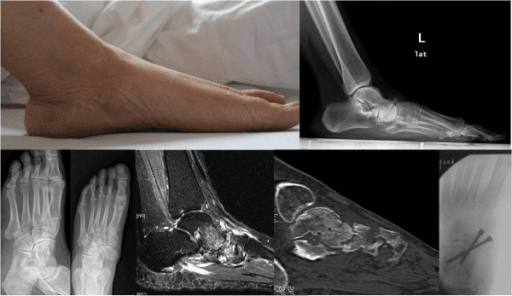
- Weightbearing radiographs are essential: comma-shaped/hourglass navicular on AP with medial protrusion; broken Meary line on lateral; varus heel on hindfoot alignment view.
- Non-operative care (rigid orthoses with medial arch support AND lateral hindfoot posting, rocker sole) is first line and frequently durable in stages 1-3.
- Surgical workhorse: talonavicular or talonavicular-cuneiform fusion with alignment correction and bone grafting; triple fusion when the subtalar joint is involved.
- “Flat arch plus varus heel in a middle-aged woman with dorsomedial midfoot pain equals Müller-Weiss until proven otherwise.
- “Orthosis prescription is the classic viva trap: medial arch support with LATERAL hindfoot posting — a standard medial-posted flatfoot orthosis worsens the varus.
- “Avoid isolated dorsal wedge excision procedures — they do not address the collapse and fail.
- “The bone is sclerotic and relatively avascular: fusion demands thorough joint preparation, compression, grafting of defects and patience with union.
Müller-Weiss produces pes planovarus — a flattened medial arch with hindfoot varus. Ordinary adult-acquired flatfoot (tibialis posterior dysfunction) produces planovalgus. Mistaking the two leads to the wrong orthosis and the wrong operation.
Köhler disease is the childhood (age roughly 4 to 7) navicular osteochondrosis — self-limiting, treated with symptomatic care or a short period of casting, and heals with normal ossification. Müller-Weiss is the adult disease with permanent structural collapse.
Non-weightbearing films underestimate collapse and alignment. Insist on standing AP, lateral and hindfoot alignment views of both feet — the disease is frequently bilateral and often initially misdiagnosed as generic midfoot arthritis.
The collapsed navicular is dense and poorly vascular. Non-union risk is real: debride to bleeding bone, graft defects (structural graft to restore medial column length where needed), compress rigidly, and extend the fusion to involved adjacent joints rather than under-treating.
Definition, Epidemiology and Pathogenesis
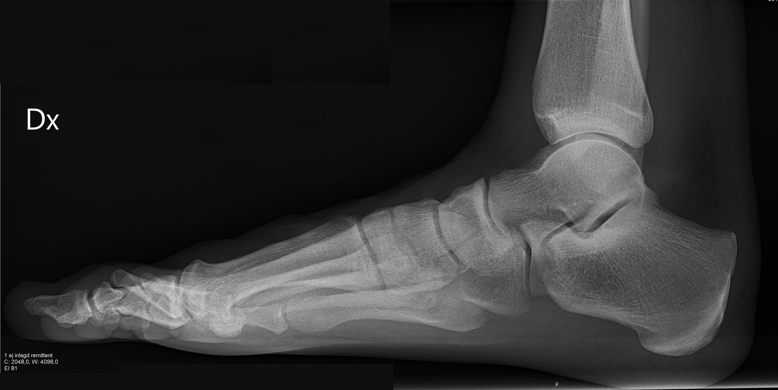
Müller-Weiss disease (described by Müller in 1927 and Weiss in 1929) is a spontaneous, adult-onset deformity of the tarsal navicular characterised by lateral compression, fragmentation and dorsomedial extrusion of the bone, with secondary peritalar malalignment and arthritis.
Epidemiology
- Presents in the fourth to sixth decades; female predominance in most published series.
- Frequently bilateral (though often asymmetric in severity and symptoms).
- Higher reported prevalence in populations with childhood nutritional deprivation or heavy barefoot childhood loading, supporting a developmental origin.
- Commonly misdiagnosed for years as "midfoot osteoarthritis" or tibialis posterior dysfunction.
The navicular is the last tarsal bone to ossify (around age 3, later in girls' relative terms but ossifying earlier in girls chronologically; delayed ossification leaves it vulnerable). Maceira and Rochera proposed that delayed ossification in childhood — from nutritional, socioeconomic or mechanical stress — leaves a soft, cartilaginous lateral navicular exposed to asymmetric compressive load. A short first ray (index minus) or forefoot morphology shifting load laterally concentrates force on the lateral navicular between the talar head and lateral cuneiform. Over decades the lateral pole is compressed and effectively "extruded" medially and dorsally: the bone becomes comma-shaped, the talar head migrates dorsolaterally past the deficient lateral navicular, and the subtalar joint rotates into varus. On this reading Müller-Weiss is a dysplasia with secondary mechanical failure, not a primary vascular event.
The Deformity Paradox — Why Pes Planovarus?
- Müller-Weiss disease
- Lateral half of navicular collapses, shortening the lateral talonavicular articulation; talar head slides dorsally and laterally, abducting the talus over the calcaneus
- Tibialis posterior dysfunction
- Spring ligament and tibialis posterior tendon failure; peritalar subluxation with forefoot abduction
- Müller-Weiss disease
- Flattened — medial column collapse through the navicular, with loss of navicular height and a broken Meary line
- Tibialis posterior dysfunction
- Flattened — medial column sag at talonavicular and naviculocuneiform level
- Müller-Weiss disease
- VARUS — subtalar complex compensates into inversion
- Tibialis posterior dysfunction
- VALGUS with a positive 'too many toes' sign
- Müller-Weiss disease
- Dorsomedial midfoot over the talonavicular joint
- Tibialis posterior dysfunction
- Posteromedial along the tibialis posterior, later lateral sinus tarsi impingement
- Müller-Weiss disease
- Often possible; the heel is already in varus
- Tibialis posterior dysfunction
- Impaired or absent; heel fails to invert
- Müller-Weiss disease
- Comma-shaped navicular, medial protrusion, lateral compression, talar head lateral migration
- Tibialis posterior dysfunction
- Talonavicular uncoverage from forefoot abduction; navicular shape normal
- Müller-Weiss disease
- Medial arch support with a LATERAL hindfoot post to correct varus
- Tibialis posterior dysfunction
- Medial arch support with a MEDIAL hindfoot post to correct valgus
- Müller-Weiss disease
- A flat arch with a VARUS heel — this combination should immediately trigger the diagnosis
- Tibialis posterior dysfunction
- A flat arch with a VALGUS heel — the far commoner, expected pattern
This is the single most examinable concept in the topic.
- The lateral half of the navicular collapses, shortening the lateral side of the talonavicular articulation.
- The talar head, losing lateral navicular support, slides dorsally and laterally relative to the navicular remnant.
- Lateral migration of the talar head effectively abducts the talus over the calcaneus; the subtalar complex compensates by rotating into inversion — hindfoot varus.
- Simultaneously the medial longitudinal arch flattens because the medial column has lost navicular height and the talus is dorsiflexed/plantarflexed abnormally relative to the first metatarsal (Meary line breaks).
Hence: flat arch + varus heel = pes planovarus, the mirror image of tibialis posterior dysfunction (flat arch + valgus heel, abducted forefoot, "too many toes" sign). Recognising the varus heel behind a flat foot should immediately trigger the diagnosis.
- Müller-Weiss disease
- Flattened (medial column collapse through navicular)
- Tibialis posterior dysfunction
- Flattened (spring ligament / tendon failure)
- Müller-Weiss disease
- VARUS
- Tibialis posterior dysfunction
- VALGUS with 'too many toes' sign
- Müller-Weiss disease
- Dorsomedial midfoot over talonavicular joint
- Tibialis posterior dysfunction
- Posteromedial along tibialis posterior, later sinus tarsi
- Müller-Weiss disease
- Often possible; heel already in varus
- Tibialis posterior dysfunction
- Impaired; heel fails to invert
- Müller-Weiss disease
- Comma-shaped navicular, medial protrusion, lateral compression
- Tibialis posterior dysfunction
- Talonavicular uncoverage from forefoot abduction, normal navicular shape
- Müller-Weiss disease
- Medial arch support with LATERAL hindfoot post
- Tibialis posterior dysfunction
- Medial arch support with MEDIAL hindfoot post
Clinical Presentation and Examination
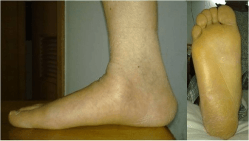
History
- Insidious, chronic dorsomedial midfoot pain, often present for years before diagnosis.
- Worse on uneven ground, prolonged standing and push-off; better in stiff-soled shoes.
- No single traumatic event (distinguishing from stress fracture and Lisfranc pathology).
- Ask about childhood nutrition/illness, occupation with prolonged loading, and contralateral symptoms (frequently bilateral).
Examination — describe it like a surgeon
- Standing inspection from behind: with the patient standing feet at shoulder width, observe the heel axis relative to the tibial axis — in Müller-Weiss the heel sits in varus despite a flattened medial arch (a Coleman-block-style assessment of hindfoot flexibility can follow: if a lateral forefoot block corrects the heel towards neutral, the hindfoot varus is forefoot-driven and flexible; a fixed varus implies structural subtalar change).
- Standing inspection from the side: loss of the medial longitudinal arch; a dorsolateral prominence over the talar head as it subluxes past the collapsed navicular.
- Palpation: patient seated, examiner's thumb over the talonavicular joint dorsally and dorsomedially — focal tenderness, sometimes a palpable medially protruding navicular fragment. Distinguish from the "N spot" (dorsal central navicular tenderness of stress fracture) and from naviculocuneiform tenderness (adjacent joint arthritis in advanced stages).
- Rigid vs flexible assessment: with the patient seated and knee flexed, grasp the heel with one hand and the midfoot with the other; passively invert/evert the subtalar joint and adduct/abduct through the transverse tarsal joints. Record whether the hindfoot varus and midfoot deformity correct passively — flexibility guides whether osteotomy adjuncts or in-situ fusion positioning are needed.
- Neurovascular and sensory examination: mandatory to exclude neuropathy (Charcot differential) — monofilament testing, vibration, pulses.
- Gait: antalgic with reduced push-off; lateral border overload callosities may be present under the fifth metatarsal from the varus heel.
Imaging and Measurements
Differential Diagnosis
- Discriminating features
- Middle-aged, often female, bilateral, pes planovarus, comma-shaped navicular
- Key investigation / trap
- Weightbearing films of both feet; varus heel behind flat arch
- Discriminating features
- Child aged roughly 4 to 7, limp, self-limiting; navicular sclerosis then normal re-ossification
- Key investigation / trap
- Do NOT conflate — different disease, symptomatic treatment only
- Discriminating features
- Athlete, activity-related pain, 'N spot' tenderness dorsally, sagittal central-third fracture
- Key investigation / trap
- CT/MRI; risk of non-union in the watershed central third
- Discriminating features
- Neuropathy (diabetes), warm swollen foot, often painless relative to destruction, rocker-bottom collapse in valgus/abduction
- Key investigation / trap
- Sensory examination mandatory; fusion in unrecognised Charcot fails
- Discriminating features
- Older patient, normal navicular shape, joint-space narrowing without lateral compression or varus
- Key investigation / trap
- Alignment usually neutral; treat as midfoot OA
Management
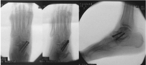
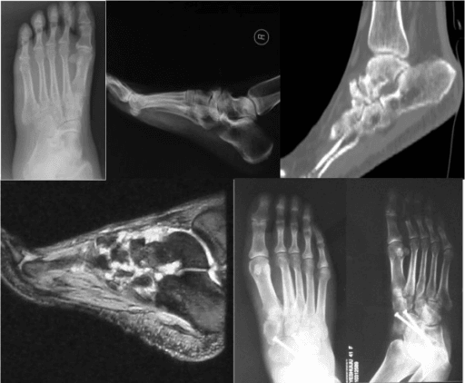
- Mechanism
- Sclerotic, avascular, fragmented navicular bone with poor biological potential; under-débridement leaves dead surfaces apposed
- Prevented by
- Debride sclerotic surfaces back to bleeding cancellous bone, drill/petal dense bone, excise non-viable fragments, compression screws talus to cuneiforms plus dorsomedial bridging plate, cancellous autograft, non-weightbearing 6 to 8 weeks then protected loading to roughly 12 weeks
- Salvage
- Revision with fresh grafting, more rigid fixation, biologic augmentation and metabolic work-up; confirm union on CT if in doubt
- Mechanism
- Fusing the transverse tarsal joints without correcting a rigid varus heel causes lateral column overload and painful gait
- Prevented by
- Set the hindfoot in about 5 degrees of valgus — never fuse in varus; add a lateralising calcaneal osteotomy (lateral closing wedge or lateral displacement) if the subtalar joint is being spared; extend to triple fusion if varus is rigid
- Salvage
- Corrective calcaneal osteotomy, or extension to triple fusion (talonavicular plus subtalar plus calcaneocuboid)
- Mechanism
- Skipping structural graft after excision of the collapsed navicular leaves the column short with recurrent dorsal talar subluxation and uncorrected Meary line
- Prevented by
- Tricortical structural (iliac crest or allograft) wedge in the navicular defect bridging talus to cuneiforms; restore length and reduce the talar head onto the reconstructed column; confirm Meary line on intra-operative imaging
- Salvage
- Revision reconstruction with a fresh structural graft to re-establish length and alignment
- Mechanism
- Naviculocuneiform involvement is frequent and under-estimated; isolated talonavicular fusion through diseased bone leaves a persistent pain source
- Prevented by
- Extend to talonavicular-cuneiform fusion as the workhorse; assess the naviculocuneiform and subtalar joints pre-operatively; diagnostic image-guided talonavicular injection to confirm the pain generator
- Salvage
- Extension of the fusion to the symptomatic adjacent joint
- Mechanism
- Isolated dorsal wedge excision of extruded fragments or dorsal cheilectomy neither restores the column nor addresses the arthritic joints
- Prevented by
- Do not perform them — reconstruct with fusion plus structural graft in refractory Maceira stage 3 to 5 disease
- Salvage
- Conversion to talonavicular-cuneiform or triple fusion
- Mechanism
- Dorsalis pedis artery and deep peroneal nerve dorsally, superficial peroneal nerve branches, tibialis posterior insertion medially/plantar; crowded incisions when combining with calcaneal osteotomy
- Prevented by
- Dorsomedial incision in the tibialis anterior / extensor hallucis longus interval, retract the bundle laterally, plan skin bridges, layered closure over capsule and below-knee backslab
- Salvage
- Nerve exploration or neuroma management; hardware removal for prominence; wound care
- Mechanism
- Load transfer to subtalar and cuneiform-metatarsal joints after transverse tarsal fusion
- Prevented by
- Counsel pre-operatively; avoid over-long constructs where not indicated; accept that midfoot stiffness is well tolerated as these joints were already stiff and painful
- Salvage
- Selective further fusion of the symptomatic joint
First line in all stages, and frequently durable in Maceira stages 1-3. Published series report the majority of patients achieving acceptable symptom control without surgery.
- Rigid custom orthoses — the prescription is the exam point: medial longitudinal arch support to unload the collapsed medial column, combined with LATERAL hindfoot posting (lateral heel wedge) to accommodate/correct the varus heel and shift load off the compressed lateral navicular. A standard flatfoot orthosis with medial posting drives the heel further into varus and worsens symptoms.
- Rocker-sole, stiff-soled footwear to offload the talonavicular joint during push-off.
- Activity modification (avoid uneven ground, prolonged standing), weight optimisation.
- Simple analgesia/NSAIDs; a period in a walking boot for flares.
- Image-guided talonavicular corticosteroid injection — diagnostic value (confirms the pain generator before fusion) and temporary therapeutic benefit.
Decision threshold for surgery: persistent disabling pain despite at least 6 months of well-executed orthotic management, or progressive collapse with advancing stage — not radiographic stage alone.
COMMAMüller-Weiss Core Features
Hook:The comma-shaped navicular writes a 'comma' in the patient's story — a long pause of misdiagnosis before the answer.
MALOrthosis Prescription
Hook:Müller-Weiss orthoses are 'MAL' — because a standard flatfoot orthosis would be bad (mal) for this foot.
Guidelines, Registries & Global Practice
- Global epidemiology: the disease is described worldwide but with striking regional clustering — higher reported prevalence in southern Europe (notably Spain, where Maceira's series originated) and in populations with historical childhood nutritional deprivation, consistent with the developmental theory. In many regions it remains under-recognised and coded simply as midfoot osteoarthritis.
- Society guidance: no major society (AAOS, NICE, BOA, EFORT) publishes a disease-specific guideline for Müller-Weiss; practice is guided by expert consensus and case series, principally Maceira's framework. General midfoot arthritis and arthrodesis principles from foot and ankle society consensus documents apply to fixation and aftercare.
- Registries: joint registries do not capture midfoot fusion outcomes in a disease-specific way, so evidence remains limited to institutional series — a fair point to make in a viva about the quality of evidence.
- Resource-setting variation: in well-resourced systems, weightbearing CT increasingly refines staging and fusion planning; in resource-limited settings, weightbearing plain films plus clinical assessment remain fully adequate for diagnosis and staging, and prolonged high-quality orthotic/footwear management is the mainstay — appropriately so, given that most patients do well without surgery. Access to custom rigid orthoses, rather than surgery, is the key resource determinant of outcome.
Controversies & Areas of Uncertainty
- Dysplasia versus osteonecrosis: histological studies from fusion specimens often show viable degenerative bone, undermining a pure osteonecrosis model, yet MRI necrosis-like change is common — most authors now accept a combined mechanical–ischaemic model.
- Extent of fusion: isolated talonavicular versus talonavicular-cuneiform versus triple fusion for the same stage remains surgeon-dependent; the trend favours extended medial column fusion because of naviculocuneiform involvement and non-union risk through the diseased navicular, but comparative data are absent.
- Role of joint-sparing surgery: calcaneal osteotomy with or without percutaneous procedures as stand-alone treatment in early flexible disease has advocates but lacks robust supporting series; fusion remains the standard for structural disease.
- When to operate: because non-operative care is so often durable, the threshold and timing for fusion — and whether earlier surgery prevents progression to stage 5 — are unresolved.
- Biologic adjuncts: vascularised grafting and orthobiologics for the sclerotic navicular are described anecdotally without comparative evidence.
Confirm protective sensation and exclude Charcot neuroarthropathy, verify the pain generator (diagnostic injection where doubt exists), obtain weightbearing imaging of both feet, and plan alignment correction — a fusion fixed in residual heel varus converts one painful foot into another.
MCQ Practice Points
Q: What hindfoot–arch combination characterises Müller-Weiss disease? A: Pes planovarus — flattened medial longitudinal arch with hindfoot varus, caused by lateral navicular collapse, dorsolateral talar head migration and subtalar rotation into varus. This is the opposite of the planovalgus of tibialis posterior dysfunction.
Q: What is Maceira's proposed aetiology? A: Delayed ossification of the lateral navicular in childhood, subjected to asymmetric compressive load (often with a short first ray / index minus), producing chronic lateral compression and adult collapse — a dysplasia with mechanical failure rather than pure primary osteonecrosis.
Q: Describe the AP radiographic hallmark. A: A comma-shaped or hourglass navicular — compressed laterally with the medial pole protruding medial to the talar head — with talonavicular uncoverage; obtained on WEIGHTBEARING films of both feet.
Q: What defines Maceira stages 2 versus 4? A: The direction of the Meary (talo-first metatarsal) line break on the lateral weightbearing view — dorsal break in stage 2 (talar head subluxing dorsally), plantar break with talar equinus and talonavicular arthritis in stage 4; stage 5 is complete collapse with the talus articulating towards the cuneiforms.
Q: How does the orthosis for Müller-Weiss differ from a standard flatfoot orthosis? A: It combines medial arch support with a LATERAL hindfoot post (lateral heel wedge) to address the varus heel, plus a rocker sole; a medially posted device worsens the varus and symptoms.
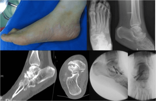
Q: What is the workhorse operation for refractory disease? A: Talonavicular or talonavicular-cuneiform fusion with excision of non-viable fragments, structural bone grafting to restore medial column length, correction of alignment and rigid compressive fixation; triple fusion if the subtalar joint is involved; isolated dorsal wedge excision procedures should be avoided.
Exam Viva Scenarios
Practise clinical reasoning and management decisions out loud
“A 52-year-old woman presents with 3 years of dorsomedial midfoot pain, worse on cobblestones. Standing examination shows a flattened medial arch but, viewed from behind, the heel is in varus. How do you proceed?”
“A 58-year-old woman with Maceira stage 4 Müller-Weiss disease has failed 12 months of orthotic management including a diagnostic talonavicular injection that gave excellent temporary relief. CT shows navicular fragmentation with talonavicular and naviculocuneiform arthritis; the subtalar joint is preserved and the heel varus corrects passively. What operation and why?”
“An examiner shows you a sclerotic, flattened navicular on a foot radiograph and asks: how does your interpretation differ if the patient is 5 years old, 25 and a distance runner, or 55?”
Core Facts
- Adult-onset navicular dysplasia/osteonecrosis; 4th-6th decades; female predominance; often bilateral
- Maceira theory: delayed lateral navicular ossification plus asymmetric load (short first ray) — lateral compression collapse
- Distinct from Köhler disease (self-limiting childhood osteochondrosis)
The Paradox
- PES PLANOVARUS: flat medial arch WITH varus heel
- Lateral navicular collapses, talar head migrates dorsolaterally, subtalar rotates into varus
- Opposite of tibialis posterior dysfunction (planovalgus)
Imaging
- Weightbearing films of BOTH feet mandatory
- AP: comma/hourglass navicular, medial protrusion, lateral compression
- Lateral: broken Meary line — dorsal break early, plantar break late (Maceira stages 1-5)
- Hindfoot alignment view: varus heel; CT for fragmentation/joint mapping; MRI for early oedema
Management
- Non-operative first and often durable (stages 1-3): rigid orthosis with medial arch support + LATERAL hindfoot post, rocker sole
- Surgery for refractory pain: talonavicular / TNC fusion with structural graft and alignment correction
- Triple fusion if subtalar involved; lateralising calcaneal osteotomy for residual varus
- Never isolated dorsal wedge excision; never fuse in varus
- Sclerotic bone: debride to bleeding bone, compress, graft, be patient with union
Evidence Base
Müller-Weiss Disease: Clinical and Biomechanical Features
- The navicular appears squeezed and eventually fragmented between the talar head and the lateral cuneiforms; because the talar head protrudes laterally, the subtalar joint remains INVERTED
- The authors state plainly that the disease 'is NOT the consequence of an osteonecrotic process but results from impaired development of the bone' - in most cases from stressing environmental factors during childhood
- Most patients are grouped by the authors as 'late collateral damage of war or poverty'; isolated cases from individual predisposing factors are rare
- IMPORTANTLY, THE ARCH IS NOT ALWAYS THE SAME: the plantar arch 'may correspond to that of a normal, cavus, or flat foot'
- Source of the five-stage radiographic classification built on the weightbearing lateral Meary line