The Strongest Evertor and the Tendon That Splits
- Origin: distal two-thirds of the lateral surface of the fibula and the adjacent intermuscular septa - deep and distal to peroneus longus.
- Insertion: styloid process (tuberosity) at the base of the fifth metatarsal, on its dorsolateral aspect.
- It lies anterior and deep in the retromalleolar groove, directly against the fibula - which is why it, and not peroneus longus, develops longitudinal splits.
- It is the strongest and most efficient evertor of the hindfoot and the direct antagonist of tibialis posterior.
- Innervated by the superficial peroneal nerve, roots L5 and S1.
- “The peroneus brevis is compressed between the peroneus longus tendon and the sharp posterolateral fibular ridge - the mechanism of the longitudinal split tear.
- “In a fifth metatarsal tuberosity avulsion the fracture is caused by the lateral band of the plantar fascia and the peroneus brevis, and it heals reliably.
- “The Jones fracture sits in a watershed zone between the metaphyseal and diaphyseal blood supply - that is the reason it does not heal.
- “Harvesting the whole peroneus brevis for a Chrisman-Snook reconstruction removes the strongest evertor - modern anatomic repair avoids this.
Overview
Peroneus brevis (fibularis brevis) is the shorter, deeper and more distal of the two lateral compartment muscles. It arises from the distal two-thirds of the lateral fibula, so its muscle belly extends further distally than that of peroneus longus, and its tendon runs anterior and deep to the longus in the retromalleolar groove, directly against the bone. It inserts on the styloid process at the base of the fifth metatarsal.
Three facts follow from that anatomy and account for almost everything clinically important about the muscle:
- Because it lies against the fibula with the longus tendon pressed over it, peroneus brevis is the tendon that develops longitudinal split tears.
- Because it inserts on the fifth metatarsal base, it is implicated in the fracture patterns of that bone and is a fixed point that must be respected in every lateral foot procedure.
- Because it is the strongest and most direct evertor, it is the muscle whose loss matters most - and yet it was historically harvested wholesale for lateral ligament reconstruction, which is exactly why modern practice avoids that.
In the retromalleolar groove on the posterior surface of the distal fibula, the two peroneal tendons share a single synovial sheath roofed by the superior peroneal retinaculum:
- Peroneus brevis lies anterior and deep, its flat tendon pressed directly against the fibular groove and its sharp posterolateral fibular ridge.
- Peroneus longus lies posterior and superficial, riding on top of the brevis.
Every time the ankle dorsiflexes and everts, the round longus tendon is driven forward against the flat brevis tendon, compressing it against the fibular ridge. Over years this produces a longitudinal split - the tendon splays open like a book, and the longus tendon can subluxate into the split.
- A shallow, flat or convex retromalleolar groove instead of the normal concavity.
- A low-lying peroneus brevis muscle belly extending distally into the groove.
- An accessory peroneus quartus muscle (present in roughly 10 to 20 per cent of feet).
- A lax or torn superior peroneal retinaculum, allowing the tendons to sublux and shear.
- Hindfoot varus, which increases the load on the peroneals with every step.
repairing a brevis split without decrowding the groove and repairing the retinaculum treats the symptom and not the cause. Correcting hindfoot varus is often the most important step of all.
Bone, Base, BehindBrevis - Three Bs
Hook:Brevis is Bone, Base and aBove the tubercle. Longus is superficial and goes beLow the tubercle.
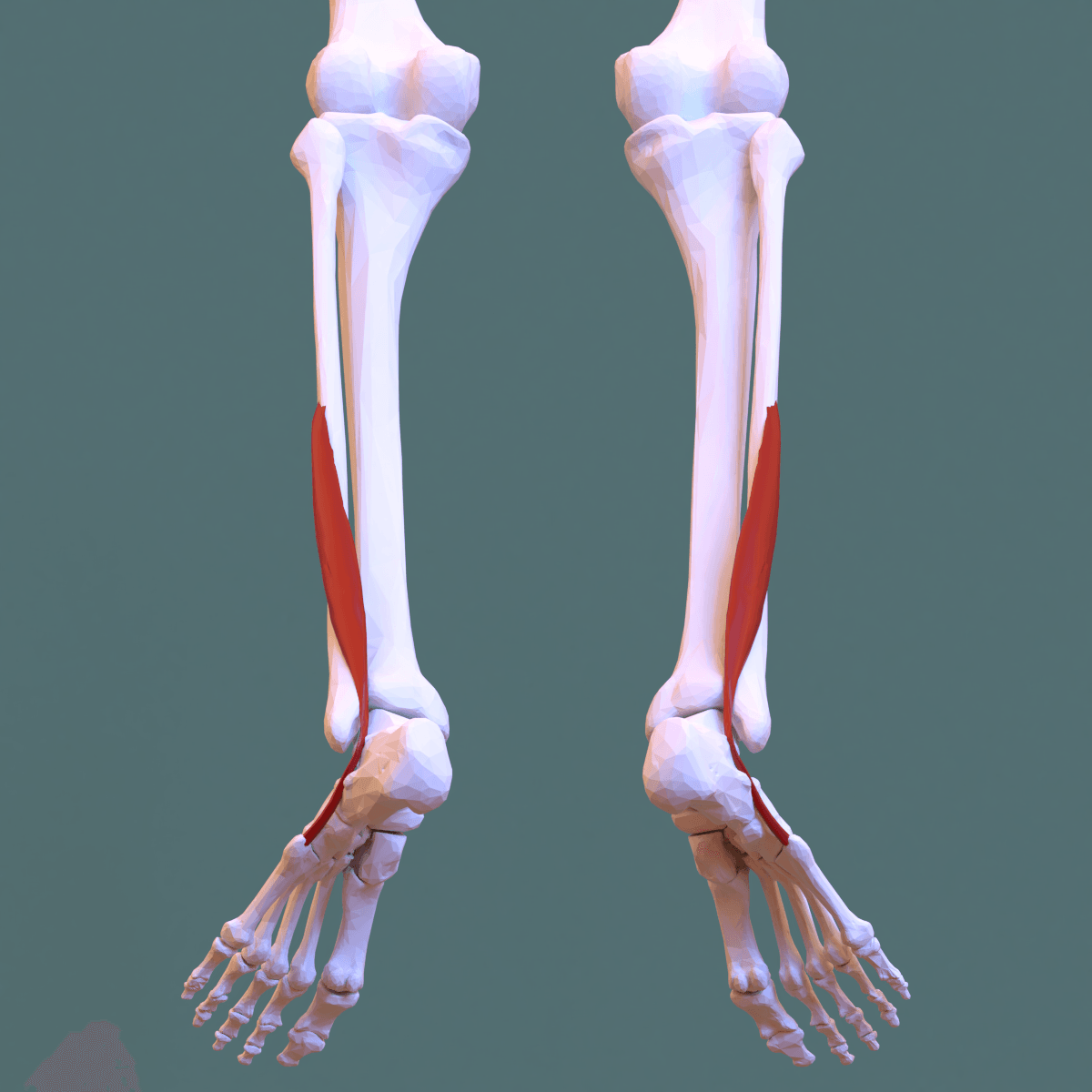
Attachments, Innervation and Relations
Origin
- Distal two-thirds of the lateral surface of the fibula, deep and distal to the origin of peroneus longus.
- Anterior and posterior intermuscular septa.
- The muscle belly extends more distally than that of peroneus longus - a fact that becomes pathological when it extends past the tip of the fibula into the retromalleolar groove (a low-lying muscle belly).
Tendon course
- Descends behind the lateral malleolus anterior and deep to peroneus longus, in the common sheath beneath the superior peroneal retinaculum, lying directly on the retromalleolar groove and the posterolateral fibular ridge.
- Turns forward around the tip of the lateral malleolus, passing superior to the peroneal tubercle of the calcaneus (peroneus longus passes inferior to it), beneath the inferior peroneal retinaculum, where the two tendons acquire separate sheaths.
- Runs forward along the lateral wall of the calcaneus and the lateral border of the cuboid to its insertion.
Insertion
- Styloid process (tuberosity) of the base of the fifth metatarsal, on its dorsolateral aspect.
- A variable expansion continues distally along the fifth metatarsal to blend with the extensor apparatus of the fifth toe - the peroneus digiti minimi slip, present in a majority of feet.
- The insertion is a fixed point of the lateral column and shares the tuberosity with the lateral band of the plantar fascia (plantar aponeurosis) and the lateral collateral ligament of the calcaneocuboid joint.
Retinacula and the groove
- Superior peroneal retinaculum (SPR): from the posterolateral ridge of the distal fibula to the lateral calcaneus and the deep fascia, roughly 1 to 2 cm wide. It is the primary restraint against peroneal tendon dislocation.
- Retromalleolar groove: concave in the majority (reported around 80 per cent), flat in about 10 to 20 per cent and convex in a small minority. The concavity is enhanced by a fibrocartilaginous ridge attached to the posterolateral fibula that forms part of the SPR attachment. A flat or convex groove predisposes to instability.
- Inferior peroneal retinaculum: attaches to the peroneal tubercle and separates the two tendons distally.
The sural nerve runs from the posterior midline of the calf, passing lateral to the Achilles, and crosses the lateral hindfoot approximately 1.5 cm posterior and inferior to the tip of the lateral malleolus, then runs anteriorly toward the base of the fifth metatarsal, which it reaches almost directly. It is therefore in the field of a peroneal tendon exposure, a calcaneal osteotomy, a lateral ligament reconstruction and a fifth metatarsal base fixation. Make full-thickness incisions straight to bone, use subperiosteal retractors, and never dissect in the subcutaneous plane in this region.
Action and Biomechanics
Actions
- Eversion (pronation) of the foot at the subtalar and transverse tarsal joints - it is the strongest and most efficient evertor, with a larger and more direct eversion moment arm than peroneus longus.
- Weak ankle plantarflexion - the tendon passes behind the ankle axis with a small moment arm.
- Abduction of the forefoot, pulling the lateral column laterally.
- Dynamic lateral ankle stabiliser - by resisting inversion it protects the anterior talofibular and calcaneofibular ligaments. Reflex peroneal contraction is a key component of the dynamic defence against an inversion sprain, and slowed peroneal reaction time is a recognised feature of chronic ankle instability.
The antagonist pairings
- Action
- Eversion of the hindfoot
- Direct antagonist
- Tibialis posterior
- Relative strength
- Tibialis posterior is roughly twice as strong
- Result if the antagonist wins
- Hindfoot varus
- Action
- Plantarflexion of the first ray
- Direct antagonist
- Tibialis anterior
- Relative strength
- Comparable, but TA weakens first in neuropathy
- Result if the antagonist wins
- Plantarflexed first ray and forefoot cavus
- Action
- Dorsiflexion and inversion
- Direct antagonist
- Peroneus longus at the first ray, triceps surae at the ankle
- Relative strength
- Plantarflexors are six times stronger than dorsiflexors
- Result if the antagonist wins
- Equinus and drop foot
- Action
- Inversion and arch support
- Direct antagonist
- Peroneus brevis
- Relative strength
- Tibialis posterior is stronger
- Result if the antagonist wins
- Planovalgus if tibialis posterior fails
Why the strength mismatch matters
- In a normal foot the balance between tibialis posterior and peroneus brevis is maintained by neural control, not by equal strength. Tibialis posterior is stronger, but it is not asked to work unopposed.
- In a length-dependent neuropathy such as Charcot-Marie-Tooth disease, the more distal and smaller muscles fail first: peroneus brevis weakens while tibialis posterior remains strong, and the inversion moment goes unopposed. Combined with a strong peroneus longus plantarflexing the first ray, this generates the classic cavovarus foot.
- Restoring eversion is therefore a therapeutic aim in cavovarus, and it is one of the two things a peroneus longus to peroneus brevis tenodesis achieves - the other being abolition of first ray plantarflexion.
The fifth metatarsal base as a mechanical node
- The tuberosity is loaded by three structures: the peroneus brevis tendon (dorsolateral), the lateral band of the plantar aponeurosis (plantar), and the lateral collateral ligament of the calcaneocuboid joint.
- In a sudden inversion with the foot plantarflexed, tension in these structures avulses the tuberosity - the zone 1 avulsion fracture. Cadaveric and clinical work implicates the lateral band of the plantar fascia at least as much as the peroneus brevis, and many authorities now regard the plantar fascia as the dominant deforming structure.
- Because it is a metaphyseal, well-vascularised, minimally displaced avulsion in cancellous bone, it heals reliably with symptomatic treatment.
- Contrast with the Jones fracture, which sits distal to this at the metaphyseal-diaphyseal junction, in a vascular watershed, in cortical bone, in a region loaded by the adduction-and-plantarflexion moment across the lateral column rather than by a simple avulsion. That is why it behaves entirely differently.
Surface Anatomy and Examination
Palpation
- The tendon is palpable behind the lateral malleolus, deep to peroneus longus, and can be traced forward above the peroneal tubercle to the base of the fifth metatarsal, which is subcutaneous and easily felt as a prominence at the mid-lateral border of the foot.
- Ask for resisted eversion with the foot in slight plantarflexion to isolate the peroneals from the extensor digitorum longus and peroneus tertius, which also evert but from a dorsiflexed position.
- Isolating brevis from longus: peroneus brevis is best assessed by resisted eversion (which it does more efficiently than longus); peroneus longus is assessed by resisted plantarflexion of the first ray. Pain or weakness in one but not the other narrows the diagnosis.
Named tests and signs
- How to perform
- Foot in slight plantarflexion, everted against resistance
- Positive finding
- Pain or weakness along the tendon behind and below the fibula
- What it means
- Peroneal tendinopathy, split tear, or superficial peroneal nerve lesion
- False positives
- Lateral ligament pain, sinus tarsi syndrome, subfibular impingement
- How to perform
- From a plantarflexed inverted position ask for active dorsiflexion and eversion while palpating behind the fibula
- Positive finding
- Palpable or audible snap as the tendons ride over the posterolateral fibula
- What it means
- Incompetent superior peroneal retinaculum
- False positives
- Voluntary subluxation in a lax patient - ask directly whether they can do it at will
- How to perform
- Direct pressure over the tendons behind the fibula with resisted eversion
- Positive finding
- Reproduces the pain
- What it means
- Tenosynovitis or an intrasheath split
- False positives
- Any lateral hindfoot pathology
- How to perform
- Ankle in slight plantarflexion, draw the hindfoot forward on a stabilised tibia
- Positive finding
- Increased translation with a soft end point
- What it means
- Anterior talofibular ligament insufficiency, commonly coexisting with peroneal pathology
- False positives
- Generalised laxity - compare both sides
- How to perform
- Invert the hindfoot with the ankle in neutral
- Positive finding
- Increased tilt compared with the other side
- What it means
- Calcaneofibular ligament insufficiency
- False positives
- Guarding, subtalar rather than tibiotalar tilt
- How to perform
- Look at the standing patient from the front; then block test
- Positive finding
- Visible medial heel pad; hindfoot varus that corrects on the block
- What it means
- Underlying cavovarus loading the peroneals - the reason the tendons fail
- False positives
- A rigid hindfoot will not correct regardless
- How to perform
- Direct palpation of the subcutaneous styloid
- Positive finding
- Focal tenderness after an inversion injury
- What it means
- Zone 1 avulsion fracture - obtain radiographs including an oblique view
- False positives
- Os vesalianum or an unfused apophysis in an adolescent
Imaging
- Radiographs: anteroposterior, lateral and oblique foot views for the fifth metatarsal base - the oblique view is essential and the fracture is often invisible on the other two. Look also for a fleck sign at the distal fibula on an ankle series, indicating retinacular avulsion.
- The two mimics of a fifth metatarsal base fracture in a child or adolescent:
- Unfused apophysis - the physeal line runs parallel to the shaft (longitudinal), whereas a fracture line runs transverse to the shaft. It appears around age 9 to 11 and fuses by around 12 to 15.
- Os vesalianum - a rounded, corticated accessory ossicle proximal to the tuberosity within the peroneus brevis tendon, present in a small percentage of feet.
- MRI: longitudinal splits appear as a C-shaped, chevron-shaped or bisected brevis tendon on axial images, sometimes with the longus tendon nestling in the split. Also assesses retinacular integrity, groove morphology, a low-lying muscle belly and a peroneus quartus.
- Ultrasound: dynamic assessment is the best modality for demonstrating subluxation, which is a movement phenomenon and can be entirely normal on a static MRI.
- CT or weightbearing CT: best for groove morphology, the peroneal tubercle, and hindfoot alignment.
Complications
- Mechanism
- Lateral hindfoot or fifth metatarsal incision crossing the nerve about 1.5 cm posteroinferior to the fibular tip
- Prevention
- Full-thickness incision to bone; identify or protect the nerve; avoid subcutaneous dissection
- Management
- Desensitisation; neuroma excision and burial in muscle if refractory
- Mechanism
- Anterolateral incision or arthroscopy portal at the fascial exit point
- Prevention
- Mark the nerve by plantarflexing and inverting the foot with the fourth toe flexed
- Management
- Neuroma excision and burial if a painful neuroma develops
- Mechanism
- Sacrificing the strongest evertor for a non-anatomic reconstruction
- Prevention
- Use anatomic Brostrom-Gould repair; if a graft is needed use hamstring or allograft
- Management
- Strengthening and bracing; revision to an anatomic reconstruction if unstable
- Mechanism
- A non-anatomic graft crossing the subtalar joint
- Prevention
- Anatomic repair or reconstruction reproducing native footprints
- Management
- Physiotherapy; revision is difficult once stiff
- Mechanism
- Retinacular repair without deepening a flat groove or correcting varus
- Prevention
- Assess groove morphology; deepen by impaction; correct hindfoot alignment
- Management
- Revision with groove deepening, retinacular reconstruction or a fibular bone block
- Mechanism
- Screw too small or too short; hindfoot varus not corrected; premature return to sport
- Prevention
- Largest-diameter screw the canal accepts, correct entry point, Coleman block test and osteotomy if indicated, radiographic union before return
- Management
- Revision with a larger screw, bone graft, and a lateralising calcaneal osteotomy
- Mechanism
- An over-long screw straightening the natural bow of the metatarsal
- Prevention
- Template the screw length; check the lateral and oblique views intra-operatively
- Management
- Exchange for a shorter screw
- Mechanism
- Tenodesing to a tendon that is itself split
- Prevention
- Inspect both tendons directly at surgery - do not rely on imaging alone
- Management
- Convert to an FDL or FHL transfer, or a staged graft reconstruction
- Mechanism
- Underlying cavovarus, subfibular impingement or sinus tarsi pathology not addressed
- Prevention
- Full alignment assessment before surgery
- Management
- Reassess alignment; osteotomy as required
- Mechanism
- Thin skin over a subcutaneous bony prominence, tension from swelling
- Prevention
- Delay surgery until swelling settles; full-thickness flaps; no undermining
- Management
- Local wound care, negative pressure dressing, flap cover if tendon or bone is exposed
Clinical Relevance
Fifth metatarsal base fractures
- Anatomical site
- Styloid process, cancellous metaphysis
- Mechanism
- Inversion with the foot plantarflexed; avulsion by the lateral plantar fascia band and peroneus brevis
- Healing
- Reliable - well vascularised
- Management
- Symptomatic: hard-soled shoe or boot, weightbearing as tolerated. Consider fixation only if there is more than 2 to 3 mm displacement with articular involvement of the cuboid-metatarsal joint, or a large displaced fragment in an athlete
- Anatomical site
- Metaphyseal-diaphyseal junction, extending into the fourth-fifth intermetatarsal articulation
- Mechanism
- Acute adduction load on a plantarflexed foot
- Healing
- Poor - vascular watershed
- Management
- Non-weightbearing cast for 6 to 8 weeks in the low-demand patient; intramedullary screw fixation in athletes and where early return is required
- Anatomical site
- Distal to the fourth-fifth articulation, in the proximal 1.5 cm of diaphysis
- Mechanism
- Repetitive load, often with hindfoot varus
- Healing
- Poor - high nonunion and refracture rate
- Management
- Intramedullary screw fixation, usually with correction of hindfoot varus; bone graft for established nonunion
- The Torg classification of zone 2 and 3 injuries grades them radiographically as acute (a sharp fracture line, no intramedullary sclerosis), delayed union (a widened fracture line with some sclerosis) and nonunion (complete medullary sclerosis). Sclerosis indicates that non-operative management is unlikely to succeed.
- Hindfoot varus is the single most important predisposing factor for zone 2 and 3 fractures and for their recurrence. A fifth metatarsal stress fracture or a refracture after screw fixation should always trigger an assessment of hindfoot alignment and a Coleman block test. Failing to correct varus means the screw will simply break or the bone will refracture.
- Screw technique: a solid or cannulated intramedullary screw of the largest diameter the canal will accept, inserted from the tip of the tuberosity in line with the medullary canal ("high and inside"), engaging the diaphysis. A screw that is too short, too narrow, or that straightens the natural curvature of the metatarsal and causes distal cortical impingement, predisposes to failure. The sural nerve is at risk at the entry point.
Peroneus brevis longitudinal split tears
- Prevalence: described in a substantial proportion of asymptomatic cadaveric specimens as well as in symptomatic patients, so a split on MRI must be correlated with symptoms and examination.
- Presentation: chronic posterolateral ankle pain and swelling, worse on uneven ground, often with a history of a "sprain that never got better". Weakness of eversion is variable.
- The Krackow and Bohne classification and the description by Sobel and colleagues stratify splits by extent, from partial-thickness fraying to a complete bisection with the longus lying between the two limbs.
- Almost always associated with at least one of: superior peroneal retinacular laxity, a low-lying muscle belly, a peroneus quartus, a flat or convex fibular groove, a hypertrophic peroneal tubercle, lateral ankle instability, or hindfoot varus.
- Treatment:
- Less than 50 per cent of the tendon involved: debride and tubularise with a running non-absorbable suture.
- Greater than 50 per cent involved, or the tendon is unusable: tenodesis to peroneus longus proximally and distally, excising the diseased segment - provided the longus is intact.
- Both tendons unusable: flexor digitorum longus or flexor hallucis longus transfer to the fifth metatarsal base, or a staged reconstruction with a silicone rod followed by an allograft or hamstring autograft.
- In every case: decrowd the groove (debulk a low-lying belly, excise a peroneus quartus), repair the superior peroneal retinaculum, deepen a flat groove, and correct hindfoot varus.
Peroneal tendon subluxation and dislocation
- Mechanism: sudden forced dorsiflexion with reflex peroneal contraction, classically a skiing injury, frequently misdiagnosed as a lateral ankle sprain.
- Pathoanatomy: the superior peroneal retinaculum strips from the posterolateral fibula with a periosteal sleeve, sometimes taking a rim of cortex - the fleck sign, diagnostic on an anteroposterior ankle radiograph.
- Eckert and Davis grades I to III describe increasing depth of that failure, from elevation of the retinaculum alone (I), through elevation with the fibrocartilaginous ridge (II), to a displaced cortical avulsion (III); a grade IV in some descriptions is avulsion from the posterior attachment.
- Treatment: cast immobilisation in slight plantarflexion and inversion for six weeks is an option acutely in a low-demand patient, but recurrence is common; surgical repair or reconstruction of the retinaculum, with fibular groove deepening if the groove is flat or convex, is standard in athletes.
- Intrasheath subluxation is a distinct entity in which the tendons swap position within an intact retinaculum, producing a painful snap without visible dislocation. It is diagnosed on dynamic ultrasound and is easily missed on static MRI.
Peroneal tendinopathy and tenosynovitis
- Overuse pain along the tendons, worse with lateral loading. Always look for the underlying cause.
- A hypertrophied peroneal tubercle (more common in cavovarus feet) causes attritional wear as the tendons ride over it and can be resected.
- Subfibular impingement in a valgus hindfoot compresses the tendons between the calcaneus and the fibula - here the cause is the opposite alignment, and correcting the flatfoot is the treatment.
Denervation and neuromuscular disease
- Peroneus brevis weakens early in Charcot-Marie-Tooth disease, and its loss against a strong tibialis posterior is one of the two engines of cavovarus.
- Common peroneal nerve palsy at the fibular neck causes complete loss of eversion together with drop foot.
- Isolated superficial peroneal nerve lesion causes loss of eversion with preserved dorsiflexion and sensory loss on the dorsum of the foot sparing the first web space.
Surgical Relevance
Peroneus brevis harvest for lateral ankle ligament reconstruction - and why it fell out of favour
The non-anatomic (tenodesis) era
- Evans procedure (1953): the whole peroneus brevis tendon is divided proximally, passed through a drill hole in the distal fibula from anteroinferior to posterosuperior, and sutured back to itself. It creates a checkrein between the fibula and the fifth metatarsal base.
- Watson-Jones procedure: the brevis is routed through the fibula and then into the talar neck, reconstructing a vector approximating the anterior talofibular ligament.
- Chrisman-Snook procedure (1969): a modification of the Elmslie procedure using a split (half) peroneus brevis tendon, routed through a drill hole in the fibula and then to the lateral calcaneus, reconstructing both the anterior talofibular and the calcaneofibular ligaments.
Why they were abandoned as first-line
- They are non-anatomic: they do not restore the native ligament footprints or the native vectors.
- They restrict subtalar motion, sometimes markedly, because the graft crosses the subtalar joint in a non-anatomic line.
- They sacrifice or weaken the strongest evertor of the foot - and eversion strength is a dynamic protector of the lateral ankle. Removing it to reconstruct a static restraint is a poor trade.
- Long-term follow-up shows higher rates of stiffness and degenerative change than anatomic repair.
Fibular groove deepening
- Indication: a flat or convex retromalleolar groove in a patient with peroneal tendon subluxation or dislocation, or recurrent instability after a previous retinacular repair.
- Techniques:
- Indirect (impaction) deepening: a drill or burr is introduced through a small cortical window at the fibular tip and the cancellous bone hollowed out; the posterior cortex is then impacted into the void with a tamp, deepening the groove while preserving the smooth cortical and fibrocartilaginous gliding surface. This is the preferred technique because it retains the natural bearing surface.
- Direct (osteoperiosteal flap) deepening: an osteoperiosteal flap is raised from the posterior fibula, cancellous bone removed beneath it, and the flap replaced.
- Simple burring of the groove is discouraged because it removes the fibrocartilage and leaves a raw cancellous surface against the tendon.
- Always combined with repair or reconstruction of the superior peroneal retinaculum, and with correction of hindfoot varus where present.
Fifth metatarsal base fixation
- Approach: a short longitudinal incision just proximal and dorsal to the tuberosity, or a percutaneous entry, in both cases with the sural nerve identified or protected.
- Entry point: at the very tip of the tuberosity, aiming "high and inside" - dorsal and medial - so the screw follows the medullary canal rather than perforating the lateral cortex.
- Screw: the largest diameter the canal will accept (commonly 4.5 to 5.5 mm), and long enough to engage good diaphyseal bone but not so long that it straightens the natural lateral bow of the metatarsal, which levers the distal fragment and causes cortical impingement or a distal fracture.
- Adjuncts: bone graft or bone marrow aspirate for a sclerotic nonunion; plantar plating is an alternative in revision.
- Do not forget the alignment: a Coleman block test and an assessment of hindfoot varus is mandatory in a recurrent or stress fracture, and a lateralising calcaneal osteotomy may be needed.
The muscle as a donor
- Tendon donor: historically a major graft source (Evans, Watson-Jones, Chrisman-Snook), now largely superseded. It remains a reasonable revision graft as a split tendon where no alternative exists.
- Recipient of a transfer: peroneus brevis is a common recipient - the peroneus longus is tenodesed to it in cavovarus and in irreparable longus tears, and a flexor digitorum longus or flexor hallucis longus transfer is anchored to the fifth metatarsal base when the brevis itself is unusable.
- Muscle flap: never used. It is small, its supply is by unnamed perforators, and its loss would remove the strongest evertor.
- A varus hindfoot overloads the peroneals with every step.
- Peek-a-boo heel and Coleman block test in every case.
- Repairing the tendon without correcting the varus guarantees recurrence.
- Flat or convex groove means instability will recur after a soft-tissue repair alone.
- Assess on axial MRI or CT.
- Deepen the groove by impaction, preserving the fibrocartilage.
- Low-lying peroneus brevis muscle belly.
- Peroneus quartus, present in 10 to 20 per cent of feet.
- Hypertrophic peroneal tubercle.
- Debulk or excise, and check the state of both tendons directly.
Guidelines, Registries & Global Practice
Anatomical variation across populations
- Peroneus quartus is present in roughly 10 to 20 per cent of feet in cadaveric series, with reported prevalence varying widely by population and by whether the definition includes small slips. Its most common form arises from the peroneus brevis and inserts on the retrotrochlear eminence of the calcaneus.
- Retromalleolar groove morphology: concave in the majority (commonly quoted around 80 per cent), flat in roughly 10 to 20 per cent, and convex in a small minority. Groove morphology is the single most important anatomical predictor of peroneal tendon instability.
- Low-lying peroneus brevis muscle belly extending distal to the tip of the fibula is common and is a recognised contributor to a crowded groove.
- Os vesalianum is an uncommon accessory ossicle within the peroneus brevis tendon proximal to the fifth metatarsal tuberosity.
- Peroneus digiti minimi slip from the brevis to the fifth toe extensor apparatus is present in the majority of feet.
Side-by-side guidance
- Position relevant to peroneus brevis
- Support anatomic lateral ligament repair (Brostrom-Gould) as first line for chronic instability, reserving tendon-sacrificing tenodesis for selected revision cases.
- Position relevant to peroneus brevis
- Emphasise assessment and correction of hindfoot alignment as an integral part of peroneal tendon and lateral ligament surgery.
- Position relevant to peroneus brevis
- For proximal fifth metatarsal fixation, emphasises the largest-diameter intramedullary screw the canal accepts, the high-and-inside entry point, and protection of the sural nerve.
- Position relevant to peroneus brevis
- Favour early intramedullary screw fixation of zone 2 and 3 fractures in elite athletes to reduce time to return and refracture, with radiographic union across all cortices before return to play.
- Position relevant to peroneus brevis
- Emphasise distinguishing the normal fifth metatarsal apophysis (longitudinal) from an avulsion fracture (transverse), and recognising Iselin disease as a traction apophysitis.
Resource-dependent practice
- Well-resourced settings: MRI to characterise the tendons and groove, dynamic ultrasound for subluxation, weightbearing CT for alignment and groove morphology, suture tape augmentation of anatomic repairs, early screw fixation for elite athletes.
- Limited-resource settings: the diagnosis of a peroneal tendon problem is clinical - tenderness and swelling behind the fibula, pain and weakness on resisted eversion, a provocable snap. The peek-a-boo heel and the Coleman block test cost nothing and answer the alignment question. Zone 1 fractures require nothing more than a hard-soled shoe. Zone 2 fractures can be managed successfully with six to eight weeks of non-weightbearing immobilisation in a non-athlete. A Brostrom-Gould repair requires only suture. Groove deepening by impaction needs only an osteotome and a tamp.
- The single most transferable message across settings is that hindfoot varus must be sought and corrected, because it is free to diagnose and it determines whether everything else will hold.
Registry and outcome signals
- There is no registry for peroneal tendon or lateral ligament surgery. Comparative cohort data consistently favour anatomic repair over tenodesis reconstruction for function and for preservation of hindfoot motion.
- Athletic cohorts consistently report that fifth metatarsal fracture refracture correlates with undersized screws, premature return to play and uncorrected hindfoot varus, rather than with the fixation principle itself.
- Series of peroneal tendon dislocation consistently report high recurrence with non-operative management in athletes and reliable results with retinacular repair combined with groove deepening where the groove is unfavourable.
MCQ Practice Points
Q: Where does peroneus brevis insert? A: The styloid process (tuberosity) at the base of the fifth metatarsal, on its dorsolateral aspect, often with an expansion to the extensor apparatus of the fifth toe.
Q: Which peroneal tendon lies against the fibula and why does it matter? A: Peroneus brevis, anterior and deep, with peroneus longus posterior and superficial. Brevis is compressed against the posterolateral fibular ridge, producing the longitudinal split tear.
Q: Which tendon passes above and which below the peroneal tubercle? A: Brevis above, longus below.
Q: What supplies peroneus brevis? A: The superficial peroneal nerve, roots L5 and S1, with motor branches in the proximal third of the leg.
Q: Which is the stronger evertor, longus or brevis? A: Peroneus brevis, with a larger and more direct eversion moment arm. It is the direct antagonist of tibialis posterior, which is roughly twice as strong.
Q: Why does the Jones fracture fail to heal? A: It lies in a vascular watershed at the metaphyseal-diaphyseal junction, where the diaphyseal nutrient supply and the metaphyseal supply meet and neither is generous.
Q: Which structure is now considered the main deforming force in a zone 1 tuberosity avulsion? A: The lateral band of the plantar aponeurosis, at least as much as the peroneus brevis tendon. It heals reliably with symptomatic treatment.
Q: How do you distinguish the fifth metatarsal apophysis from a fracture? A: The apophyseal line runs parallel to the shaft; a fracture line runs transverse to it. The apophysis appears at around 9 to 11 years and fuses by 12 to 15.
Q: What is peroneus quartus and why does it matter? A: An accessory muscle present in roughly 10 to 20 per cent of feet, usually arising from peroneus brevis and inserting on the retrotrochlear eminence of the calcaneus. It crowds the retromalleolar groove and predisposes to brevis tears and subluxation.
Q: What is the preferred technique for fibular groove deepening? A: Indirect (impaction) deepening, hollowing the cancellous bone through a cortical window and impacting the posterior cortex, which preserves the smooth cortical and fibrocartilaginous gliding surface. Simple burring is discouraged.
Q: Why is the Chrisman-Snook procedure no longer first line? A: It is non-anatomic, restricts subtalar motion, is associated with more degenerative change, and sacrifices the strongest evertor. Anatomic Brostrom-Gould repair is the modern standard.
Q: Where does the sural nerve cross the lateral hindfoot? A: Approximately 1.5 cm posterior and inferior to the tip of the lateral malleolus, running anteriorly toward the base of the fifth metatarsal.
Exam Viva Scenarios
Practise clinical reasoning and management decisions out loud
“A 22-year-old semi-professional footballer had an intramedullary screw for a Jones fracture nine months ago. He returned to play at four months and has re-fractured at the same level. Radiographs show a broken 4.0 mm screw and a fracture line with surrounding sclerosis. What went wrong and what do you do now?”
“A 34-year-old woman has had posterolateral ankle pain and swelling for 14 months after an inversion injury. She feels the ankle giving way on uneven ground. Examination shows tenderness and fullness behind the lateral malleolus, pain on resisted eversion, a positive anterior drawer, and when you look at her standing from the front you can see the medial heel pad on that side. How do you proceed?”
“An 11-year-old boy has lateral foot pain after an inversion injury while playing. He is tender over the base of the fifth metatarsal. A radiograph shows a lucency at the base of the fifth metatarsal running parallel to the shaft. The emergency department has diagnosed a fracture. Do you agree?”
Anatomy
- Origin: distal two-thirds of the lateral fibula and intermuscular septa
- Insertion: styloid process of the fifth metatarsal base, dorsolateral
- Nerve: superficial peroneal, L5-S1
- Anterior and deep in the retromalleolar groove - against the bone
- Passes superior to the peroneal tubercle (longus inferior)
Function
- Strongest and most direct evertor of the hindfoot
- Direct antagonist of tibialis posterior, which is roughly twice as strong
- Dynamic protector of the lateral ligaments against inversion
- Weakens early in Charcot-Marie-Tooth - a driver of hindfoot varus
Fifth Metatarsal Zones
- Zone 1: tuberosity avulsion - heals reliably, symptomatic treatment
- Zone 2: Jones fracture at the metaphyseal-diaphyseal junction - vascular watershed
- Zone 3: proximal diaphyseal stress fracture - highest nonunion risk
- Torg: medullary sclerosis predicts failure of non-operative care
- Always assess hindfoot varus in zone 2 and 3 injuries
Tendon Pathology
- Longitudinal split at the fibular groove - compressed by the longus
- Crowding: low-lying belly, peroneus quartus, lax retinaculum, flat groove
- Less than 50% torn = tubularise; more than 50% = tenodese to an intact longus
- Fleck sign on the ankle radiograph = retinacular avulsion
- Deepen a flat groove by impaction, preserving the fibrocartilage
Surgery
- Sural nerve about 1.5 cm posteroinferior to the fibular tip, running to the 5th MT base
- SPN pierces the fascia 10-12 cm above the lateral malleolus
- Brostrom-Gould, not Chrisman-Snook, is first line for instability
- Never sacrifice the whole brevis in a foot with weak eversion
- Correct hindfoot varus or the repair will fail
Evidence Base
The Dynamics of Peroneus Brevis Tendon Splits
- Investigation of the mechanism of peroneus brevis splits, with a technique of diagnosis and a proposed classification
- Concluded that splits result from a dynamic mechanical insult at the fibular groove, not from primary ischaemia
- Laxity of the superior peroneal retinaculum combined with mechanical compression by the peroneus longus causes the brevis to splay out and split over the sharp posterior edge of the fibula
- Named the contributing anatomical factors: a shallow or congenitally convex fibular groove, an anomalous low-lying peroneus brevis muscle belly, and a peroneus quartus tendon
Longitudinal Attrition of the Peroneus Brevis Tendon in the Fibular Groove
- 124 fresh cadaveric ankles from 65 cadavers dissected under loupe magnification
- Attrition of the peroneus brevis was found in 14 ankles (11.3%) - and in NO specimen was the peroneus longus involved
- Change ranged from simple splaying of the tendon in the fibular groove to frank longitudinal splits with fraying of the remaining halves
- Longitudinal ruptures averaged 1.9 cm in length (range 1 to 4 cm)
Fractures of the Base of the Fifth Metatarsal Distal to the Tuberosity
- 46 fractures distal to the tuberosity treated between 1973 and 1982, followed for a mean of 40 months
- Three radiographic types: acute (narrow line, no sclerosis), delayed union (widened line with sclerosis), and nonunion (medullary canal obliterated by sclerotic bone)
- Of 25 acute fractures, 14 of the 15 placed in a non-weightbearing toe-to-knee cast healed in a mean of 7 weeks, but only 4 of the 10 managed with weightbearing methods united
- Of 9 nonunions treated by medullary curettage and bone grafting, 8 healed in a mean of 3 months
Jones Fractures and Related Fractures of the Proximal Fifth Metatarsal
- Distinguished at least three fracture types in the proximal fifth metatarsal: the Jones fracture, the proximal diaphyseal stress fracture, and the tuberosity avulsion fracture
- Emphasised that the diaphyseal stress fracture is commonly confused with the Jones fracture, obscuring vital differences in prognosis and treatment
- Reviewed the anatomical, biomechanical and vascular characteristics that explain their differing healing potential
- Concluded that treatment guidelines are controversial and frequently need individualisation, and that most such fractures heal without surgery
The Intraosseous Blood Supply of the Fifth Metatarsal
- 10 fresh-frozen specimens injected with India ink or barium sulfate to map the intraosseous supply
- The tuberosity is supplied by numerous metaphyseal vessels entering its non-articular surfaces in a random radiate pattern
- The proximal diaphysis is supplied primarily by the nutrient artery via longitudinal intramedullary branches
- The two supplies meet just distal to the tuberosity, precisely the region of poor prognosis for fracture healing, suggesting relative ischaemia underlies delayed union and nonunion
Reconstruction of Lateral Ligament Tears of the Ankle
- Described a modification of the Elmslie procedure using a split peroneus brevis tendon
- Reconstructs both the anterior talofibular and calcaneofibular ligaments
- Reported good stability in the small series presented
- Became the standard non-anatomic reconstruction for decades
Anatomical Reconstruction Versus Tenodesis for Chronic Anterolateral Ankle Instability
- Retrospective multicentre comparison of 106 anatomical reconstructions against 110 tenodeses, at mean follow-up of about 5 years
- The tenodesis group had significantly more reoperations, a smaller range of ankle motion, and more medially located osteophytes
- On stress radiographs the tenodesis group had significantly greater talar tilt and greater anterior talar translation
- There was NO difference in mean Karlsson functional score between the groups - the advantage of anatomical repair lay in motion, stability and degenerative change rather than in the mean functional score
Early and Late Repair of Lateral Ligament of the Ankle
- Instrumented testing of the anterior talofibular and calcaneofibular ligaments in 25 healthy adults aged 15 to 30
- Stability of the ankle depends primarily on the integrity of the anterior talofibular ligament; anterior translation exceeding 4 mm gives a positive anterior drawer, and normal talar tilt ranged up to 18 degrees
- Repair, whether early or late, is achieved by suturing the ligament remnant that is always present
- The anterior talofibular repair is then reinforced by overlapping the nearby lateral talocalcaneal ligament together with the marginal ankle retinaculum - the augmentation now known as the Gould modification