The Workhorse Autograft of the Knee
- Pes anserinus from anterior to posterior and superficial to deep is sartorius, gracilis, semitendinosus — Say Grace before Tea.
- Three pes tendons, three different nerves: sartorius (femoral, L2-L4), gracilis (obturator, L2-L4), semitendinosus (tibial division of sciatic, L5-S2).
- An accessory insertion runs from the semitendinosus tendon toward the medial head of gastrocnemius; cadaveric work found it in 77 per cent of knees.
- Failure to release that band before advancing the stripper is the classic cause of premature graft amputation.
- The infrapatellar branch of the saphenous nerve crosses the harvest field transversely and is the commonest iatrogenic injury of the approach.
- “Semitendinosus has a tendinous raphe across its mid-belly and receives two separate motor branches, one above and one below it.
- “Regeneration of the harvested tendon is common but far from universal: in one two-year MRI study only 35 per cent of patients had regenerated both semitendinosus and gracilis.
- “The measurable long-term deficit after hamstring harvest is knee flexion strength at deep flexion angles greater than 70-90 degrees.
- “A four-strand graft less than 8 mm in diameter in a young patient is associated with higher early revision rates.
Overview
Semitendinosus is the superficial and posterior of the two medial hamstrings. Its name describes its architecture: the muscle belly occupies the proximal half and gives way, at about the mid-thigh, to a long, round, cord-like tendon that runs almost half the length of the thigh. That geometry — a long, uniform, tubular free tendon with a single distal attachment — is exactly what makes it the most useful autograft in the knee.
The muscle carries a distinguishing internal feature: an oblique tendinous raphe running across the mid-belly, which divides the muscle into a proximal and a distal portion. Each portion has its own motor branch from the tibial division of the sciatic nerve. This is not merely a curiosity: it explains why a partial denervation can weaken one half of the muscle without abolishing its function, and it is a favourite question in a nerve-injury viva.
The examiner's favourite paradox: we harvest a structure that protects the ligament we are reconstructing.
- The medial hamstrings pull the tibia posteriorly on the femur. That vector is the same direction as the ACL's restraint of anterior tibial translation, so hamstring contraction unloads the ACL and the ACL graft.
- This is the biomechanical basis for hamstring-dominant rehabilitation after reconstruction and for the hamstring-strengthening core of neuromuscular ACL prevention programmes.
- Semitendinosus additionally internally rotates the tibia, opposing the anterolateral rotatory subluxation of the pivot shift, and as a pes tendon it is a secondary restraint to valgus.
- The trade-off: harvest removes part of that protection and leaves a measurable deep-flexion knee flexion deficit that persists for years. The counterargument is that the tendon often regenerates and the functional deficit at everyday flexion angles is small — but regeneration cannot be assumed.
- Where the deficit does matter: sports demanding force at deep knee flexion — wrestling, sprinting from a deep start position, and the deep-flexion positions of some martial arts — and in patients who will need the tendon later for a different reconstruction.
Say Grace before TeaPes Anserinus Order
Hook:Anterior to posterior AND superficial to deep — the order is the same either way. Three tendons, three different nerves.
FOTThree Pes Tendons, Three Nerves
Hook:Three tendons sharing one insertion but supplied by three different nerves from two different plexus divisions — the classic pes anserinus question.
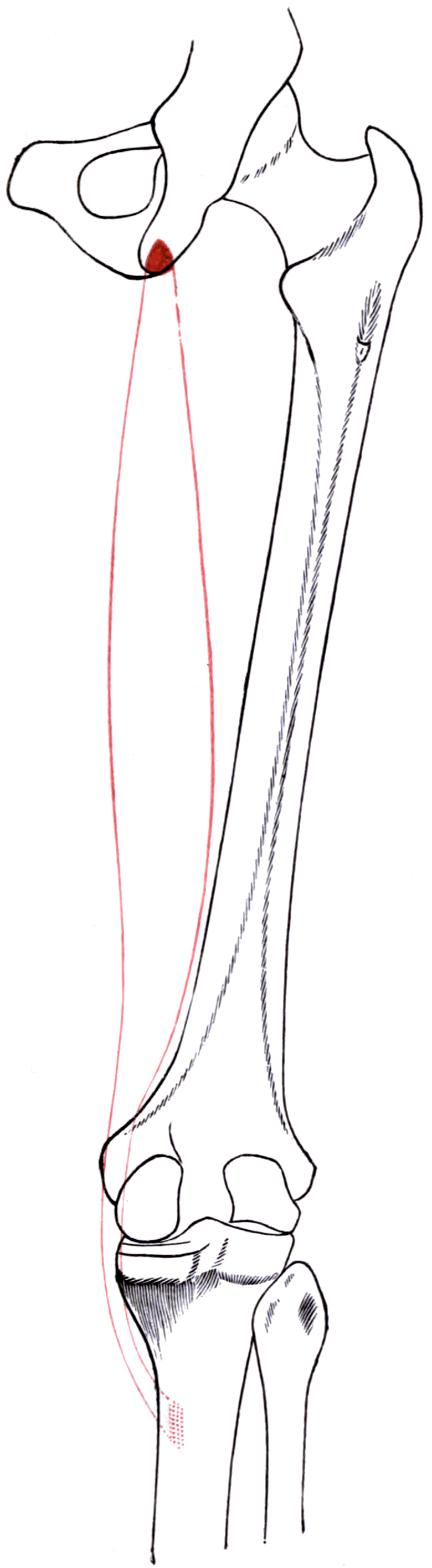
Attachments, Innervation and Relations
Proximal Origin
- Site: the posteromedial aspect of the ischial tuberosity, as part of the conjoint tendon shared with the long head of biceps femoris.
- Relationship: the conjoint footprint lies posteromedial; the semimembranosus footprint lies anterolateral to it. The two are contiguous but distinct on the tuberosity.
- Surgical consequence: semitendinosus and biceps long head avulse together because they share a tendon. A "two-tendon avulsion" therefore usually means the conjoint tendon plus a partial semimembranosus, and a "three-tendon avulsion" means the conjoint tendon has separated from semimembranosus as well.
The Tendinous Raphe
- An oblique fibrous intersection crosses the muscle belly at about the level of the mid-thigh, running from proximal-medial to distal-lateral.
- It divides semitendinosus into two functional compartments, each with an independent motor nerve.
- Identification value: at open dissection or in an MRI cross-section, the raphe is the reliable feature distinguishing semitendinosus from gracilis and semimembranosus.
Distal Insertion
- The tendon becomes free at roughly the junction of the middle and distal thirds of the thigh, runs distally and anteriorly around the medial femoral condyle, and inserts into the superomedial surface of the tibia as the deepest layer of the pes anserinus.
- Precise footprint: on the anteromedial tibia, roughly 2-3 cm distal to the medial joint line and just medial to the tibial tuberosity, beneath the sartorius fascia.
- The pes anserinus is the conjoined insertion of sartorius (most superficial and anterior), gracilis (middle) and semitendinosus (deepest and most posterior), resembling a goose's foot.
- Layer note: the pes tendons are superficial to the superficial MCL, with the pes anserine bursa interposed between them. The bursa is the anatomical basis for pes anserine bursitis.
Harvestable Tendon
- Length: typically 24-30 cm of usable tendon from a standard harvest — longer than gracilis, which yields about 20-26 cm.
- Diameter: a doubled semitendinosus alone gives roughly 6-8 mm; a quadrupled semitendinosus alone or a doubled semitendinosus-gracilis construct gives 7-10 mm.
- Fascial expansions: the tendon gives an expansion into the crural fascia distally and, critically, one or more fascial bands to the medial head of gastrocnemius proximally.
This is the single most examinable technical hazard of hamstring harvest.
- An accessory insertion (fascial expansion) runs from the deep surface of the semitendinosus tendon posteriorly and proximally toward the medial head of gastrocnemius, arising a few centimetres proximal to the tibial insertion. Cadaveric work found it in 77 per cent of knees, and the tendon is additionally ensheathed in a dense investing fascial layer that impedes stripping.
- If the stripper is advanced before these bands are divided, it follows the band instead of the tendon and either (a) cuts the graft short — premature amputation — leaving an unusably short graft, or (b) travels toward the medial gastrocnemius and the popliteal fossa.
- Technique to avoid it: deliver the tendon, place a whip-stitch, apply firm distal traction, then run a finger or a right-angled clamp along the deep and posterior surface of the tendon toward the popliteal fossa and sharply divide every band under direct vision with tenotomy scissors.
- Advance the stripper along the axis of the tendon with the knee flexed 60-90 degrees and the hip flexed, keeping constant tension on the tendon so the stripper is guided, not steered.
- Never force the stripper. Resistance means an undivided band. Withdraw, re-palpate, and divide it.
Action and Biomechanics
Actions by Plane
- Hip: extension (and a minor contribution to hip adduction through its medial line of pull).
- Knee: flexion — the dominant action — and internal rotation of the flexed tibia.
- Rotatory control: with gracilis and sartorius, it contributes to the medial dynamic sling that resists tibial external rotation and valgus.
Moment Arms and Length-Tension
- As a biarticular muscle it is placed on maximal stretch by hip flexion combined with knee extension — the late-swing position in sprinting, and the mechanism of hamstring strain.
- The knee flexion moment arm rises from extension to about 30-50 degrees and is broadly maintained thereafter.
- The tibial internal rotation moment arm rises steeply with flexion, which is why the rotatory contribution is negligible near extension and substantial at 90 degrees. This is also why the post-harvest deficit is deep-flexion specific.
Synergists and Antagonists
- Synergists: semimembranosus, biceps femoris long head, gracilis, sartorius, popliteus, gastrocnemius.
- Antagonists: quadriceps at the knee, iliopsoas and rectus femoris at the hip, biceps femoris for tibial rotation.
What Happens When It Is Removed
- Immediately after harvest: knee flexion strength is reduced, most measurably at deep flexion; hip extension power is essentially maintained because semimembranosus and biceps compensate.
- At 6-12 months: overall knee flexion torque is close to the contralateral side at mid-range, but a deep-flexion deficit persists.
- Muscle morphology: the semitendinosus belly shortens and its musculotendinous junction migrates proximally; compensatory hypertrophy occurs in the biceps femoris and semimembranosus.
- Regeneration: a regenerated tendon-like structure is demonstrable on MRI in many patients, but not all — one two-year study found both semitendinosus and gracilis had regenerated in only 35 per cent. Where it does form, it typically inserts more proximally, so it does not fully restore the original moment arm.
Graft diameter is the modifiable predictor of failure and is one of the most heavily examined numbers in ACL surgery. Quote it, but quote what sits beside it.
- Grafts of 8 mm diameter or less in young patients are associated with higher early revision. Magnussen's unadjusted rates: 1.7 per cent above 8 mm, 6.5 per cent at 7.5-8 mm, 13.6 per cent at 7 mm or less.
- AGE IS THE STRONGER SIGNAL, AND IT IS NOT MODIFIABLE. In the same series revision was 0.7 per cent at 20 years or over against 14.3 per cent under 20 — a twentyfold gap. In the multivariable model, age under 20 carried an odds ratio of 18.97 while decreased graft size carried 2.20, with a confidence interval of 1.00 to 4.85 and p = 0.05. The graft-size interval touches unity. Sixteen of the eighteen revisions were in patients under 20 with grafts of 8 mm or less: 16.4 per cent in that group.
- The honest reading: diameter is worth optimising because you can change it, not because it is the dominant risk. The dominant risk is being young, and no choice of graft removes it — which is the point to make when a 16-year-old and a parent ask what the failure rate is.
- Options when a doubled semitendinosus-gracilis construct measures small: quadruple or quintuple the semitendinosus, add the gracilis, use an all-inside quadrupled technique, augment with allograft, or change graft source entirely.
- Predicting it pre-operatively: height and weight correlate weakly with graft diameter; pre-operative MRI cross-sectional area of the tendons predicts better and is used in some units to plan the graft source.
- Always have a plan B on the table before you divide the tendon — this is what the examiner is testing.
Surface Anatomy and Examination
Palpation
- Position the patient prone with the knee flexed to 90 degrees and ask for resisted flexion.
- The most prominent, most posterior, round cord at the posteromedial knee is the semitendinosus tendon. Immediately anterior and medial to it, a thinner, flatter, more anterior ribbon is the gracilis. Deep and slightly lateral to semitendinosus, the broad blunt structure is semimembranosus.
- A useful trick for the harvest: ask a supine patient to flex the knee against resistance with the hip flexed and externally rotated (the "figure-of-four" position). The gracilis and semitendinosus tendons stand out as two distinct cords beneath the sartorial fascia and can be marked on the skin.
- The pes insertion is palpable on the anteromedial tibia about 2-3 cm distal to the joint line; the pes anserine bursa lies just deep to it, about 4-5 cm distal to the medial joint line.
Named Clinical Tests
- How to perform
- Prone, knee 90 degrees, foot turned in, examiner resists flexion
- Positive finding
- Pain or weakness compared with external rotation position
- What it means
- Medial hamstring (semitendinosus / semimembranosus) involvement
- False positive
- Pain from a posterior horn medial meniscal tear or popliteal cyst
- How to perform
- Direct palpation 4-5 cm distal to the medial joint line, anteromedially
- Positive finding
- Localised tenderness with or without swelling
- What it means
- Pes anserine bursitis or tendinopathy
- False positive
- Medial tibial stress reaction; medial compartment osteoarthritis
- How to perform
- Supine, hip and knee maximally flexed, then the knee is slowly extended
- Positive finding
- Ischial pain as the knee approaches extension
- What it means
- Proximal hamstring tendinopathy at the conjoint origin
- False positive
- Lumbar radiculopathy; ischiofemoral impingement
- How to perform
- Prone, examiner passively flexes the knee fully, patient holds it
- Positive finding
- Inability to hold the flexed position
- What it means
- Significant hamstring weakness or avulsion
- False positive
- Pain inhibition; quadriceps contracture limiting passive flexion
- How to perform
- Isokinetic or manual flexion strength tested at greater than 70 degrees
- Positive finding
- Deficit compared with the contralateral limb
- What it means
- The characteristic post-harvest donor deficit
- False positive
- Generalised deconditioning after any knee surgery
- How to perform
- Percuss the sartorial fascia transversely, distal to the medial joint line
- Positive finding
- Radiating dysaesthesia into the anterolateral proximal leg
- What it means
- Infrapatellar branch neuroma after harvest or arthroscopy
- False positive
- Sensitised skin over any healing incision
Grading and Interpretation
- Manual grading uses the standard 0-5 scale, but after harvest the deficit is angle-specific, so a manual test at 45 degrees will miss it. Test at 90 degrees or more, or use isokinetic testing where available.
- Functional grading matters more than manual grading for return to sport: single-leg bridge repetitions, Nordic hamstring performance, and hop testing symmetry.
Pitfalls
- Do not confuse tendon absence with weakness. After harvest the cord is absent on palpation but the muscle still contracts, and a regenerated tendon may reappear over the following year or two.
- Pain over the pes after ACL reconstruction is more often incisional or infrapatellar branch neuropathic pain than true pes bursitis.
- Numbness in a patch below and lateral to the incision is the infrapatellar branch, not the saphenous trunk. Saphenous trunk injury numbs the medial leg down to the medial malleolus.
Complications
Donor-Site Morbidity
- Deep-flexion knee flexion weakness — the reproducible, angle-specific deficit. Counsel athletes whose sport demands force in deep flexion.
- Hip extension weakness — measurable in some studies but rarely functionally important, since semimembranosus and biceps compensate.
- Altered muscle architecture — proximal migration of the musculotendinous junction, reduced belly volume, compensatory hypertrophy of the other hamstrings, persisting for at least two years.
- Reduced future graft availability — a real problem in the young patient facing revision or a multiligament reconstruction.
Neurological
- Infrapatellar branch of the saphenous nerve injury — numbness and hypersensitivity over the anterolateral proximal leg, and occasionally a painful neuroma requiring excision and burial in muscle. The commonest complication of the harvest and a mandatory consent point.
- Saphenous nerve trunk injury — numbness along the medial leg to the medial malleolus; far less common but far more troublesome.
- Complications from the stripper reaching the popliteal fossa — tibial nerve and popliteal vessel injury; extremely rare but catastrophic and entirely preventable.
Technical
- Premature graft amputation — as above. Always consent for and have available an alternative graft.
- Undersized graft — a four-strand construct measuring 7 mm or less in a young patient. Augment or change source rather than accept it.
- Graft slippage on the back table — inadequate whip-stitching; secure both ends before tensioning.
Local Wound Problems
- Haematoma in the posteromedial calf and thigh from the harvest bed. Reduced by closing the sartorial fascia and by careful haemostasis of the crural fascial vessels.
- Infection at the donor site is uncommon but shares the same significance as an intra-articular infection because the tunnels communicate.
Preventing Each
- Mechanism
- Stripper follows an undivided gastrocnemius band
- Prevention
- Digital and sharp release of all bands under vision before stripping
- Mechanism
- Vertical incision crossing the transverse nerve
- Prevention
- Oblique incision parallel to the nerve; blunt subcutaneous dissection
- Mechanism
- Stripper driven posterolaterally with the knee extended
- Prevention
- Harvest at 90 degrees flexion, hip flexed and externally rotated; never force
- Mechanism
- Assuming diameter from patient size
- Prevention
- Pre-operative planning, consider MRI cross-sectional area, plan extra strands
- Mechanism
- Open harvest bed and unclosed sartorial fascia
- Prevention
- Close the fascia, achieve haemostasis, consider a compressive dressing
- Mechanism
- Harvesting both tendons when one would do
- Prevention
- Consider quadrupled semitendinosus alone and spare the gracilis in young patients
Clinical Relevance
Indications for Harvest
- ACL reconstruction — the commonest indication worldwide; doubled semitendinosus-gracilis or quadrupled/quintupled semitendinosus alone.
- MCL and posteromedial corner reconstruction — semitendinosus, sometimes left attached distally at the pes as a natural tibial fixation.
- MPFL reconstruction — usually gracilis, but semitendinosus is used when a larger graft is required or the gracilis is inadequate.
- PCL and posterolateral corner reconstruction, multiligament knee reconstruction, and ankle lateral ligament reconstruction.
- Paediatric and adolescent ACL reconstruction — a physeal-sparing or transphyseal soft-tissue graft avoids the bone plug of a patellar tendon graft crossing the physis.
Step-by-Step Technique
- Position: supine, knee flexed to about 90 degrees, hip flexed and externally rotated (figure-of-four) to bring the tendons subcutaneous and away from the popliteal vessels.
- Incision: a 2-3 cm oblique or vertical incision over the pes, roughly 2-3 fingerbreadths distal to the medial joint line and 1-2 cm medial to the tibial tuberosity. An oblique incision parallel to the infrapatellar branch reduces the risk of nerve division.
- Expose the sartorial fascia; identify the gracilis as the more proximal, thinner cord and the semitendinosus as the distal, thicker cord palpable through the fascia.
- Incise the sartorial fascia along the superior border of the gracilis (an "L" or transverse incision) and deliver both tendons with a right-angled clamp. Preserve the fascial flap for closure.
- Whip-stitch the free end of each tendon after detaching it from the tibia, or detach it later — practice varies.
- Release the fascial bands. With firm distal traction on the tendon, sweep a finger along its deep and posterior surface toward the popliteal fossa and sharply divide the accessory insertion and the dense investing fascia a few centimetres proximal to the tibial insertion.
- Pass the closed-loop stripper over the tendon, keep it coaxial with the tendon under constant tension, and advance with a smooth push toward the ischium. Never force it.
- Detach the tibial insertion subperiosteally if this has not already been done, preserving a periosteal cuff for repair.
- Prepare the graft on the back table: strip muscle, whip-stitch both ends, measure length and diameter, pre-tension.
- Close the sartorial fascia to reduce the risk of a symptomatic donor-site defect and haematoma.
Yields
- Typical diameter
- 6-7 mm
- Typical length available
- 12-15 cm doubled
- Comment
- Usually too small alone in an adult
- Typical diameter
- 7-9 mm
- Typical length available
- 6-8 cm quadrupled
- Comment
- Spares the gracilis; needs a tendon 26 cm or longer
- Typical diameter
- 7-9 mm
- Typical length available
- 9-11 cm
- Comment
- The classic four-strand hamstring graft
- Typical diameter
- 9-11 mm
- Typical length available
- 6-7 cm
- Comment
- All-inside technique with adjustable-loop fixation
- Typical diameter
- 6-7 mm single strand
- Typical length available
- 20-24 cm free proximal end
- Comment
- MCL, posteromedial corner and some MPFL techniques
Cuts the infrapatellar branch.
- The nerve runs transversely across the field.
- A vertical incision crosses it at right angles and divides it.
- Use an oblique incision parallel to the nerve's course, and dissect bluntly in the subcutaneous plane before incising the sartorial fascia.
- Consent must include numbness over the anterolateral proximal leg — it is common enough to be expected, not exceptional.
Either amputates the graft or reaches the popliteal fossa.
- Resistance always means an undivided fascial band, never a reason to push harder.
- Withdraw, apply distal traction, palpate the deep surface, divide the band sharply under vision, and re-advance.
- Keep the knee at 90 degrees of flexion so the neurovascular bundle falls away from the stripper's path.
Surgical Relevance
Structures at Risk with Distances
- Location relative to a landmark
- Crosses the sartorial fascia transversely, approximately 1-4 cm distal to the medial joint line
- How to protect it
- Oblique incision parallel to the nerve; blunt subcutaneous dissection
- Location relative to a landmark
- Deep to sartorius with the great saphenous vein, at the level of the adductor canal exit
- How to protect it
- Do not dissect deep to sartorius proximally; stay in the tendon plane
- Location relative to a landmark
- Arise from the deep surface of the tendon approximately 5-7 cm proximal to the tibial insertion
- How to protect it
- Sharp division under direct vision before advancing the stripper
- Location relative to a landmark
- In the popliteal fossa, moving posteriorly and away from the field with knee flexion
- How to protect it
- Harvest at 90 degrees of knee flexion; never force the stripper
- Location relative to a landmark
- Subcutaneous, medial, crossing the harvest incision
- How to protect it
- Identify and retract; ligate side branches rather than avulsing them
- Location relative to a landmark
- Deep to the pes tendons, separated by the pes anserine bursa
- How to protect it
- Do not strip its tibial insertion when raising the periosteal cuff
- Location relative to a landmark
- Approximately 2-3 cm proximal to the pes insertion
- How to protect it
- Keep the incision distal; a proximal incision risks the joint capsule
Approaches That Use the Tendon
- Medial approach to the knee and the posteromedial corner: the pes tendons are retracted distally or reflected, exposing the superficial MCL and the posteromedial capsule beneath.
- Tibial plateau fixation (medial and posteromedial): the pes insertion is elevated with a periosteal cuff and repaired at closure.
- Medial parapatellar and subvastus arthrotomies for knee arthroplasty do not disturb the pes but the incision may cross the infrapatellar branch, producing the familiar lateral incisional numbness.
Semitendinosus as a Reconstruction Tool
- ACL: as a free four-strand, quadrupled or quintupled graft.
- MCL and posteromedial corner: frequently left attached distally at the pes, then routed proximally to a femoral tunnel at the sMCL isometric point — a technique that gives immediate distal fixation and preserves the tendon's own blood supply at the tibia.
- MPFL: most units use gracilis, but semitendinosus works well when a larger graft is needed. Two key numbers: the femoral attachment (Schöttle point) and the fact that the graft should be tensioned at 30 degrees of flexion with minimal tension, because over-tensioning an MPFL graft causes medial patellofemoral overload.
- Posterolateral corner: used for the fibular-based Larson sling or the anatomical popliteofibular and fibular collateral ligament reconstruction.
- Ankle: lateral ligament reconstruction and, in some techniques, spring ligament and deltoid reconstruction.
- Chronic patellar and Achilles tendon reconstruction: as an augmentation graft.
Choosing Between Graft Sources
- Advantages
- Small incision, low anterior knee pain, versatile, adjustable diameter
- Disadvantages
- Deep-flexion strength deficit, slower tendon-bone healing, size uncertainty, infrapatellar branch injury
- Best-suited patient
- Most patients; particularly kneeling occupations and paediatric transphyseal reconstruction
- Advantages
- Bone-to-bone healing, predictable diameter, historically lowest laxity
- Disadvantages
- Anterior knee pain, kneeling pain, patellar fracture and patellar tendon rupture risk
- Best-suited patient
- High-demand pivoting athlete who does not kneel
- Advantages
- Large cross-sectional area, versatile, low donor pain in recent series
- Disadvantages
- Larger incision, less long-term data than the other two
- Best-suited patient
- Revision surgery; patient with small hamstrings
- Advantages
- No donor morbidity, unlimited supply, shorter operating time
- Disadvantages
- Higher failure rate in young active patients, cost, availability, processing effects
- Best-suited patient
- Older, lower-demand patients and multiligament reconstruction
Guidelines, Registries & Global Practice
Registry Signals on Graft Choice
- National ligament registries (Scandinavian registries, the UK National Ligament Registry, and the Australian and New Zealand registries) consistently report that hamstring autograft is the most commonly used graft in most countries, while showing a higher revision rate for hamstring autograft than for bone-patellar tendon-bone autograft, particularly in young patients.
- The counterpoint repeatedly made in the same datasets is that patellar tendon grafts carry more anterior knee and kneeling pain, so the choice is a trade-off rather than a hierarchy — and that graft diameter and technique are at least as influential as graft type.
- Registries also demonstrate that allograft has a substantially higher failure rate in young, high-demand patients, which is the strongest practice signal against allograft in that group.
Variation in Described Technique
- Position on hamstring harvest
- Evidence-based ACL guidance supports autograft over allograft in young active patients and recognises hamstring and patellar tendon grafts as both acceptable.
- Position on hamstring harvest
- Emphasises shared decision-making on graft choice, prompt specialist assessment of the unstable knee, and prehabilitation before reconstruction.
- Position on hamstring harvest
- Supports individualised graft selection with attention to graft diameter and to adding lateral extra-articular procedures in high-risk patients.
- Position on hamstring harvest
- In fracture practice, emphasises elevating and repairing the pes insertion with a periosteal cuff when exposing the medial tibial plateau.
- Position on hamstring harvest
- Favours soft-tissue hamstring grafts for transphyseal and physeal-sparing reconstruction to avoid a bone block crossing the physis.
Anatomical Variation Across Populations
- Tendon length and cross-sectional area vary with body size, and mean harvested semitendinosus length is reported as shorter in some East and South Asian cohorts than in Northern European cohorts. This is clinically important because a shorter tendon limits the number of strands and therefore the achievable graft diameter.
- The consequence in practice is that units serving smaller-statured populations more often need to quadruple or quintuple the semitendinosus, harvest the gracilis routinely, or plan a quadriceps tendon graft to reach an adequate diameter.
- The fascial bands to the medial gastrocnemius are described as present in essentially all specimens across populations; their number varies from one to three.
High- and Limited-Resource Practice
- Well-resourced settings: pre-operative MRI estimation of tendon cross-sectional area, adjustable-loop suspensory fixation permitting short grafts, and ready availability of alternative autografts and allografts.
- Limited-resource settings: hamstring autograft is the graft of choice worldwide precisely because it needs only a tendon stripper and a whip-stitch, no bone plugs, no allograft bank and minimal implant cost. Interference screw or suspensory fixation with a simple button is entirely adequate. The technical priorities are unchanged: an adequately sized incision, release of the fascial bands, and a knee at 90 degrees.
MCQ Practice Points
Q: Name the pes anserinus tendons from superficial to deep. A: Sartorius, gracilis, semitendinosus. The same order applies anterior to posterior. Say Grace before Tea.
Q: Which nerves supply the three pes tendons? A: Femoral (L2-L4) to sartorius, obturator (L2-L4) to gracilis, tibial division of sciatic (L5-S2) to semitendinosus. Three tendons, one insertion, three nerves.
Q: What causes premature graft amputation during hamstring harvest? A: An undivided accessory insertion running toward the medial head of gastrocnemius, plus the dense investing fascia — the accessory insertion was present in 77 per cent of knees in cadaveric work. Divide them sharply before advancing the stripper.
Q: What graft diameter is associated with higher early revision after hamstring ACL reconstruction? A: 8 mm or less, particularly in patients under about 20 years of age.
Q: What internal feature distinguishes semitendinosus? A: An oblique tendinous raphe across the mid-belly, with a separate motor branch entering above and below it.
Q: A patient has numbness over the anterolateral proximal leg after hamstring harvest. Which nerve? A: The infrapatellar branch of the saphenous nerve. If the medial leg down to the medial malleolus were numb, that would be the saphenous trunk.
Q: When does a harvested semitendinosus regenerate? A: Often, but not reliably — one two-year MRI study found both tendons had regenerated in only 35 per cent. Where it does regenerate the insertion is more proximal and non-anatomical, which is why a deep-flexion strength deficit persists.
Q: At what knee angle is the post-harvest deficit greatest? A: Deep flexion, greater than about 70-90 degrees. Testing at mid-range will miss it.
Q: Why harvest with the knee flexed to 90 degrees? A: Flexion moves the popliteal neurovascular bundle posteriorly and away from the axis of the stripper, and it brings the tendons subcutaneous. Extension does the opposite.
Q: In MCL reconstruction, why is semitendinosus often left attached distally? A: The intact pes insertion provides immediate, biologically vascularised tibial fixation, so only the femoral end needs a tunnel and fixation device.
Exam Viva Scenarios
Practise clinical reasoning and management decisions out loud
“You are harvesting a semitendinosus tendon for an ACL reconstruction. The tendon is delivered and whip-stitched, but as you advance the closed-loop stripper you meet firm resistance about 6 cm in. What is happening and what do you do?”
“Six weeks after a hamstring ACL reconstruction a 22-year-old reports a patch of numbness over the anterolateral aspect of the proximal leg, lateral to his medial incision, and a sharp electric pain when he kneels on it. What has happened and how do you manage it?”
“A 21-year-old rugby player ruptures his hamstring-autograft ACL reconstruction 18 months after surgery. He asks whether his hamstring has grown back and whether you can use it again. How do you counsel and plan?”
Core Anatomy
- Origin: ischial tuberosity via conjoint tendon with biceps long head
- Insertion: deepest layer of pes anserinus, anteromedial tibia
- Nerve: tibial division of sciatic, L5-S2, two branches around the raphe
- Pes order superficial to deep: sartorius, gracilis, semitendinosus
Harvest Numbers
- Tendon length 24-30 cm; gracilis 20-26 cm
- Accessory insertion toward medial gastrocnemius in 77 per cent of knees
- Target graft diameter greater than 8 mm in young patients
- Harvest with knee flexed 90 degrees, hip flexed and externally rotated
Complications
- Premature amputation from an undivided accessory insertion
- Infrapatellar branch of saphenous nerve — commonest injury
- Popliteal vessel injury if the stripper is forced posterolaterally
- Persistent deep-flexion knee flexion weakness
Uses
- ACL: four-strand, quadrupled or quintupled
- MCL and posteromedial corner: often left attached distally
- MPFL, PCL, posterolateral corner, ankle ligament reconstruction
- Never harvest semimembranosus instead
Evidence Base
Anatomic Considerations in Harvesting the Semitendinosus and Gracilis Tendons and a Technique of Harvest
- Cadaveric dissection of fresh-frozen adult knees to define the anatomy relevant to hamstring tendon harvest
- An accessory insertion of the semitendinosus tendon was present in 77 per cent of knees and should be identified and transected to avoid tendon damage at harvest
- The tendons are ensheathed in a dense fascial layer that may impede tendon stripping
- The superficial medial collateral ligament lies deep to the tendons and should not be disturbed
- Knee flexion may reduce the risk of injury to the saphenous nerve as it crosses the gracilis tendon
Regeneration of the Semitendinosus and Gracilis Tendons Following Their Transection for Repair of the Anterior Cruciate Ligament
- Apparent regeneration of the semitendinosus and gracilis tendons was noted during routine follow-up of 225 patients
- Four patients underwent detailed assessment with MRI, electromyography, strength testing and clinical examination
- The tendons appeared to regrow and were probably functional
- This was the first structured demonstration of donor tendon regeneration after hamstring harvest
Graft Size and Patient Age Are Predictors of Early Revision After ACL Reconstruction with Hamstring Autograft
- 256 of 338 consecutive primary hamstring autograft ACL reconstructions evaluated
- Revision was performed in 1.7 per cent of grafts greater than 8 mm, 6.5 per cent of 7.5 to 8 mm grafts and 13.6 per cent of grafts 7 mm or less
- Revision rate was 0.7 per cent in patients aged 20 years or over versus 14.3 per cent in those under 20
- 16 of 18 revisions occurred in patients under 20 years with grafts 8 mm or less, a revision rate of 16.4 per cent in that group
- Age under 20 years and decreased graft size were independent predictors of early revision
Morphologic Characteristics and Strength of the Hamstring Muscles Remain Altered at 2 Years After Use of a Hamstring Tendon Graft in ACL Reconstruction
- 20 participants assessed with bilateral MRI and isokinetic strength testing at least 2 years after hamstring graft ACL reconstruction
- Only 35 per cent of patients showed regeneration of both the semitendinosus and the gracilis tendons
- Deficits in donor muscle size were greater where the tendon had not regenerated
- Combined hamstring volume was reduced by 12 per cent on the surgical side, with a 7 per cent larger biceps femoris
- Loss of semitendinosus and gracilis volume, peak cross-sectional area and length correlated with the deficit in knee flexion strength
Anatomical Bases for Minimizing Sensory Disturbance After Arthroscopically-Assisted ACL Reconstruction Using Medial Hamstring Tendons
- 13 patients with sensory disturbance examined neurologically and 51 lower limbs from 26 cadavers dissected
- The affected region was supplied by branches of both the medial femoral cutaneous nerve and the saphenous nerve, with complementary territories
- At least one branch of both nerves crossed the longitudinal harvest incision in 80 per cent of limbs
- Anatomical variation was such that no completely safe zone could be identified
- An oblique incision for tendon harvest was recommended in preference to the typical longitudinal incision
Donor-Site Morbidity and Anterior Knee Problems After Anterior Cruciate Ligament Reconstruction Using Autografts
- Review of donor-site morbidity after patellar tendon and hamstring autograft ACL reconstruction
- Loss of anterior sensitivity from injury to the infrapatellar nerve IN CONJUNCTION WITH PATELLAR TENDON HARVEST correlated with donor-site discomfort and inability to kneel or knee-walk
- Hamstring tendon autograft caused less donor-site morbidity and fewer anterior knee problems than patellar tendon autograft
- There appeared to be regrowth of the hamstring tendons within 2 years of harvest
- Efforts should be made to spare the infrapatellar nerve during patellar tendon harvest; reharvesting the patellar tendon is not recommended