A tongue-like anterior projection from the vertebral body on the lateral radiograph — the signature of storage disease, but not only storage disease
- Beaking is a lateral-radiograph finding: an anterior tongue of bone projecting from an otherwise ovoid or flattened vertebral body, with an anterosuperior defect above it.
- Central (mid-body) anterior beak with generalised platyspondyly = Morquio syndrome (MPS IV) until disproved.
- Anteroinferior beak with an anterosuperior notch and an ovoid/hook vertebra = Hurler (MPS I H) and Hunter (MPS II).
- A single beaked vertebra at the thoracolumbar junction with gibbus, in an otherwise normal child, still demands screening — it is often the FIRST sign of MPS or achondroplasia.
- Every beaked-vertebra diagnosis obliges a lateral cervical spine in flexion/extension and MRI of the craniocervical junction before any anaesthetic: odontoid hypoplasia and cord compression kill.
- Hypothyroidism (cretinism) and Down syndrome produce beaking without any mucopolysacchariduria — the beak is not pathognomonic of storage disease.
- “Describe the level of the beak on the body, not just its presence: it is the whole answer.
- “Hypotonia of any cause (Down, neuromuscular) gives anteroinferior beaking with normal bone architecture.
- “Achondroplasia: beaking plus posterior vertebral scalloping plus decreasing interpedicular distance caudally.
- “Beaked vertebra + gibbus + short stature + corneal clouding + normal intellect = Morquio; add intellectual impairment and hepatosplenomegaly and think Hurler.
- “In an infant under 1 year, mild anterior beaking at L1–L2 may be physiological if the child is untested for sitting — reassess when upright.
Recognising the Pattern
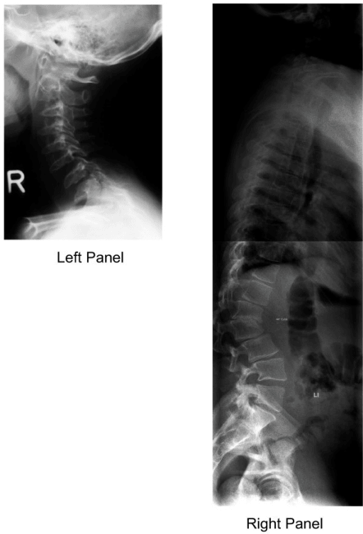
Definition. On the lateral radiograph the anterior margin of the vertebral body carries a tongue-like or hook-like projection of bone, with a corresponding defect, a notch, at the corner above or below it. The mechanism is failure of ossification of the anterosuperior corner of the body: the bone that did ossify protrudes as a beak while the deficient corner sits back.
Confirming it is real. Before the word goes in a report, check the film, the paired finding, the number of levels and the vertebral height:
- It must be seen on a true lateral with the vertebral end plates superimposed. An oblique projection creates a spurious anterior step.
- A beak requires a notch or deficiency at the opposite corner. A projection with no corresponding defect is usually an osteophyte or a limbus fragment.
- Storage disorders beak at multiple levels, maximal at the thoracolumbar junction (T12–L2) where the gibbus lies, so assess more than one level.
- Platyspondyly, loss of height with normal or increased disc height, changes the answer entirely.
Where on the body. This is the discriminator, and it must be stated out loud. A central or mid-body beak projects from the middle third of the anterior surface and is the pattern of Morquio (MPS IV). An anteroinferior beak projects from the lower third with an anterosuperior notch above it, giving the classic hook or ovoid vertebra, and is the pattern of Hurler (MPS I), Hunter (MPS II), Maroteaux–Lamy (MPS VI), hypothyroidism and hypotonia.
The words to use in a viva. "This is a lateral radiograph of the thoracolumbar spine in a skeletally immature patient. There is anterior vertebral beaking, maximal at T12 and L1, arising from the inferior third of the anterior vertebral body with an anterosuperior defect. There is an associated focal kyphosis, and the vertebral bodies are ovoid rather than rectangular. This is dysostosis multiplex, and my leading diagnosis is a mucopolysaccharidosis — the inferior position of the beak favours Hurler or Hunter over Morquio. I would want the rest of a skeletal survey, and critically a lateral cervical spine to assess the odontoid."
Mimics. A true beak is continuous with the vertebral body. The false positives, each with the feature that gives it away:
- Limbus vertebra — a discrete triangular ossicle at the anterosuperior corner, corticated on all sides and separated from the body by a lucent cleft, from herniation of nuclear material through the ring apophysis before fusion. Adolescent or adult, single level, no dysplastic features.
- Anterior osteophyte — degenerative, adult, arises at the end plate margin at the discovertebral junction, no notch.
- Scheuermann disease — anterior wedging with irregular end plates and Schmorl nodes, adolescent, no beak, no notch.
- Vertebral fracture or burst injury — anterior cortical buckling with retropulsion and a trauma history; the "beak" is a displaced fragment.
- Projectional artefact — a rotated or off-lateral film. Repeat the view before diagnosing a syndrome.
- Physiological infantile ovoid vertebra — the very young infant has ovoid bodies with a mild anterior step; it resolves with weight bearing.
Next Investigation
Skeletal survey (skull, chest, spine, pelvis, hands) confirms or excludes dysostosis multiplex. It changes management by converting a single odd vertebra into a systemic diagnosis and triggering metabolic referral.
Lateral cervical spine in flexion and extension assesses odontoid hypoplasia, os odontoideum and atlantoaxial instability; the lesion to exclude is odontoid hypoplasia or os odontoideum with an increased atlantodental interval and myelopathic signs. This is the investigation that changes management most: it dictates anaesthetic airway planning, activity restriction and the need for occipitocervical fusion.
MRI of the whole spine and craniocervical junction shows cord compression at C1–C2 from soft-tissue glycosaminoglycan deposition, which radiographs miss entirely, thoracolumbar cord tethering at the gibbus, and canal stenosis. Cord signal change mandates surgical decompression.
Urinary glycosaminoglycan screen is the screening test for the mucopolysaccharidoses, and a negative screen does not end the differential. It is normal in GM1 gangliosidosis and in mucolipidosis II, and it can be falsely negative in dilute urine or in Morquio if keratan sulphate is not specifically assayed.
Specific leucocyte or plasma enzyme assay, or a genetic panel, is definitive. It distinguishes Hurler (alpha-L-iduronidase) from Hunter (iduronate-2-sulphatase) from Morquio (GALNS), and it determines eligibility for enzyme replacement therapy or haematopoietic stem cell transplantation.
Thyroid function (TSH and free T4) is mandatory when bone age is grossly delayed and the epiphyses are fragmented. Hypothyroidism is the only cause on this list reversed by a tablet, and reversed only if treated early.
When nothing further is needed. An isolated corticated anterosuperior ossicle in a normally proportioned adolescent or adult is a limbus vertebra: reassure, do not scan, do not screen. Likewise, a single ovoid vertebra in a healthy non-sitting infant with an otherwise normal survey can simply be re-imaged once upright.
The Differential
Each cause carries a discriminating feature, on the film or in the child, and a test that confirms it.
- Typical age / setting
- Diagnosed 1–3 yrs; short-trunk dwarfism
- Discriminating feature
- CENTRAL anterior beak on a universally PLATYSPONDYLIC body; odontoid hypoplasia; normal intelligence; no corneal clouding early; genu valgum
- What confirms it
- Urinary keratan sulphate; GALNS (IVA) or beta-galactosidase (IVB) enzyme assay
- Typical age / setting
- Onset less than 2 yrs; coarse facies, hepatosplenomegaly
- Discriminating feature
- ANTEROINFERIOR beak with anterosuperior notch; oar-shaped ribs; J-shaped sella; intellectual regression; corneal clouding
- What confirms it
- Urinary dermatan and heparan sulphate; alpha-L-iduronidase assay; IDUA gene
- Typical age / setting
- Boys, X-linked, 2–4 yrs
- Discriminating feature
- Radiographically indistinguishable from Hurler (inferior beak) but NO corneal clouding and slower course; pebbly ivory skin lesions over the scapulae
- What confirms it
- Iduronate-2-sulphatase assay; male sex plus clear corneas
- Typical age / setting
- Untreated neonate/infant; now rare with screening
- Discriminating feature
- Inferior beak PLUS delayed and FRAGMENTED (stippled) epiphyseal ossification with multiple ossification centres; wormian bones; markedly retarded bone age
- What confirms it
- TSH and free T4; heel-prick screening result
- Typical age / setting
- Recognised at birth; rhizomelic short limbs
- Discriminating feature
- Beak plus POSTERIOR vertebral scalloping and interpedicular distance that NARROWS from L1 to L5; champagne-glass pelvis; trident hand
- What confirms it
- Clinical phenotype plus FGFR3 mutation testing
- Typical age / setting
- Infant with hypotonia and known karyotype
- Discriminating feature
- Anteroinferior beak with generalised hypotonia; 11 pairs of ribs; hypoplastic iliac wings with flat acetabular angles; no dysostosis multiplex
- What confirms it
- Karyotype; the beak needs no separate workup
- Typical age / setting
- Any age; non-sitting child
- Discriminating feature
- Inferior beaking with normal bone architecture and normal facies; disuse osteopenia and slender diaphyses; beaking improves with upright posture
- What confirms it
- Neurological examination and cause-specific testing; no GAG abnormality
- Typical age / setting
- Normal at birth, short stature from 2–3 yrs
- Discriminating feature
- Anterior tongue-like beak with irregular end plates, NORMAL skull and NORMAL facies, marked ligamentous laxity and epiphyseal + metaphyseal irregularity
- What confirms it
- COMP gene mutation; normal urinary GAGs
- Typical age / setting
- Childhood; severe skeletal disease
- Discriminating feature
- Full dysostosis multiplex with inferior beak but NORMAL intelligence (unlike Hurler) plus corneal clouding (unlike Hunter)
- What confirms it
- Arylsulphatase B assay; urinary dermatan sulphate
- Typical age / setting
- Infant, neurodegenerative, cherry-red macular spot
- Discriminating feature
- Dysostosis multiplex identical to Hurler but with early cherry-red spot and rapid neurological decline in the first year; NORMAL urinary GAGs
- What confirms it
- Beta-galactosidase assay; fundoscopy
- Typical age / setting
- Neonate/young infant, severe
- Discriminating feature
- Beaking present at BIRTH with periosteal cloaking and gingival hyperplasia; urinary GAGs NORMAL but plasma lysosomal enzymes markedly ELEVATED
- What confirms it
- Raised plasma lysosomal enzyme levels; GNPTAB mutation
- Typical age / setting
- Short trunk from birth; myopia, cleft palate
- Discriminating feature
- Platyspondyly with anterior beak but ABSENT/delayed ossification of pubis, femoral heads and calcaneus at birth; no visceromegaly, no GAGs
- What confirms it
- Radiographs of the pelvis at birth; COL2A1 mutation
Narrowing It Down
- 1Step 1 — Is it real, and is it one level or many?
Confirm the beak on a TRUE lateral and look for the corresponding notch; then count the involved levels and check for a thoracolumbar gibbus.
A SINGLE corticated fragment in an adolescent is a limbus vertebra and the algorithm stops here. MULTIPLE beaked levels with a gibbus means a generalised dysplasia and the workup begins.
- 2Step 2 — Where on the body does the beak arise?
Decide whether the beak springs from the MIDDLE third of the anterior vertebral body or from the INFERIOR third with an anterosuperior notch.
Central beak on a flattened body points hard at Morquio (MPS IV) and short-trunk dwarfism. Anteroinferior beak on an ovoid body points at Hurler, Hunter, Maroteaux–Lamy, hypothyroidism or hypotonia. This single observation does more work than any other step.
- 3Step 3 — Is there generalised platyspondyly?
Measure vertebral body height across the whole visible spine rather than judging the beaked level alone.
Universal loss of height widens the differential to Morquio and spondyloepiphyseal dysplasia congenita. Preserved height with an ovoid shape favours MPS I/II or a non-storage cause such as hypotonia.
- 4Step 4 — Are the other bones dysplastic (dysostosis multiplex)?
Obtain a skeletal survey and look specifically for oar-shaped ribs (wide anteriorly, narrow posteriorly), proximal pointing of the metacarpals, a J-shaped sella, thick clavicles and flared iliac wings with tapered inferior bodies.
Their presence makes a storage disorder near certain and commits you to enzyme and genetic testing. Their absence redirects to hypotonia, Down syndrome or pseudoachondroplasia.
- 5Step 5 — What is the systemic phenotype?
Examine for facial coarseness, corneal clarity, organomegaly, hernias, genu valgum and joint laxity, and take a developmental history.
Coarse facies, hepatosplenomegaly, hernias and intellectual regression = Hurler. The same picture in a boy with CLEAR corneas = Hunter. Short trunk, corneal clouding, genu valgum but NORMAL intellect = Morquio. Cherry-red spot with rapid neurodegeneration = GM1 gangliosidosis. Delayed bone age with fragmented epiphyses and a large fontanelle = hypothyroidism.
- 6Step 6 — What is the bone age and the epiphyseal appearance?
Request a bone age and inspect the epiphyses for stippling and fragmentation; send TSH and free T4 at the same time rather than waiting for the film.
Grossly retarded bone age with stippled, fragmented epiphyses is hypothyroidism until thyroid function proves otherwise — and it is the one cause on this list that is fully treatable, so a delay here is the costliest error in the algorithm.
- 7Step 7 — Regardless of the answer, image the craniocervical junction
Obtain flexion/extension lateral cervical radiographs and MRI of the craniocervical junction BEFORE any general anaesthetic, and make a difficult-airway plan alongside it.
Atlantoaxial instability occurs in Morquio, MPS I, MPS VI and Down syndrome: from odontoid hypoplasia in the dysplasias, and from ligamentous laxity rather than odontoid dysplasia in Down syndrome. Cord compression and airway obstruction are the two mechanisms that kill these children, and both are anticipated rather than discovered.
MCQ Practice Points
Q: A 3-year-old has short-trunk dwarfism, corneal clouding, genu valgum and normal intelligence. The lateral spine shows platyspondyly with a central anterior beak. Diagnosis?
A: Morquio syndrome (MPS IV). The combination of a central beak, generalised platyspondyly and preserved intellect is the exam signature. Urinary keratan sulphate and GALNS enzyme assay confirm.
Q: Which position of the anterior beak is associated with Hurler and Hunter syndromes?
A: Anteroinferior, with a corresponding anterosuperior notch, producing the hook-shaped or ovoid vertebra. Central beaking is Morquio.
Q: A beaked vertebra is found in a child with grossly delayed bone age and fragmented, stippled epiphyses. What must you exclude first?
A: Congenital hypothyroidism. It is the only cause on the list fully reversible with treatment, and epiphyseal dysgenesis with markedly retarded bone age is its discriminator. Check TSH and free T4.
Q: A child with anterior beaking also has posterior vertebral scalloping and interpedicular distances that decrease from L1 to L5. Diagnosis?
A: Achondroplasia. Narrowing caudal interpedicular distance is the classic discriminator and explains the symptomatic spinal stenosis these patients develop in early adulthood.
Q: A child with dysostosis multiplex has NORMAL urinary glycosaminoglycans. Which two diagnoses remain?
A: GM1 gangliosidosis (cherry-red macular spot, rapid neurodegeneration, beta-galactosidase deficiency) and mucolipidosis II / I-cell disease (present at birth, gingival hyperplasia, markedly elevated plasma lysosomal enzymes).
Q: Before any general anaesthetic in a child with beaked vertebrae and dysostosis multiplex, what single investigation is mandatory?
A: Craniocervical junction imaging — flexion/extension lateral cervical radiographs and MRI — to exclude odontoid hypoplasia and atlantoaxial instability with cord compression, alongside a difficult-airway plan.
Exam Viva Scenarios
Practise clinical reasoning and management decisions out loud
“You are shown a lateral thoracolumbar radiograph of a 4-year-old with disproportionate short stature. There is generalised platyspondyly and a beak arising from the middle of the anterior vertebral body at several levels. The parents report the child is doing well at nursery and is bright.”
“You are shown a lateral lumbar radiograph of an 18-month-old referred with a lump on the back. There is a focal kyphosis at T12–L1 with an ovoid vertebra bearing an anteroinferior beak and an anterosuperior notch. The GP wonders about tuberculosis.”
“You are shown a lateral lumbar radiograph of a 19-year-old footballer with back pain. There is a small triangular fragment at the anterosuperior corner of L4, separated from the body by a thin lucent line. The rest of the spine is normal and the patient is of normal stature.”
Define it
- Tongue-like anterior projection from the vertebral body with a corresponding notch at the opposite corner
- Requires a true lateral; maximal at the thoracolumbar junction (T12–L2)
- Usually multilevel and accompanied by a gibbus in dysplasia
The one discriminator
- CENTRAL beak + platyspondyly = Morquio (MPS IV)
- ANTEROINFERIOR beak + ovoid body = Hurler (MPS I), Hunter (MPS II), MPS VI, hypothyroidism, hypotonia
Separating the storage disorders
- Hurler: coarse facies, hepatosplenomegaly, corneal clouding, intellectual regression
- Hunter: X-linked male, CLEAR corneas, pebbly skin lesions, slower course
- Morquio: normal intellect, short trunk, genu valgum, keratan sulphate
- MPS VI: severe skeletal disease, corneal clouding, NORMAL intellect
- GM1 gangliosidosis and mucolipidosis II: dysostosis multiplex with NORMAL urinary GAGs
Non-storage causes
- Achondroplasia — posterior scalloping, caudally narrowing interpedicular distance
- Congenital hypothyroidism — fragmented epiphyses, grossly delayed bone age (treatable)
- Down syndrome — hypotonia, 11 rib pairs, hypoplastic iliac wings
- Neuromuscular hypotonia / non-ambulation — normal facies and architecture
- Pseudoachondroplasia and SED congenita — normal skull and facies
Mimics to exclude
- Limbus vertebra — corticated separate ossicle, single level, adolescent/adult, no workup needed
- Anterior osteophyte — degenerative adult, no notch
- Scheuermann disease — wedging, irregular end plates, Schmorl nodes
- Fracture — trauma, uncorticated fragment, retropulsion
- Rotated projection — repeat the film
Investigations in order
- Skeletal survey for dysostosis multiplex
- Flexion/extension cervical laterals + craniocervical MRI (before any anaesthetic)
- Urinary GAGs, then specific enzyme assay or gene panel
- TSH and free T4 if bone age is grossly delayed
- No imaging at all if the finding is an isolated limbus vertebra
What changes management
- Atlantoaxial instability or cord signal change → occipitocervical fusion, activity restriction, airway planning
- Confirmed Hurler in infancy → time-critical haematopoietic stem cell transplantation
- Confirmed MPS I/II/VI → enzyme replacement therapy eligibility
- Confirmed hypothyroidism → thyroxine, with skeletal recovery if early
- Progressive thoracolumbar gibbus → bracing or instrumented fusion
Evidence Base
A pictorial review of the radiographic skeletal findings in Morquio syndrome (mucopolysaccharidosis type IV)
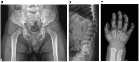
- Morquio syndrome (MPS IVA: N-acetylgalactosamine-6-sulfate sulfatase deficiency; IVB: beta-galactosidase) produces the dysostosis multiplex pattern with vertebral deformity, paddle-shaped ribs, odontoid hypoplasia and steep acetabula
- The axial skeleton - specifically the vertebrae - is most commonly affected, and myelopathy from spinal cord compression and airway obstruction are the critical causes of morbidity and mortality
- Radiological overlap with other storage disorders makes timely imaging and radiological expertise central to avoiding diagnostic delay
Mucopolysaccharidoses: overview of neuroimaging manifestations
- The MPS disorders share dysostosis multiplex of the skull and spine with short-trunk dwarfism; Hunter (MPS II) is X-linked, the rest autosomal recessive
- Central nervous system complications include compressive myelopathy at the craniocervical junction, hydrocephalus and white-matter injury
- Imaging has a crucial role in monitoring craniocervical junction stenosis and cord compression, allowing timely intervention before permanent damage
Review of cervical spine anomalies in genetic syndromes
- Pseudoachondroplasia and Morquio syndrome cause C1-C2 instability related to odontoid dysplasia (hypoplasia and os odontoideum); Morquio adds soft-tissue glycosaminoglycan deposition causing stenosis and myelopathy
- Down syndrome causes C1-C2 and occiput-C1 instability from ligamentous laxity; spondyloepiphyseal dysplasia congenita carries a high myelopathy risk from atlantoaxial instability plus stenosis
- These potentially debilitating cervical anomalies are easily overlooked among the systemic features of the syndromes and demand active vigilance