Five observations — distribution, joint space, erosions, bone density, new bone — name the disease before you name the patient
- Uniform (concentric) joint space loss suggests an inflammatory pannus or infection; non-uniform loss along the weight-bearing or load-bearing surface suggests osteoarthritis.
- Periarticular osteopenia is the earliest radiographic sign of rheumatoid arthritis, and bone density is characteristically PRESERVED in the seronegative spondyloarthritides and in gout - but read it as a pointer, not a rule: osteopenia is often absent in established or treated rheumatoid disease, and it is among the least reproducible judgements on a hand film.
- Erosions plus preserved bone density plus new bone formation equals seronegative spondyloarthropathy — psoriatic or reactive arthritis.
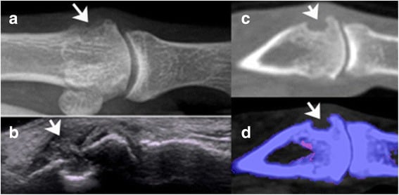
- Gout preserves the joint space until very late and produces para-articular punched-out erosions with overhanging edges and a soft-tissue tophus.
- A monoarticular aggressive arthropathy in a child, diabetic, immunosuppressed or intravenous drug user is septic until aspirated.
- Calcium pyrophosphate deposition produces chondrocalcinosis and an osteoarthritis-like picture in joints osteoarthritis normally spares: radiocarpal, patellofemoral, elbow, shoulder.
- “Say the distribution first: 'symmetrical polyarthropathy of the metacarpophalangeal joints and wrists' already excludes half the differential.
- “Distal interphalangeal disease means osteoarthritis, psoriatic arthritis or erosive osteoarthritis — never classical rheumatoid arthritis.
- “Bone density is free information: preserved density in an erosive arthritis points away from rheumatoid arthritis.
- “Never diagnose 'joint space narrowing' on a non-weight-bearing knee film — always ask for a standing anteroposterior or Rosenberg view.
Recognising the Pattern
What "arthritis on a radiograph" means. An arthropathy is present when the radiograph shows abnormality of the joint space, the subchondral bone, the joint margins or the surrounding soft tissues, in a distribution centred on the articulation. A single one of these findings in isolation is weak evidence; two or more make the diagnosis secure.
The five observations. Make them in this order every time, and say the distribution first: "symmetrical polyarthropathy of the metacarpophalangeal joints and wrists" already excludes half the differential.
- Distribution — which joints, and is it symmetrical? Axial or appendicular, large joint or small, proximal or distal within the hand or foot.
- Joint space — uniform (concentric, across the whole articular surface) or non-uniform (focal, in the load-bearing zone).
- Erosions — present or absent; if present, marginal, para-articular, central or subchondral.
- Bone density — periarticular osteopenia, generalised osteopenia, or preserved.
- New bone and soft tissue — osteophytes, enthesophytes, periostitis, ankylosis, subchondral sclerosis, soft-tissue swelling, calcification, tophi.
Confirming each finding is real. Joint space loss must be reproduced on an orthogonal view or under load. An erosion must show a cortical break with loss of the thin white subchondral line, seen on two views; a single-view "erosion" at an overlapping bone edge is usually projectional. Periarticular osteopenia is compared with the diaphysis of the same bone, not with the film next to it.
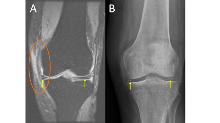
Load the knee before commenting on it. Supine films unload the joint and can make a bone-on-bone knee look preserved. A weight-bearing anteroposterior view and a Rosenberg view (posteroanterior, 45 degrees of flexion) are mandatory before any comment on knee joint space. Say so out loud.
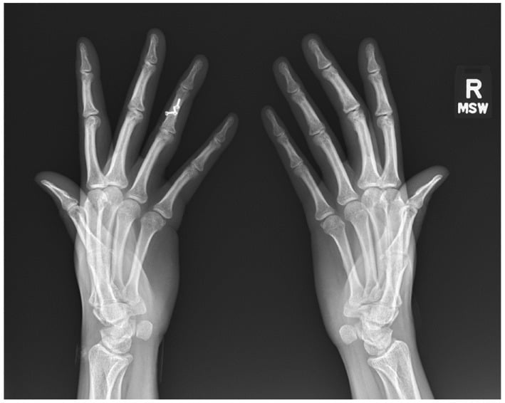
The description, spoken. The five observations run in the same order in the spoken answer: "This is a posteroanterior radiograph of both hands. There is a symmetrical polyarthropathy involving the metacarpophalangeal joints, proximal interphalangeal joints and both wrists, with sparing of the distal interphalangeal joints. There is uniform joint space narrowing, marginal erosions at the radial aspects of the second and third metacarpal heads, and periarticular osteopenia. There is ulnar deviation at the metacarpophalangeal joints. The appearances are those of rheumatoid arthritis."
Mimics. Before committing, exclude the false positives:
- Positioning and obliquity — an obliqued metacarpophalangeal joint appears narrowed, and the joint space of a rotated hip appears lost.
- Nutrient foramina and vascular channels — smooth and corticated with a sclerotic margin; erosions are not corticated early.
- Bare-area normality — the physiological bare area at the second metacarpal head can look scalloped on a single view.
- Neuropathic joint versus infection — both show destruction; the neuropathic joint is typically dense, deranged, debris-laden and relatively painless.
- Postoperative appearance and particle disease — periprosthetic osteolysis mimics erosive arthropathy but centres on the implant, not the articular surface.
- Amyloid and haemophilic arthropathy — well-defined subchondral cysts may mimic erosions.
- Osteonecrosis — subchondral collapse with a preserved joint space early; the joint space is the discriminator.
A-B-C-D-E-SABCDES — the systematic review of any arthritis film
Narrowing It Down
The emergency first. Before reasoning through the pattern, ask whether this is aggressive and monoarticular: rapid destruction over days to weeks, an effusion, and ill-defined subchondral bone on both sides of a single joint. If it is, the joint is septic until aspiration proves otherwise, whatever the film shows, so aspirate before you finish reasoning. Indolent monoarticular disease over months with juxta-articular osteoporosis suggests tuberculosis; a painless, disorganised, dense joint suggests neuropathic arthropathy.
Plain films are normal in the first 7 to 10 days of septic arthritis; the film lags the infection. A hot swollen joint with a normal radiograph is still septic until aspiration decides, and being reassured by the film is the single commonest viva failure point.
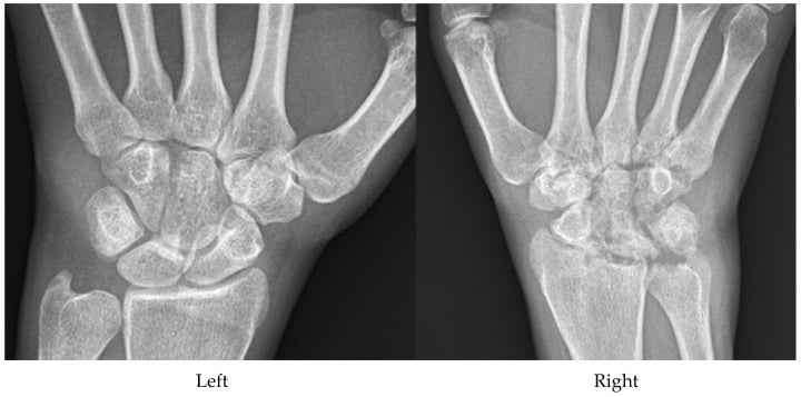
Distribution and symmetry. Read the whole film for which joints are involved and whether the two sides match before examining any single joint in detail. Distal interphalangeal disease means osteoarthritis, psoriatic arthritis or erosive osteoarthritis, never classical rheumatoid arthritis. The distribution alone points to a diagnosis:
- Symmetrical small joints of the hands and wrists with distal interphalangeal sparing — rheumatoid arthritis
- Asymmetric, with distal interphalangeal involvement in a ray pattern — psoriatic arthritis
- Distal interphalangeal and first carpometacarpal joints only — osteoarthritis
- Asymmetric lower limb with enthesitis — reactive arthritis
- Bilateral symmetrical sacroiliitis — ankylosing spondylitis or inflammatory bowel disease
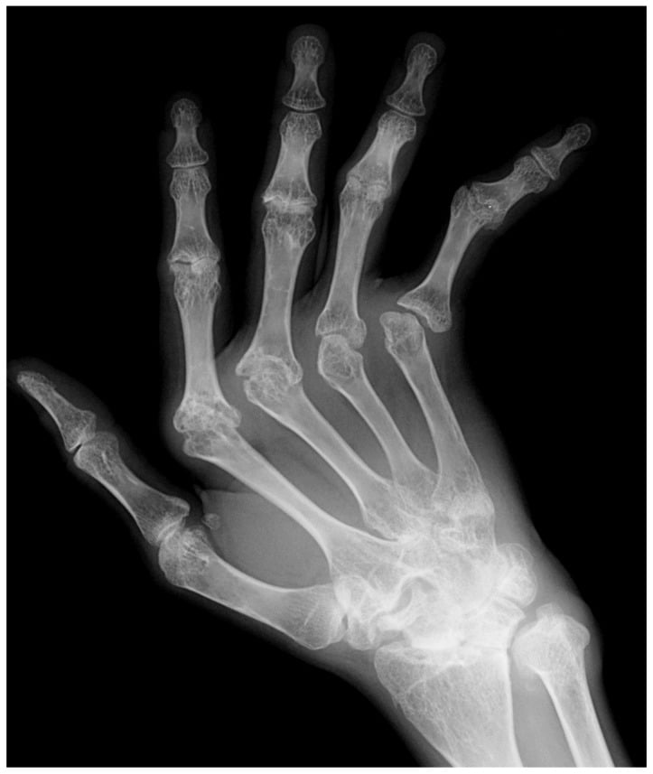
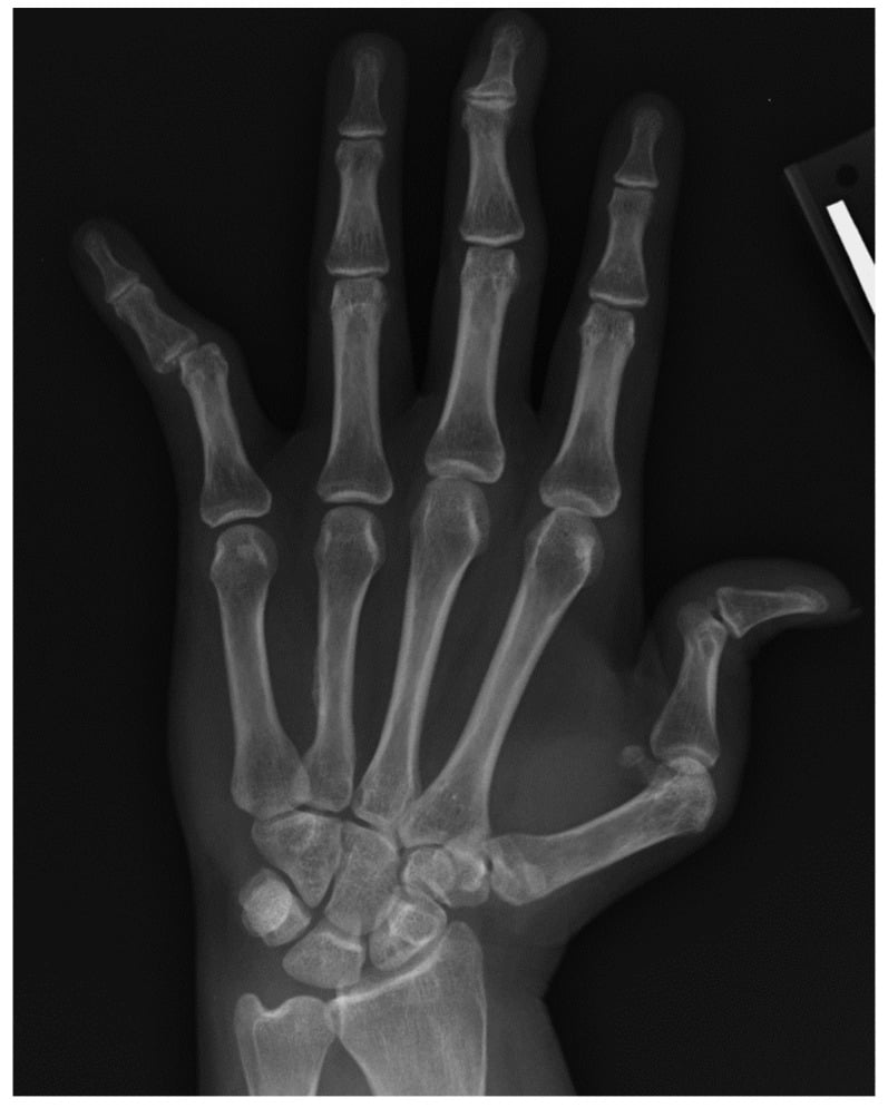
Joint space. Compare cartilage thickness across every compartment of the joint. Uniform, concentric loss means the whole synovium is diseased: inflammatory arthritis or infection. Non-uniform loss confined to the load-bearing compartment (the medial knee, the superolateral hip) means mechanical cartilage wear: osteoarthritis. At the hip, decide the direction of femoral head migration: an axially migrated head is inflammatory, a superolaterally migrated head is degenerative.
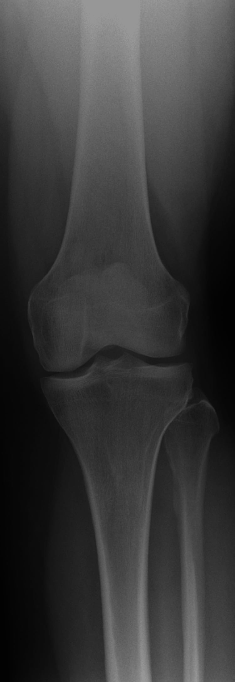
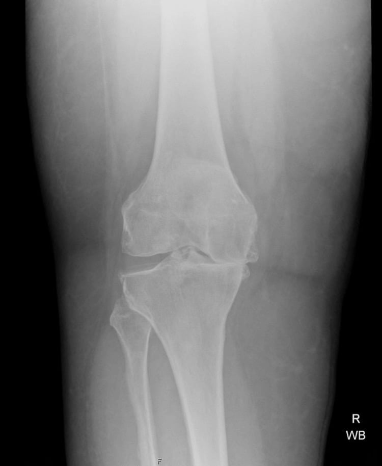
Erosions. Find each erosion and classify it by position (marginal at the bare area, para-articular, central) and by whether the surrounding bone is being resorbed or laid down. Each type has an owner:
- Marginal at the bare area with osteopenia — rheumatoid arthritis
- Para-articular with overhanging edges and a preserved joint space — gout
- Central, producing the "gull-wing" contour — erosive osteoarthritis
- Erosions with fluffy periosteal new bone — psoriatic or reactive arthritis
- Pressure erosions on both sides of the joint with a soft-tissue mass — pigmented villonodular synovitis or amyloid
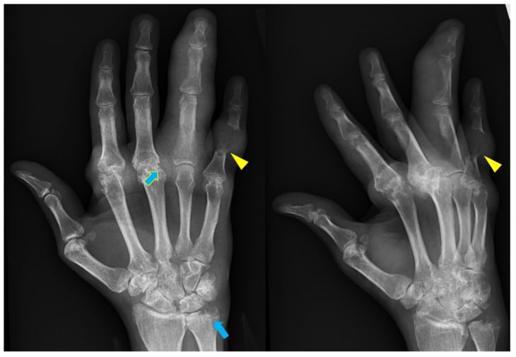
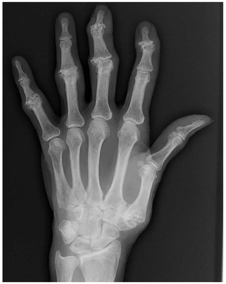
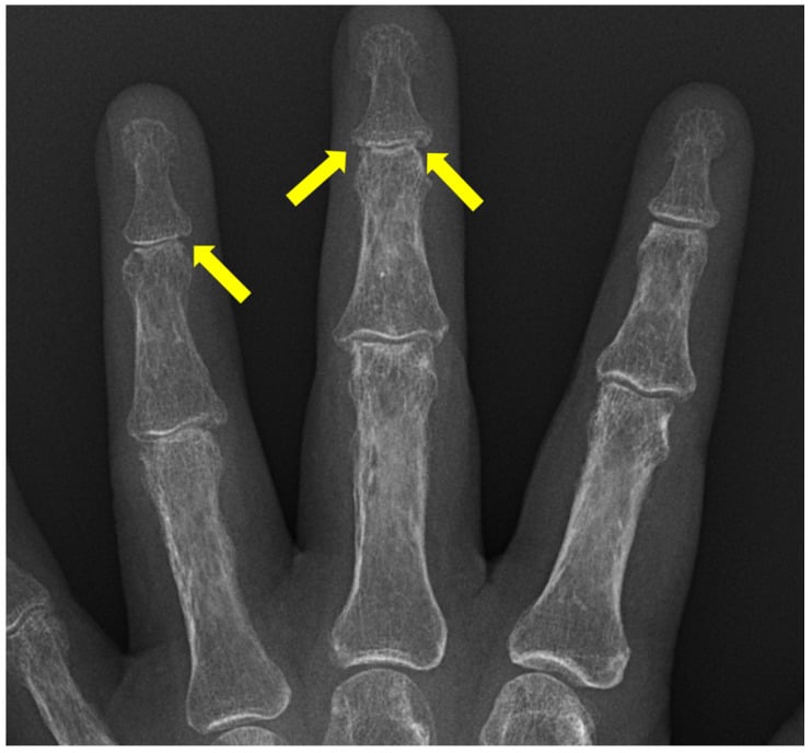
Bone density. Check the other hand as well as the diaphysis. Periarticular osteopenia narrows the list to rheumatoid arthritis, juvenile idiopathic arthritis or infection; preserved density in the presence of erosions shifts it strongly towards seronegative spondyloarthropathy, gout or pigmented villonodular synovitis. Preserved density does not exclude rheumatoid arthritis: osteopenia is an early sign, frequently absent in established or treated disease, and one of the least reproducible judgements on a hand film. Say which way it points, not what it rules out.
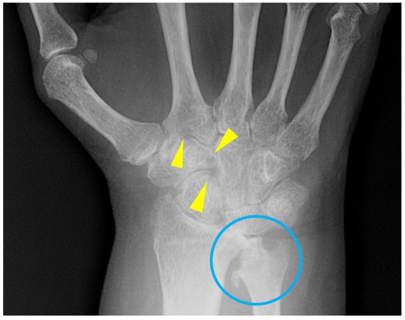
New bone, calcification and the soft tissues. Ask specifically whether the disease is laying bone down as well as taking it away, and inspect the soft tissues and cartilage for calcification. Osteophytes are degenerative; enthesophytes and periostitis are seronegative; chondrocalcinosis is calcium pyrophosphate deposition; a dense lobulated soft-tissue mass over an erosion is a tophus. Syndesmophytes separate ankylosing spondylitis (thin, marginal, symmetrical) from diffuse idiopathic skeletal hyperostosis (flowing, right-sided, joint spaces preserved).
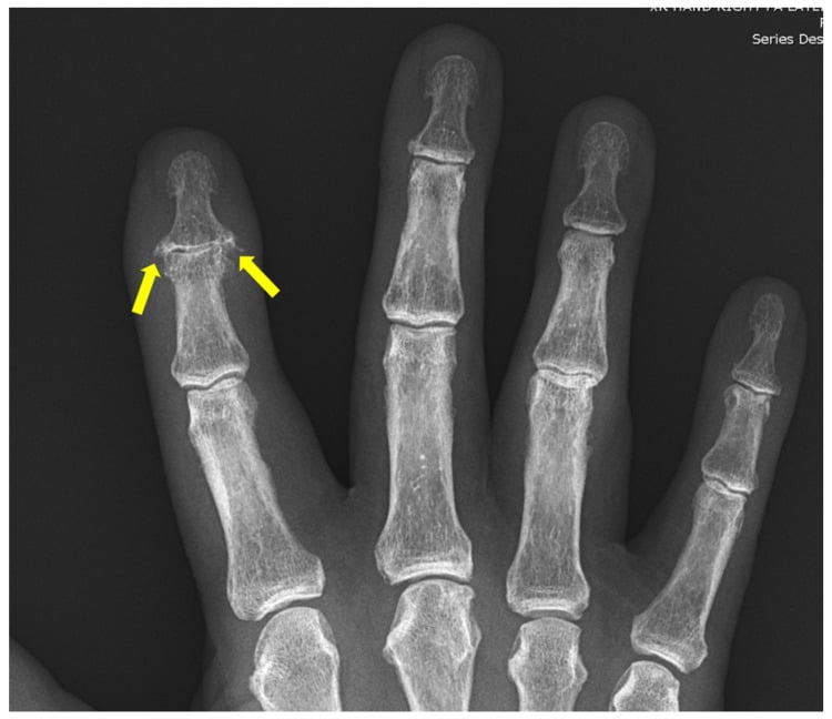
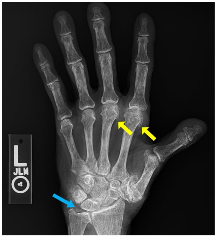
The family follows from two questions asked together. Is bone being eroded, and is bone simultaneously being laid down? New bone without erosion is degenerative. Erosion with osteopenia and no new bone is inflammatory resorptive (rheumatoid arthritis and juvenile idiopathic arthritis); erosion with new bone and preserved density is inflammatory proliferative (the seronegative spondyloarthropathies). A spared joint space beside the erosion or chondrocalcinosis is deposition; aggressive destruction across both sides of one joint is infective; destruction in a dense joint the patient does not defend is neuropathic.
Age and systemic context. Put the film back into the patient. Under 16 with epiphyseal overgrowth is juvenile idiopathic arthritis. Chondrocalcinosis in a knee that otherwise looks osteoarthritic changes the diagnosis to calcium pyrophosphate arthropathy, and in a patient under 55 mandates a search for haemochromatosis, hyperparathyroidism and hypomagnesaemia. Any erosive arthropathy of the hands in which rheumatoid arthritis is suspected mandates a lateral flexion cervical spine film before general anaesthesia: ask about the neck before an anaesthetist does.
Next Investigation
The pattern on the film decides the next test.
- Next test
- Urgent joint aspiration before antibiotics, plus C-reactive protein, white cell count and blood cultures
- What it shows
- Cell count and differential, Gram stain, culture, crystal microscopy under polarised light
- What changes management
- Purulent aspirate mandates theatre for washout within hours; crystals with a negative culture allow medical management
- Next test
- Ultrasound with power Doppler, or magnetic resonance imaging of hands and wrists
- What it shows
- Synovitis, tenosynovitis, bone marrow oedema and erosions months before they appear on plain film
- What changes management
- Confirms inflammatory disease and triggers early disease-modifying therapy — the window that prevents erosion
- Next test
- Lateral cervical spine radiographs in flexion and extension
- What it shows
- Atlantoaxial subluxation — an anterior atlantodental interval greater than 3 millimetres in adults is abnormal
- What changes management
- Alters intubation technique and may mandate awake fibreoptic intubation or surgical stabilisation
- Next test
- Magnetic resonance imaging of sacroiliac joints with short tau inversion recovery sequences
- What it shows
- Subchondral bone marrow oedema — inflammatory sacroiliitis before structural change
- What changes management
- Confirms diagnosis years earlier and justifies biologic therapy
- Next test
- Dual-energy computed tomography
- What it shows
- Colour-coded monosodium urate deposition in tophi and tendons
- What changes management
- Avoids an unnecessary washout and starts urate-lowering therapy
- Next test
- Serum ferritin and transferrin saturation, calcium, parathyroid hormone, magnesium, alkaline phosphatase
- What it shows
- Haemochromatosis, hyperparathyroidism, hypophosphatasia or hypomagnesaemia
- What changes management
- Treats the systemic disease; haemochromatosis needs venesection and hepatology referral
- Next test
- NO further imaging
- What it shows
- The radiograph is the diagnostic test; magnetic resonance imaging adds meniscal findings that do not change management in established osteoarthritis
- What changes management
- Proceed to non-operative management or arthroplasty planning — long-leg alignment films only if osteotomy is considered
Order of operations in a hot joint: aspirate, then antibiotics. Antibiotics given before aspiration sterilise the sample and lose the organism, and no imaging modality recovers that information.
The Differential
Each cause with the setting it arrives in, the feature that discriminates it and the test that confirms it.
- Typical age / setting
- Any age; neonates, immunosuppressed, intravenous drug use, prosthetic joint, recent injection
- Discriminating feature
- Rapid uniform joint space loss over days with ill-defined destruction of subchondral bone on BOTH sides of the joint; effusion and soft-tissue swelling; film may be normal in the first week
- What confirms it
- Joint aspiration: white cell count typically greater than 50,000 per cubic millimetre with neutrophil predominance, Gram stain and culture
- Typical age / setting
- Endemic exposure, immunosuppression; any age, often hip or knee, monoarticular
- Discriminating feature
- Phemister triad: juxta-articular osteoporosis, peripherally sited erosions and gradual joint space narrowing — indolent over months, out of proportion to symptoms
- What confirms it
- Synovial biopsy with acid-fast culture and nucleic acid amplification; magnetic resonance imaging shows rim-enhancing collections
- Typical age / setting
- Diabetes (midfoot), syringomyelia (shoulder), tabes/spinal cord injury; adults
- Discriminating feature
- The 5 Ds — density increased, debris, dislocation, disorganisation, destruction — with disproportionately little pain and a deranged but dense joint
- What confirms it
- Clinical sensory examination plus magnetic resonance imaging to exclude superimposed osteomyelitis; check glycated haemoglobin
- Typical age / setting
- Boys and young men with factor VIII or IX deficiency; knee, elbow, ankle
- Discriminating feature
- Dense effusion from haemosiderin, widened intercondylar notch, squared patella, epiphyseal overgrowth with early physeal fusion
- What confirms it
- Coagulation factor assay; magnetic resonance imaging shows blooming haemosiderin on gradient echo
- Typical age / setting
- Over 50 years; knee, hip, first carpometacarpal, distal interphalangeal, facet joints
- Discriminating feature
- NON-uniform joint space loss in the load-bearing compartment with subchondral sclerosis, osteophytes and subchondral cysts; NO erosions, NO osteopenia
- What confirms it
- Weight-bearing anteroposterior and Rosenberg knee views, or standing pelvis — no further imaging usually needed
- Typical age / setting
- 30 to 50 years, female predominance; hands, wrists, forefoot, cervical spine
- Discriminating feature
- Symmetrical, uniform joint space loss, MARGINAL erosions at the bare area, periarticular OSTEOPENIA, distal interphalangeal sparing, ulnar deviation, ulnar styloid erosion
- What confirms it
- Anti-cyclic citrullinated peptide antibody and rheumatoid factor; ultrasound or magnetic resonance imaging power Doppler synovitis; flexion-extension cervical spine views
- Typical age / setting
- 20 to 50 years; distal interphalangeal joints, ray distribution, asymmetric, sacroiliac joints
- Discriminating feature
- Erosions WITH proliferative fluffy new bone; 'pencil-in-cup' deformity, acro-osteolysis, periostitis along the shaft, PRESERVED bone density, nail changes
- What confirms it
- Skin and nail examination; asymmetric sacroiliitis on pelvic radiograph or magnetic resonance imaging
- Typical age / setting
- Men over 40, postmenopausal women, diuretics, renal impairment; first metatarsophalangeal joint, midfoot, elbow olecranon
- Discriminating feature
- PRESERVED joint space until late, para-articular punched-out erosions with sclerotic margins and overhanging edges, dense soft-tissue tophus, no osteopenia
- What confirms it
- Aspiration with negatively birefringent needle-shaped urate crystals; dual-energy computed tomography shows colour-coded urate
- Typical age / setting
- Over 60 years, or under 55 with a metabolic cause; knee, wrist, symphysis pubis
- Discriminating feature
- Chondrocalcinosis in fibrocartilage plus a degenerative pattern in joints osteoarthritis spares — radiocarpal, patellofemoral, elbow, shoulder; scaphoid–lunate advanced collapse pattern
- What confirms it
- Aspiration with positively birefringent rhomboid crystals; screen for ferritin, calcium, parathyroid hormone and magnesium if under 55
- Typical age / setting
- Men aged 15 to 35; sacroiliac joints, spine, hips, shoulders
- Discriminating feature
- SYMMETRICAL sacroiliitis progressing to fusion; thin marginal syndesmophytes producing a bamboo spine; squared vertebral bodies; concentric hip joint space loss
- What confirms it
- Human leukocyte antigen B27, magnetic resonance imaging of sacroiliac joints showing bone marrow oedema; whole-spine imaging before any manipulation
- Typical age / setting
- Young adults, 1 to 4 weeks after enteric or genitourinary infection; lower limb, calcaneus
- Discriminating feature
- Asymmetric lower-limb oligoarthritis with fluffy plantar and Achilles enthesophytes and asymmetric sacroiliitis; preserved density
- What confirms it
- Stool or urethral pathogen identification; human leukocyte antigen B27; calcaneal lateral radiograph
- Typical age / setting
- Postmenopausal women; distal and proximal interphalangeal joints only
- Discriminating feature
- CENTRAL erosions producing the 'gull-wing' subchondral contour with osteophytes and preserved bone density — erosions plus osteoarthritic new bone in the same joint
- What confirms it
- Posteroanterior hand radiograph pattern is diagnostic; rheumatoid serology negative
- Typical age / setting
- Under 16 years; knee, wrist, cervical spine, temporomandibular joint
- Discriminating feature
- Epiphyseal overgrowth and ballooning with gracile diaphyses, early physeal fusion, carpal crowding and ankylosis; erosions appear late
- What confirms it
- Ophthalmology slit-lamp screening, antinuclear antibody; magnetic resonance imaging for synovitis
- Typical age / setting
- 20 to 40 years; monoarticular, knee then hip
- Discriminating feature
- Dense lobulated soft-tissue mass with pressure erosions on BOTH sides of the joint, preserved joint space and NO calcification and NO osteopenia
- What confirms it
- Magnetic resonance imaging showing low signal on all sequences with gradient echo blooming; synovial biopsy
MCQ Practice Points
Q: Which single radiographic feature best distinguishes rheumatoid arthritis from psoriatic arthritis? A: Bone density. Rheumatoid arthritis shows periarticular osteopenia; psoriatic arthritis shows preserved bone density with proliferative new bone. Distribution helps too — rheumatoid spares the distal interphalangeal joints, psoriatic targets them.
Q: A 70-year-old has knee osteoarthritis with chondrocalcinosis of the meniscus and severe radiocarpal narrowing. What is the diagnosis and what does the wrist add? A: Calcium pyrophosphate deposition disease. The radiocarpal joint (and patellofemoral, elbow and glenohumeral joints) are sites primary osteoarthritis normally spares — degenerative change there implies a deposition arthropathy.
Q: What differentiates the syndesmophytes of ankylosing spondylitis from diffuse idiopathic skeletal hyperostosis? A: Ankylosing spondylitis produces thin, vertical, marginal, symmetrical syndesmophytes with sacroiliac fusion and squared vertebrae. Diffuse idiopathic skeletal hyperostosis produces flowing, thick, non-marginal ossification over four or more contiguous vertebrae, typically right-sided in the thoracic spine, with preserved disc heights and normal sacroiliac joints.
Q: A hip radiograph shows axial (medial) migration of the femoral head with uniform joint space loss. Is this degenerative? A: No. Uniform loss with axial or medial migration and protrusio indicates an inflammatory arthropathy such as rheumatoid arthritis or ankylosing spondylitis. Primary osteoarthritis produces superolateral migration with non-uniform loss, subchondral sclerosis, cysts and osteophytes.
Q: In a child with a swollen knee, gracile diaphyses, ballooned epiphyses and carpal crowding on hand films — diagnosis and mandatory referral? A: Juvenile idiopathic arthritis. Mandatory ophthalmology slit-lamp screening for asymptomatic chronic anterior uveitis, highest risk in antinuclear antibody positive oligoarticular disease, which can cause blindness before the eye becomes symptomatic.
Exam Viva Scenarios
Practise clinical reasoning and management decisions out loud
“You are shown this posteroanterior radiograph of both hands in a 44-year-old woman with 6 months of morning stiffness lasting 2 hours. Describe the film and give your diagnosis.”
“You are shown this anteroposterior radiograph of a knee in a 62-year-old man with diabetes, a swollen warm knee and a C-reactive protein of 180 milligrams per litre. There is marked joint space loss with ill-defined subchondral bone on both femoral and tibial sides.”
“You are shown this radiograph of a foot in a 55-year-old man with recurrent painful first toe swelling. There is a well-defined para-articular erosion at the medial first metatarsal head with a sclerotic margin and an overhanging edge, with preserved joint space and an adjacent dense soft-tissue mass.”
The Five Observations
- Distribution and symmetry — do this first, out loud
- Joint space: uniform equals inflammatory or septic; non-uniform equals degenerative
- Erosions: marginal, para-articular, central, or proliferative
- Bone density: osteopenic versus preserved
- New bone and soft tissue: osteophyte, enthesophyte, syndesmophyte, tophus, chondrocalcinosis
Erosion Types to Disease
- Marginal at bare area plus osteopenia equals rheumatoid arthritis
- Para-articular, overhanging edge, preserved space equals gout
- Central gull-wing at interphalangeal joints equals erosive osteoarthritis
- Erosion plus fluffy periostitis equals psoriatic or reactive arthritis
- Bilateral pressure erosion with soft-tissue mass equals pigmented villonodular synovitis or amyloid
Distribution Shortcuts
- Distal interphalangeal disease: osteoarthritis, psoriatic, erosive osteoarthritis — never classical rheumatoid
- Metacarpophalangeal and wrist, symmetrical: rheumatoid arthritis
- First metatarsophalangeal joint, episodic: gout
- Radiocarpal, patellofemoral, elbow, shoulder degeneration: calcium pyrophosphate
- Sacroiliac joints symmetrical: ankylosing spondylitis; asymmetric: psoriatic or reactive
Must-Not-Miss
- Septic arthritis — normal film does not exclude it; aspirate before antibiotics
- Tuberculous arthritis — Phemister triad, indolent course
- Neuropathic joint — the 5 Ds, dense and disorganised, painless
- Atlantoaxial instability in rheumatoid arthritis before any anaesthetic
- Chondrocalcinosis under 55 — screen for haemochromatosis and hyperparathyroidism
Technique Rules
- Never comment on knee joint space without weight-bearing anteroposterior plus Rosenberg views
- Confirm every erosion on two orthogonal views
- Compare periarticular density with the diaphysis of the same bone
- Established osteoarthritis on adequate films needs no further imaging
- Long-leg alignment films only when osteotomy is being considered
Evidence Base
Septic arthritis
- Distribution is typically monoarticular with a swollen, erythematous, painful joint; infection reaches the joint by haematogenous spread, a contiguous source, direct implantation or postoperative contamination
- Radiographic abnormalities - soft-tissue swelling, joint space loss, periarticular osteopenia and central or marginal osseous erosions - may be DELAYED following the clinical onset of infection, so a normal film does not exclude it
- Early diagnostic arthrocentesis is important to prevent articular destruction, ankylosis and osteomyelitis
Advanced imaging of gout
- Characteristic of gout are well-defined, punched-out erosions with overhanging edges, preservation of the joint space until late, and lack of periarticular osteopenia
- Involvement is asymmetrical with soft-tissue nodules (tophi) and intraosseous calcifications
- On MRI tophi have low signal on T1 and T2 with a variable enhancement pattern
Early-stage rheumatoid arthritis: prospective study of the effectiveness of MR imaging for diagnosis
- Fifty patients with polyarthralgia suspected of early rheumatoid arthritis underwent gadolinium-enhanced MR of the hands; 48 completed a mean follow-up of 776 days to a final diagnosis (26 rheumatoid, 22 non-rheumatoid)
- The pre-specified MR criterion was BILATERAL enhancement in both wrists and/or the MCP and/or PIP joints
- The criterion was correct in 25 of the 26 patients with rheumatoid arthritis, with 3 false positives among the 22 without
- Compared against the traditional-format and classification-tree American Rheumatism Association criteria, MR detected 7 and 6 ADDITIONAL true rheumatoid patients respectively
- Early diagnosis allows disease-modifying treatment to start before irreversible erosive damage