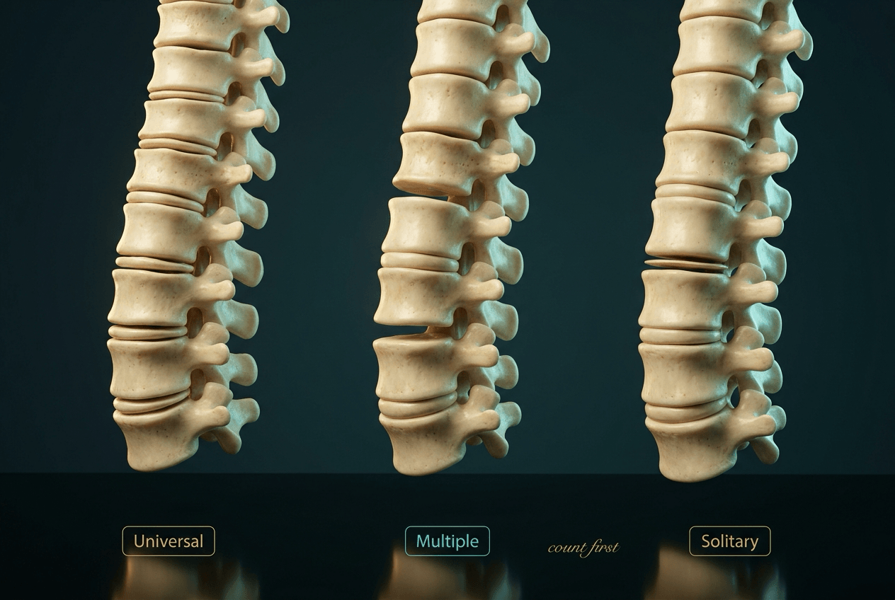
Flattened vertebral bodies — the single most useful branch point is solitary versus universal
- Platyspondyly means reduced vertebral body height relative to its anteroposterior and transverse dimensions — measure, do not eyeball.
- Universal platyspondyly (every vertebra) is nearly always a skeletal dysplasia or a metabolic/haemoglobinopathic process; solitary platyspondyly is nearly always neoplastic, histiocytic or infective.
- Vertebra plana in a child under 10 with preserved adjacent discs, no paravertebral mass and intact posterior elements is Langerhans cell histiocytosis until proven otherwise, and it usually reconstitutes height with growth — but hold the phrase 'until proven otherwise' seriously. In Baky's 27 children, only 12 had eosinophilic granuloma and SIX had a different neoplasm including leukaemia and rhabdomyosarcoma, and the degree of collapse, its symmetry and the level carried NO discriminating value.
- Vertebral body shape is a free diagnostic clue: anterior beaking (central in Morquio, inferior in Hurler), central tongue in Kniest, H-shaped/step endplates in sickle cell, cupid's bow as a normal variant.
- In an adult, a new flat vertebra is malignancy or osteoporosis until MRI says otherwise — a benign osteoporotic fracture shows a linear fluid/marrow oedema band, malignancy shows complete marrow replacement plus pedicle involvement.
- “"Universal, solitary, or multiple-but-not-all" is the opening sentence that structures the whole viva.
- “Posterior element involvement and a soft tissue mass push you away from LCH and towards Ewing sarcoma, lymphoma or infection.
- “Osteogenesis imperfecta gives biconcave codfish vertebrae with generally thin cortices; osteoporosis of any cause can mimic this.
- “Thanatophoric dysplasia gives extreme flattening with wide discs — the classic 'H' or inverted-U vertebrae on the neonatal lateral radiograph.
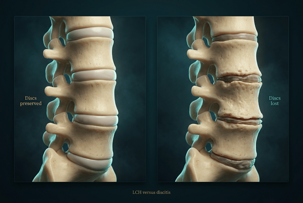
Preserved or even increased disc height with a wafer-thin body is the LCH/tumour pattern. If disc height is lost with endplate destruction, you are describing discitis–osteomyelitis and the whole differential changes.
An off-lateral or steeply angled beam, scoliotic rotation and simple magnification all make bodies look short. Confirm on a true lateral, and confirm on more than one level before you say platyspondyly.
Trainees reflexively answer "LCH" to any flat vertebra. In a patient over 50, myeloma, metastasis and osteoporotic insufficiency fracture dominate — LCH is a rare adult diagnosis.
A flat vertebra with retropulsion or epidural soft tissue is a neurosurgical problem before it is a diagnostic one. Examine for a level, examine perineal sensation, and image the whole spine urgently.
Recognising the Pattern
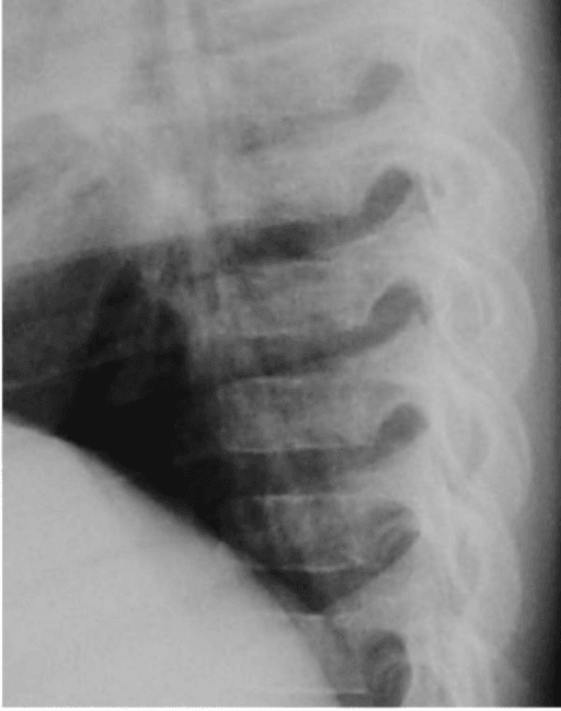
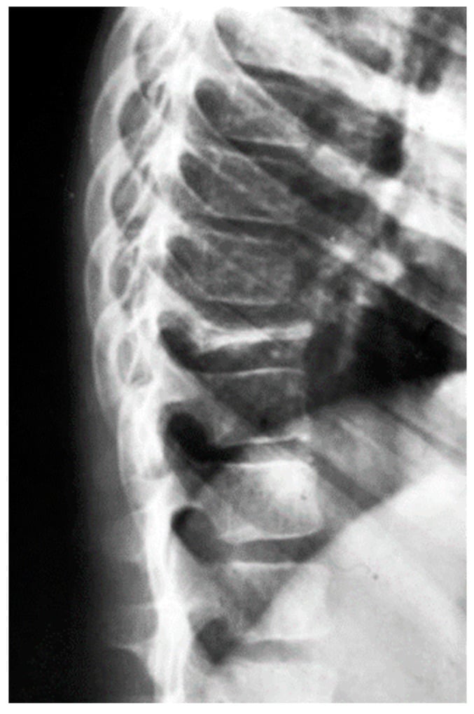
Definition. Platyspondyly is reduction in vertebral body height in the craniocaudal axis relative to its own anteroposterior and transverse dimensions. It is a shape diagnosis, not a density diagnosis. The extreme form, in which the body is reduced to a thin sclerotic wafer, is termed vertebra plana (the "coin-on-edge" or "silver dollar" vertebra).
Confirming it is genuinely present
- Use a true lateral radiograph. On a rotated film, both pedicles will not be superimposed and heights are unreliable.
- Compare the body height with the height of the adjacent bodies and with the AP diameter of the same body. In the normal thoracolumbar spine, body height and AP depth are broadly similar; a body markedly shorter than it is deep is flattened.
- Check at least three consecutive levels before declaring universal platyspondyly, and check both the cervical and lumbar segments if available.
- Assess the disc spaces in the same breath: relatively increased disc height with a flat body indicates the bone has collapsed with a healthy disc, whereas narrowed discs with irregular endplates indicate the disc is the diseased structure.
Saying it out loud in a viva
"This is a lateral radiograph of the thoracolumbar spine. There is flattening of the vertebral body height — platyspondyly. It involves [a single level / multiple non-contiguous levels / every visualised level]. The adjacent intervertebral disc spaces are [preserved / increased / narrowed]. The posterior elements are [intact / involved]. There is [no] paravertebral soft tissue swelling. The endplates are [smooth and parallel / irregular and eroded / anteriorly beaked], and overall bone density is [normal / reduced / increased]. My differential is led by the fact that the change is [solitary / universal]..."
Mimics and false positives
- Why it fools you
- Foreshortening of body height
- How to exclude
- Repeat true lateral; check pedicle superimposition
- Why it fools you
- Physiologically short with anterior notch
- How to exclude
- Normal for age; height restores with ossification
- Why it fools you
- Bilobed inferior endplate concavity on the AP view
- How to exclude
- Normal variant at L3–L5; body height is normal on lateral
- Why it fools you
- Focal loss of central height
- How to exclude
- Focal, subchondral, with sclerotic rim; anterior and posterior height preserved
- Why it fools you
- Anterosuperior corner defect resembles collapse
- How to exclude
- Corner fragment with corticated margins, body height otherwise normal
- Why it fools you
- Anterior wedging read as flattening
- How to exclude
- Requires wedging of 5 degrees or more at three contiguous levels with endplate irregularity
Next Investigation
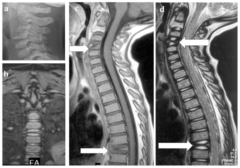
The one MRI feature that most reliably separates benign osteoporotic from malignant collapse is the preserved normal fatty marrow signal within the fractured body on T1 — malignancy replaces it completely. Add pedicle signal change and a convex posterior cortex and you are looking at tumour.
The Differential
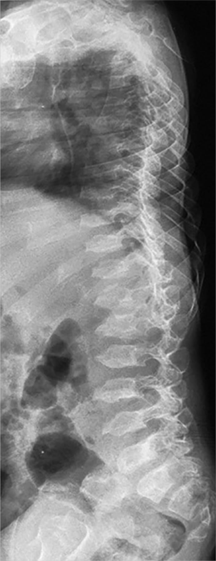
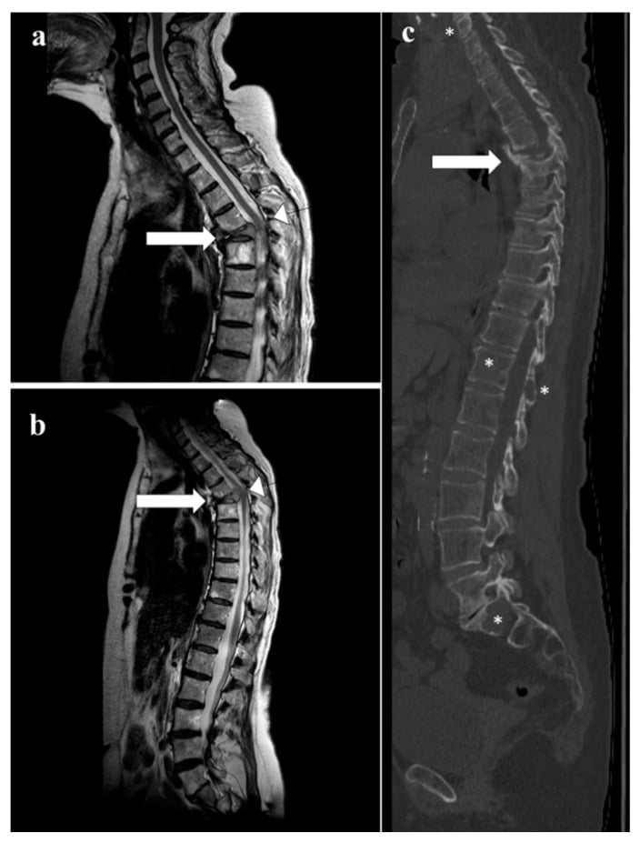
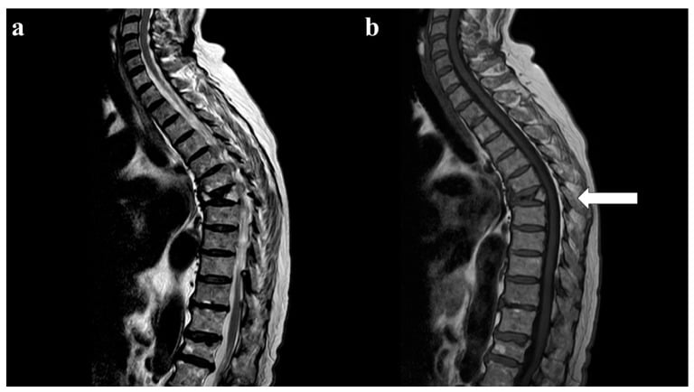
- Typical age / setting
- Over 50; known primary — breast, lung, prostate, renal, thyroid
- Discriminating feature
- Pedicle destruction (winking owl on AP) with collapse; posterior body convexity; often multiple non-contiguous levels
- What confirms it
- MRI shows complete marrow replacement and epidural soft tissue; CT-guided biopsy; staging CT and bone scan
- Typical age / setting
- Over 55; back pain, anaemia, renal impairment, raised ESR
- Discriminating feature
- Diffuse osteopenia out of keeping with age plus multiple collapses; solitary plasmacytoma gives an expansile 'mini-brain' vertebra on CT
- What confirms it
- Serum and urine electrophoresis, free light chains, whole-body MRI or low-dose whole-body CT, marrow biopsy
- Typical age / setting
- Peak 5–10 years; thoracic more than lumbar
- Discriminating feature
- Classic vertebra plana with preserved or increased adjacent disc height, no paravertebral mass, posterior elements spared
- What confirms it
- Skeletal survey for lytic skull/long bone lesions; MRI; biopsy if atypical — CD1a and langerin positive
- Typical age / setting
- Any age; diabetes, IV drug use, recent bacteraemia
- Discriminating feature
- Disc space loss precedes and accompanies collapse, with endplate erosion on both sides of a disc
- What confirms it
- MRI: T2 hyperintense disc, endplate oedema, paravertebral or epidural abscess; blood cultures, CRP, biopsy
- Typical age / setting
- Any age; endemic exposure, immunosuppression
- Discriminating feature
- Relative disc preservation with large calcified paraspinal cold abscess, multilevel and subligamentous spread, gibbus
- What confirms it
- MRI plus tissue for AFB, culture and PCR; anterior column destruction with skip lesions
- Typical age / setting
- Children 2–10 (ALL); adults (lymphoma)
- Discriminating feature
- Diffuse osteopenia with multiple collapses plus metaphyseal lucent bands in long bones; systemic upset and cytopenias
- What confirms it
- Full blood count and film, marrow aspirate; MRI shows diffuse low T1 marrow
- Typical age / setting
- Postmenopausal women; steroid use
- Discriminating feature
- Anterior wedging with preserved posterior cortex, no pedicle involvement, band-like oedema only
- What confirms it
- MRI band of oedema paralleling the endplate; DEXA; benign fractures resolve on follow-up imaging
- Typical age / setting
- Childhood; blue sclerae, multiple long bone fractures, wormian bones
- Discriminating feature
- Universal biconcave 'codfish' vertebrae with generalised gracile thin cortices
- What confirms it
- Clinical phenotype plus family history; COL1A1/COL1A2 testing; DEXA
- Typical age / setting
- African/Mediterranean ancestry; painful crises
- Discriminating feature
- H-shaped or 'Lincoln log' step-off central endplate depression from endplate infarction, uniform across many levels
- What confirms it
- Haemoglobin electrophoresis; MRI shows marrow reconversion and infarcts
- Typical age / setting
- Toddler onward; coarse facies, corneal clouding, short trunk
- Discriminating feature
- Morquio: universal platyspondyly with *central* anterior beak and odontoid hypoplasia. Hurler: *inferior* anterior beak with thoracolumbar gibbus
- What confirms it
- Urinary glycosaminoglycans, enzyme assay; flexion–extension views for atlantoaxial instability
- Typical age / setting
- Congenita from birth; tarda in boys aged 5–10, X-linked
- Discriminating feature
- Universal platyspondyly with 'hump-back' posterior mound and hypoplastic dens; tarda shows posterior humping with narrowed discs and early hip arthrosis
- What confirms it
- Skeletal survey; short trunk with normal limb length; genetic testing (COL2A1, TRAPPC2)
- Typical age / setting
- Neonate; lethal, narrow thorax, telephone-receiver femora
- Discriminating feature
- Extreme universal flattening with markedly widened discs — inverted-U or H-shaped vertebrae
- What confirms it
- Neonatal skeletal survey; FGFR3 mutation
- Typical age / setting
- Early childhood, short trunk
- Discriminating feature
- Kniest: coronal cleft and central tongue-like anterior projection. DMC: lacy iliac crests with double-humped endplates
- What confirms it
- Skeletal survey pattern; COL2A1 (Kniest), DYM (DMC)
- Typical age / setting
- Childhood to young adult; hepatosplenomegaly, thrombocytopenia
- Discriminating feature
- Vertebral collapse plus Erlenmeyer flask distal femora and avascular necrosis
- What confirms it
- Beta-glucocerebrosidase assay, GBA genotyping
- Typical age / setting
- History of spinal radiotherapy or long-term glucocorticoid
- Discriminating feature
- Flattening confined precisely to the treatment field with a sharp cut-off at the field edge; fatty marrow on T1
- What confirms it
- Radiotherapy planning records; MRI shows uniform high T1 fatty marrow within the port
Narrowing It Down
- 1Step 1 — Is it universal, multiple, or solitary?
Count the flattened vertebrae on a whole-spine film before looking at any single level in detail.
Universal (every level) means a constitutional process: skeletal dysplasia, osteogenesis imperfecta, mucopolysaccharidosis, sickle cell. Multiple non-contiguous levels in an adult means metastasis, myeloma or osteoporosis. Solitary means neoplasm, LCH or infection until proven otherwise. This single question removes two-thirds of the list.
- 2Step 2 — How old is the patient?
Pair the distribution with the decade before generating a differential.
Under 10 with a solitary vertebra plana: LCH first, then Ewing sarcoma, leukaemia and infection — but Baky's series is the caution, with only 12 of 27 proving to be eosinophilic granuloma. Aged 10-30: infection, Ewing sarcoma, aneurysmal bone cyst, giant cell tumour. Over 50: metastasis, myeloma, osteoporotic insufficiency fracture. Universal flattening in infancy points to a lethal or severe dysplasia; at 5-10 years in a boy, to SED tarda.
- 3Step 3 — What have the discs done?
Measure the disc height above and below the collapsed body and compare it with uninvolved levels.
Preserved or increased disc height with a collapsed body means the disc is healthy and the bone failed - LCH, tumour, osteoporotic fracture. Narrowed disc with irregular destroyed endplates on both sides means pyogenic discitis. Relative disc preservation with a large calcified paraspinal mass means tuberculosis.
- 4Step 4 — Are the posterior elements and pedicles involved?
Look specifically at the pedicles on the AP film and at the posterior elements on axial imaging.
Sparing of the posterior elements supports LCH. Pedicle destruction supports metastasis. Posterior element involvement with an expansile lesion suggests aneurysmal bone cyst or osteoblastoma; with a permeative pattern and soft tissue mass, Ewing sarcoma or lymphoma.
- 5Step 5 — Is there a paravertebral or epidural soft tissue mass?
Assess the paravertebral soft tissues and the epidural space on MRI, and grade any cord compression.
None with a wafer vertebra reinforces LCH. A small rim of soft tissue can occur in LCH but a bulky mass mandates biopsy for tumour or infection. Epidural extension with cord signal change converts this into an emergency: urgent whole-spine MRI, dexamethasone and a combined oncology-spinal surgery discussion.
- 6Step 6 — What is the shape of the residual body?
Read the contour on the lateral film, not only the height.
Central anterior beak equals Morquio. Inferior anterior beak equals Hurler. Anterior tongue-like projection equals Kniest. Posterior hump equals SED. Central step-off H-shape equals sickle cell. Biconcave codfish equals osteoporosis or osteogenesis imperfecta. Shape is the free diagnosis in a dysplasia viva.
- 7Step 7 — What do the rest of the skeleton and the systemic features say?
Order the skeletal survey and look outside the spine; examine for short trunk versus short limbs, and check the blood count and renal function.
Lytic skull lesions and diabetes insipidus favour LCH. Erlenmeyer flask femora favour Gaucher. Metaphyseal lucent bands and cytopenias favour leukaemia. Short trunk with normal limb length favours SED or Morquio; short limbs with a narrow chest favour thanatophoric dysplasia. Bone pain with anaemia and renal failure favours myeloma.
Exam Viva Scenarios
Exam Viva Scenarios
Practise clinical reasoning and management decisions out loud
“You are shown this lateral thoracic radiograph of a 6-year-old boy with 3 months of back pain and a mild thoracic kyphosis. T7 is reduced to a thin dense wafer. The adjacent disc spaces look increased. He is neurologically intact.”
“You are shown this lateral lumbar radiograph of a 68-year-old woman with 6 weeks of night pain. L1 and L4 are flattened; the L4 pedicle is not visible on the AP view. She is otherwise well.”
“You are shown this lateral spine radiograph of a 4-year-old with disproportionate short stature. Every vertebral body is flattened with an anterior beak, and the trunk is short with normal limb length.”
MCQ Practice Points
Q: A 5-year-old has an isolated vertebra plana with preserved disc spaces and no soft tissue mass. Most likely diagnosis?
A: Langerhans cell histiocytosis. Solitary extreme flattening with preserved or increased adjacent disc height, sparing of the posterior elements and no paravertebral mass is characteristic, and vertebral height usually reconstitutes with growth. Say MOST LIKELY, not certain: in Baky's 27 children only 12 had eosinophilic granuloma and six had another neoplasm, and neither the degree of collapse nor its symmetry nor the level separated them. Five of seventeen biopsies were also nondiagnostic, so neither the film nor the needle closes the case on its own.
Q: Which single radiographic sign most strongly suggests metastatic rather than osteoporotic collapse?
A: Destruction of the pedicle — the "winking owl" sign on the AP radiograph — together with a convex posterior vertebral cortex. Osteoporotic fractures spare the pedicles.
Q: Central anterior vertebral beaking in a short-trunked child with corneal clouding indicates which condition, and what is the key preoperative concern?
A: Morquio syndrome (MPS IV). The key concern is odontoid hypoplasia with atlantoaxial instability and the risk of cord injury at intubation; obtain cervical radiographs with flexion-extension views and MRI before anaesthesia. Remember the beak position: CENTRAL in Morquio, INFERIOR in Hurler.
Q: A patient with recurrent painful crises has multiple vertebrae with central endplate depressions producing a step-like configuration. Diagnosis?
A: Sickle cell disease — the H-shaped or "Lincoln log" vertebra, caused by infarction of the central portion of the endplate growth zone while the periphery, supplied differently, keeps growing. Confirm with haemoglobin electrophoresis.
Q: Which feature best distinguishes pyogenic discitis from tuberculous spondylitis on imaging?
A: What the disc does. Pyogenic infection destroys it early with rapid loss of disc height and bilateral endplate erosion; tuberculosis relatively PRESERVES the disc, involves multiple levels with subligamentous skip spread, and produces large, often calcified paraspinal cold abscesses with a gibbus deformity. Note the overlap with the tumour pattern — preserved disc height also characterises LCH and neoplastic collapse, so disc preservation narrows to "not pyogenic" rather than to a diagnosis.
FETISHCauses of vertebra plana
Describe it in this order
- Universal, multiple non-contiguous, or solitary
- Adjacent disc height: preserved / increased / lost
- Posterior elements and pedicles: intact or destroyed
- Paravertebral or epidural soft tissue: present or absent
- Residual body shape and overall bone density
Shape equals diagnosis
- Central anterior beak — Morquio (MPS IV)
- Inferior anterior beak with gibbus — Hurler (MPS I)
- Anterior tongue-like projection with coronal cleft — Kniest
- Posterior hump with hypoplastic dens — spondyloepiphyseal dysplasia
- H-shaped central step-off — sickle cell endplate infarction
- Biconcave codfish — osteoporosis or osteogenesis imperfecta
- Inverted-U with wide discs in a neonate — thanatophoric dysplasia
Must-not-miss list
- Metastasis — pedicle destruction, multiple levels, age over 50
- Myeloma — diffuse osteopenia, electrophoresis and light chains
- Leukaemia in a child — metaphyseal bands, cytopenias
- Pyogenic discitis — disc destruction, raised CRP, blood cultures
- Tuberculosis — calcified paraspinal abscess, skip lesions, gibbus
- Cord compression — a deficit outranks the diagnosis
Benign versus malignant collapse on MRI
- Benign: band-like oedema parallel to endplate, preserved fatty marrow, fluid cleft, no pedicle change
- Malignant: complete T1 marrow replacement, convex posterior cortex, pedicle signal change, epidural or paravertebral mass
- Diffusion and chemical-shift imaging support but do not replace these findings
Investigation ladder
- True lateral plus AP full spine radiographs first
- Whole-spine MRI with contrast for any solitary or destructive lesion
- CT for cortical detail, matrix and biopsy planning
- Skeletal survey in children; whole-body low-dose CT or MRI in adults
- Bloods: FBC, ESR/CRP, calcium, ALP, renal, electrophoresis, light chains, PSA, Hb electrophoresis
- Biopsy for histology AND microbiology, with an excisable tract, after MDT discussion
When to stop investigating
- Classic paediatric vertebra plana with corroborating survey, no mass, no deficit — brace and observe
- Genetically confirmed dysplasia — surveillance for instability, not repeat diagnostic imaging
- Typical osteoporotic wedge fracture with no red flags — analgesia and bone protection
Evidence Base
Vertebra Plana in Children May Result from Etiologies Other Than Eosinophilic Granuloma
- Every patient under 18 whose Mayo record contained the term vertebra plana between 1976 and 2017, excluding trauma - 27 children, mean age 9 years
- ONLY 12 OF 27 had eosinophilic granuloma. SIX of 27 had a different neoplasm: acute lymphoblastic leukaemia, primary germ cell tumour, giant cell tumour, rhabdomyosarcoma and teratoma
- 17 were biopsied - 6 confirmed eosinophilic granuloma, 6 showed another cause, and FIVE WERE NONDIAGNOSTIC
- THE RADIOGRAPHIC DISCRIMINATORS DID NOT DISCRIMINATE: neither the degree of collapse (less than 50 per cent versus 50-100 per cent), nor symmetry, nor spinal location, nor pain distinguished eosinophilic granuloma from the other diagnoses
- The authors conclude that children with vertebra plana and back pain need a comprehensive work-up and potentially tissue biopsy
Vertebra Plana: A Narrative Clinical and Imaging Overview among Possible Differential Diagnoses
- Narrative review of 602 articles from an orthopaedic oncology unit, reporting demographics, presentation, imaging and final diagnosis
- States the conclusion directly: vertebra plana is NOT a pathognomonic feature of Langerhans cell histiocytosis
- Offers the mnemonic HEIGHT OF HOMO for the differential - Histiocytosis, Ewing sarcoma, Infection, Giant cell tumour, Haematologic neoplasms, Tuberculosis, Osteogenesis imperfecta, Fracture, Haemangioma, Osteoblastoma, Metastasis, Osteomyelitis (chronic)
- The list spans oncological, infective, metabolic and traumatic causes, so the differential cannot be narrowed by category before imaging