Popliteal fossa swelling — the discriminators that separate benign from limb- and life-threatening
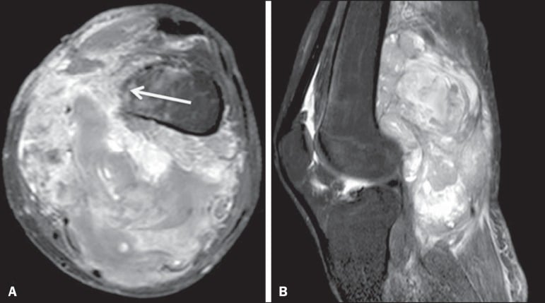
- A popliteal mass that is solid on ultrasound is a sarcoma until proven otherwise — refer to a sarcoma unit before any biopsy.
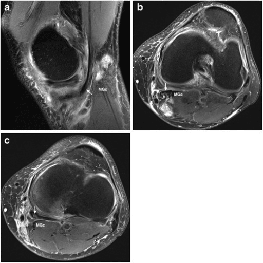
- A Baker's cyst arises between the medial head of gastrocnemius and the semimembranosus tendon, and communicates with the joint — it is medial and it points inferomedially.
- A ruptured Baker's cyst produces a painful, swollen, warm calf that is clinically indistinguishable from DVT ('pseudothrombophlebitis') — ultrasound must exclude both, not one.
- Popliteal artery aneurysm is bilateral in a large proportion of cases and is associated with abdominal aortic aneurysm — examine the other knee and the abdomen.
- In a child, a popliteal cyst is usually primary, does not communicate with the joint in the same way, and resolves spontaneously — do not aspirate it.
- Any popliteal mass with night pain, size increasing, or firmness deep to the fascia warrants MRI with contrast, not reassurance.
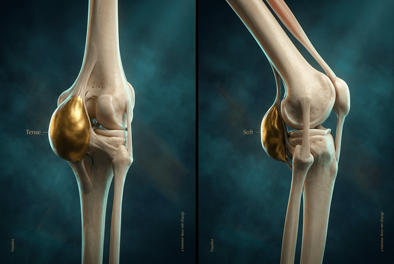
- “Foucher's sign: a Baker's cyst becomes tense in full extension and soft in 45 degrees of flexion — a solid tumour does not change.
- “Adult Baker's cyst is a symptom, not a diagnosis — look for the intra-articular cause (meniscal tear, osteoarthritis, inflammatory arthritis).
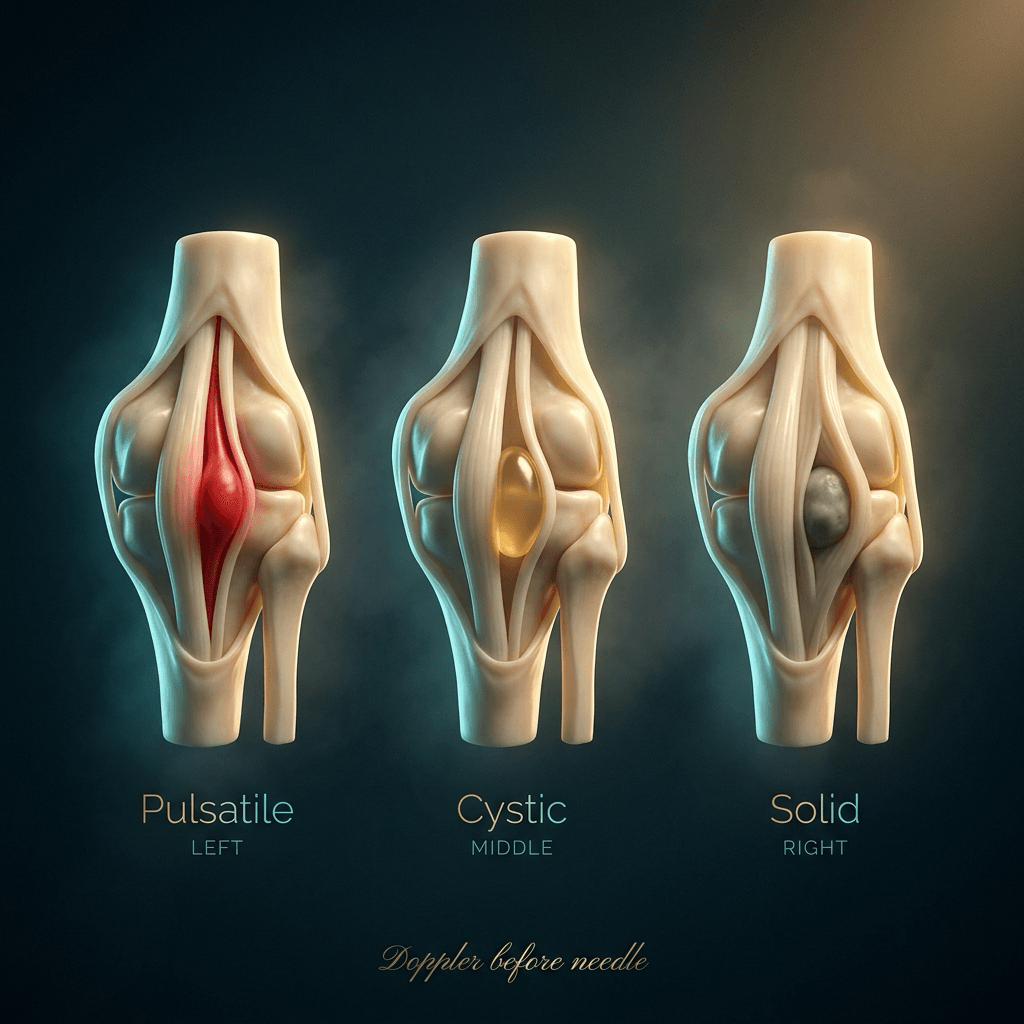
- “Never aspirate a pulsatile popliteal mass — Doppler before needle.
- “Synovial sarcoma is the classic 'small, slow-growing, painful' periarticular mass mistaken for a cyst for years.
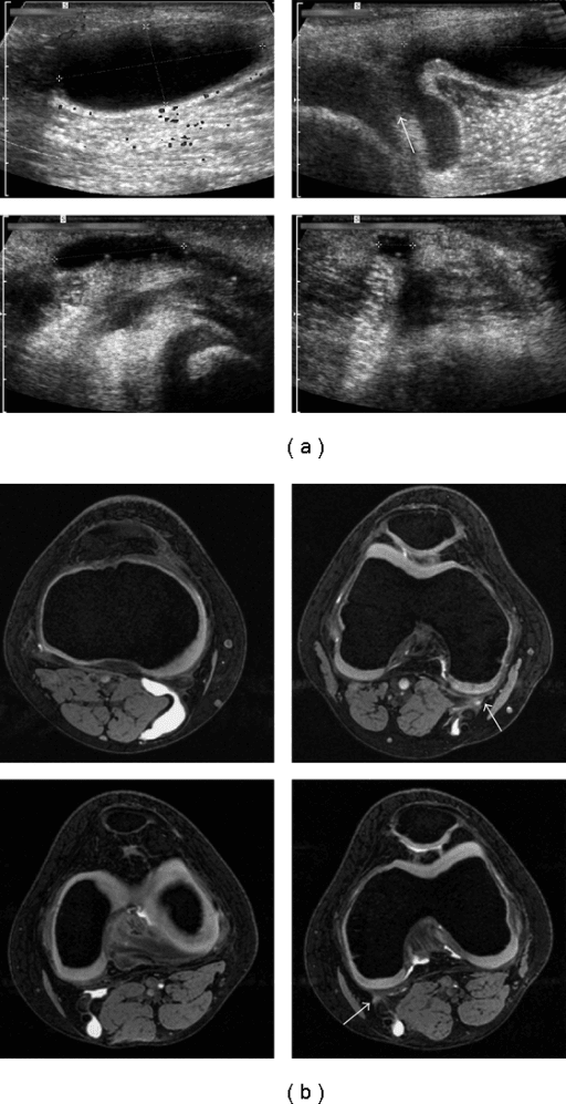
A cyst is anechoic, compressible and has a neck between medial gastrocnemius and semimembranosus. Anything solid, heterogeneous or lacking that neck is a different diagnosis. Roughly one in ten popliteal soft tissue masses referred as "cyst" is a tumour.
Aspirating a popliteal artery aneurysm or a vascular sarcoma is a catastrophe. Ultrasound with colour Doppler costs minutes. Needle after imaging, never before.
A percutaneous or open biopsy performed locally through a transverse incision contaminates all three compartments and may convert a limb-sparing resection into an amputation. Biopsy is a sarcoma-unit decision.
A negative D-dimer does not exclude a ruptured cyst, and a positive Doppler for DVT does not exclude the mass that caused venous compression. Extrinsic compression by a mass is a recognised cause of popliteal DVT.
Recognising the Pattern
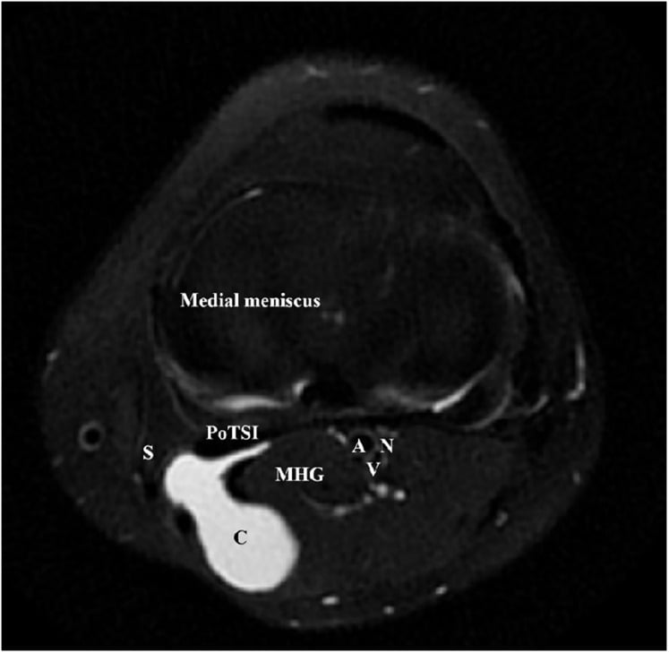
The finding. A palpable or imaged swelling within the popliteal fossa — the diamond bounded superiorly by biceps femoris laterally and semimembranosus/semitendinosus medially, and inferiorly by the two heads of gastrocnemius. Contents in order from superficial to deep and lateral to medial: tibial nerve, popliteal vein, popliteal artery (the mnemonic order from posterior to anterior).
Confirming the pattern is genuinely present.
- Examine with the patient prone and the knee in full extension, then at 45 degrees. Foucher's sign: a genuine Baker's cyst is tense in extension and softens with flexion, because the valve-like neck closes.
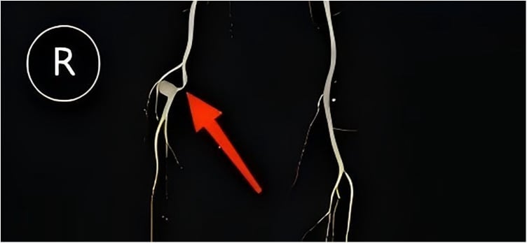
- Determine whether the mass is medial or central. The gastrocnemio-semimembranosus bursa is medial. A midline pulsatile mass is arterial until disproven.
- Test compressibility, transillumination, pulsatility and fixity to deep fascia. Fixed, firm and deep to fascia is the sarcoma triad.
- Assess the joint itself: effusion, joint line tenderness, range, and signs of inflammatory arthritis. An adult cyst without an intra-articular cause is unusual and should raise suspicion.
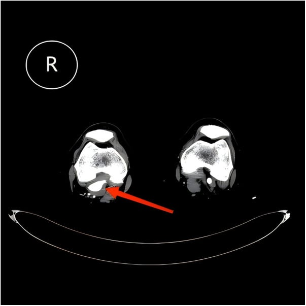
Saying it out loud in a viva. "This is a well-defined mass in the popliteal fossa. I would describe its position relative to the midline, whether it is cystic or solid on examination, whether it is pulsatile, whether it is fixed to deep fascia, and its relationship to the neurovascular bundle. On this radiograph I am looking for calcification, phleboliths, a soft tissue shadow, periosteal reaction on the posterior femoral or tibial cortex, and any bony destruction. My first investigation is an ultrasound with colour Doppler to answer three questions: cystic or solid, vascular or avascular, and is there a coexisting deep vein thrombosis."
What mimics the pattern (false positives).
- Prominent medial head of gastrocnemius or muscle hernia in a lean athlete — reduces with relaxation.
- Meniscal (parameniscal) cyst presenting posteromedially — tracks from the joint line, not the bursa.
- Ganglion of the proximal tibiofibular joint — posterolateral, may cause common peroneal palsy with foot drop.
- Distal femoral or proximal tibial osteochondroma with an overlying bursa — the bone is the mass; the "cyst" is reactive.
- Fabella or a fabella-related bursitis — a sesamoid in lateral gastrocnemius, present in a minority of knees, easily mistaken for a loose body.
- Post-traumatic haematoma or medial gastrocnemius tear ("tennis leg") in a middle-aged squash player.
Next Investigation
The Differential
- Typical age / setting
- Synovial sarcoma 15-40 years; UPS/myxofibrosarcoma 55-80 years
- Discriminating feature
- Solid, firm, deep to deep fascia, greater than 5 cm, enlarging; synovial sarcoma is often painful and may be small with calcification on plain film
- What confirms it
- MRI with gadolinium showing a solid enhancing mass; core biopsy through a planned longitudinal tract at a sarcoma unit
- Typical age / setting
- Men over 60 years, smokers, often with abdominal aortic aneurysm
- Discriminating feature
- Pulsatile and expansile mass in the midline; may present with acute ischaemia or distal embolic 'trash foot' rather than a lump
- What confirms it
- Duplex ultrasound showing focal dilatation greater than 2 cm or 1.5 times the adjacent artery; CT angiography for planning
- Typical age / setting
- Any age; post-operative, immobilised, malignancy, long-haul travel
- Discriminating feature
- Diffuse calf swelling and tenderness rather than a discrete lump; asymmetry of calf circumference measured 10 cm below tibial tuberosity
- What confirms it
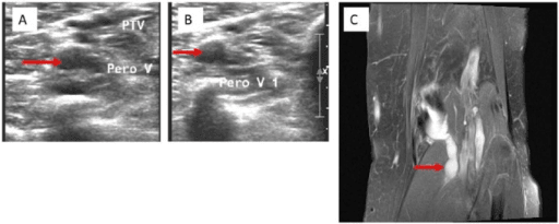
- Compression ultrasound of femoral and popliteal veins — non-compressible vein with absent flow
- Typical age / setting
- Osteosarcoma 10-25 years; Ewing 5-25 years
- Discriminating feature
- Bone-centred mass with night pain and systemic upset; aggressive periosteal reaction (Codman triangle, sunburst, onion-skin) on plain film
- What confirms it
- Plain radiograph then MRI of the whole bone; staging CT chest and bone scan before biopsy
- Typical age / setting
- 40-70 years with osteoarthritis, meniscal tear or inflammatory arthritis
- Discriminating feature
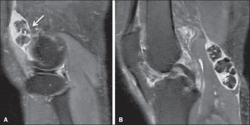
- Medial, arises between medial gastrocnemius and semimembranosus, compressible, positive Foucher's sign, accompanied by joint effusion
- What confirms it
- Ultrasound showing anechoic collection with a neck communicating with the joint; MRI if the underlying joint pathology is unclear
- Typical age / setting
- Same population, sudden onset after activity
- Discriminating feature
- Sudden calf pain with a 'crescent sign' of ecchymosis around the medial malleolus a few days later; the popliteal lump may have disappeared
- What confirms it
- Ultrasound showing fluid dissecting into the calf plus a compressible popliteal vein excluding DVT
- Typical age / setting
- Children 4-8 years, boys more than girls
- Discriminating feature
- Painless, no effusion, no intra-articular pathology, often noticed by a parent; does not fluctuate with joint symptoms
- What confirms it
- Ultrasound alone; observation is definitive management as most resolve within a few years
- Typical age / setting
- 20-50 years, sporting population
- Discriminating feature
- Sits at or just above the joint line, tender over the meniscus, associated with mechanical symptoms and a horizontal cleavage tear
- What confirms it
- MRI showing a fluid collection contiguous with a meniscal tear
- Typical age / setting
- Middle-aged recreational athletes, sudden push-off
- Discriminating feature
- Acute audible 'pop' with immediate pain at the musculotendinous junction; the mass is a haematoma below the fossa, not within it
- What confirms it
- Ultrasound showing fluid between medial gastrocnemius and soleus with normal deep veins
- Typical age / setting
- 30-60 years
- Discriminating feature
- Posterolateral position with common peroneal nerve symptoms — foot drop, dorsal foot paraesthesia, Tinel's over the fibular neck
- What confirms it
- MRI showing a lobulated cyst tracking along the peroneal nerve from the tibiofibular joint
- Typical age / setting
- 20-50 years, monoarticular
- Discriminating feature
- Recurrent haemarthrosis with brown-stained aspirate; boggy diffuse swelling rather than a discrete cyst
- What confirms it
- MRI showing blooming artefact on gradient echo from haemosiderin
- Typical age / setting
- 30-50 years
- Discriminating feature
- Multiple similar-sized calcified loose bodies within a distended posterior recess, mechanical locking
- What confirms it
- Radiograph showing multiple ring-and-arc calcifications; MRI confirms intrasynovial origin
- Typical age / setting
- Any age; malformation often present since childhood
- Discriminating feature
- Soft, compressible, enlarges on dependency or Valsalva; phleboliths on plain radiograph are pathognomonic of venous malformation
- What confirms it
- Duplex ultrasound and MRI; venous aneurysm is a source of pulmonary embolism and needs vascular referral
- Typical age / setting
- 10-25 years, may be multiple in hereditary multiple exostoses
- Discriminating feature
- Hard, bony, immobile mass continuous with the femur; may cause a reactive bursa or pseudoaneurysm of the popliteal artery
- What confirms it
- Radiograph showing cortical and medullary continuity; MRI if the cartilage cap exceeds 2 cm in an adult (malignant transformation)
Narrowing It Down
- 1Step 1 — Is it pulsatile?
Palpate for EXPANSILE pulsation and auscultate — then, in the same examination, feel the contralateral popliteal fossa and the abdominal aorta.
A pulsatile mass is a popliteal artery aneurysm until duplex says otherwise, and this question comes first because it changes the next action from 'reassure' to 'do not touch, vascular referral'. The two extra examinations are not optional: popliteal aneurysms are bilateral in 50 to 75 per cent, and 33 to 43 per cent have a coexisting abdominal aortic aneurysm.
- 2Step 2 — Is it cystic or solid?
Get an ultrasound - it answers this in minutes and is the correct first test.
Anechoic, compressible, no internal vascularity is a cyst. Internal echoes with vascularity, or a mass that does not compress, is solid. A solid mass deep to deep fascia, larger than 5 cm, or enlarging triggers the sarcoma pathway: MRI with contrast and referral BEFORE biopsy.
- 3Step 3 — Where exactly is it?
Localise the mass precisely against the anatomy rather than calling it 'popliteal'.
Medial, between medial gastrocnemius and semimembranosus, pointing inferomedially: Baker's cyst. At the joint line posteromedially: parameniscal cyst. Posterolateral near the fibular neck with peroneal symptoms: tibiofibular ganglion. Central and deep: vascular or nerve sheath. Below the fossa in the calf: gastrocnemius tear.
- 4Step 4 — How old is the patient and is there joint disease?
Examine the knee itself, not only the swelling behind it.
A child with a painless cyst and a normal joint has a primary popliteal cyst - observe. An adult with a cyst almost always has an intra-articular cause: effusion, meniscal tear, osteoarthritis, rheumatoid or crystal arthropathy. Treating the cyst without treating the joint guarantees recurrence - and note that the cyst is a marker of the joint rather than a source of pain in its own right, being present in 28 per cent of PAINLESS osteoarthritic knees against 33 per cent of painful ones (Hill 2001).
- 5Step 5 — Is the calf involved and is the leg red and swollen?
Request ultrasound that specifically compresses the femoral and popliteal VEINS as well as imaging the fossa.
An acute painful swollen calf puts deep vein thrombosis and ruptured Baker's cyst on the table together, and they can coexist. Do not anticoagulate a ruptured cyst; do not discharge an undiagnosed DVT.
- 6Step 6 — Are there red flags for malignancy?
Ask about night pain, rest pain, systemic upset and weight loss, and measure the mass; note whether it is deep to fascia, growing, or recurrent after previous excision.
Any one of these mandates MRI with gadolinium of the whole compartment plus staging, REGARDLESS of how cystic it looks - myxoid sarcomas can appear near-cystic on ultrasound and on T2. Excise a sarcoma unplanned and salvage is possible but conditional: outcomes matched primary resection only in patients who reached a sarcoma centre and were liberally re-excised, and residual disease found at that re-excision predicted metastasis (Fiore 2006).
- 7Step 7 — Does the plain film add anything?
Take orthogonal radiographs and look specifically for calcification pattern and cortical continuity.
Phleboliths mean venous malformation. Ring-and-arc calcification means synovial chondromatosis or a chondroid tumour. Amorphous calcification in a young adult means synovial sarcoma. Cortical continuity means osteochondroma. Aggressive periosteal reaction or bone destruction means primary bone sarcoma. A normal radiograph never excludes a soft-tissue sarcoma.
MCQ Practice Points
Q: Between which two structures does a Baker's cyst arise?
A: The medial head of gastrocnemius and the semimembranosus tendon — the gastrocnemio-semimembranosus bursa. This is why the cyst is medial and points inferomedially. A laterally placed cyst is not a Baker's cyst and demands imaging.
Q: What is Foucher's sign and what does a negative sign suggest?
A: A Baker's cyst becomes tense on full knee extension and softens at about 45 degrees of flexion, because the valvular neck closes in extension. A mass whose consistency does not change with knee position is more likely solid — tumour, thrombosed aneurysm or ganglion.
Q: Which four features of a soft tissue mass mandate referral to a sarcoma service?
A: Size greater than 5 cm, deep to the deep fascia, increasing in size, and painful. Any one of these is sufficient. Recurrence after previous excision is a fifth. A mass may be mobile, painless and still malignant.
Q: A 6-year-old boy has a painless 3 cm swelling behind the knee, no effusion, full range of motion. Management?
A: Ultrasound to confirm a simple cyst, then reassurance and observation. Paediatric popliteal cysts are usually primary, not associated with intra-articular pathology, and the majority resolve spontaneously. Excision has a high recurrence rate and is reserved for large, symptomatic or atypical lesions.
Q: Which popliteal mass classically causes foot drop and why?
A: A ganglion arising from the proximal tibiofibular joint. It tracks along the articular branch of the common peroneal nerve, which winds around the fibular neck, producing progressive foot drop and dorsolateral foot numbness. Treatment must include disconnection of the articular branch and the joint connection or it recurs.
Q: An adult presents with a recurrent popliteal cyst after two previous aspirations. What has been missed?
A: The intra-articular cause. Secondary Baker's cysts are driven by joint effusion from meniscal tears, osteoarthritis or inflammatory arthritis. Address the joint — arthroscopic meniscal treatment, joint replacement or disease-modifying therapy — and the cyst resolves. Isolated cyst excision without treating the joint has a high recurrence rate.
A BAD MASSPopliteal Mass — 'A BAD MASS'
Exam Viva Scenarios
Practise clinical reasoning and management decisions out loud
“You are shown a photograph and a lateral knee radiograph of a 32-year-old woman with an 18-month history of a firm, mildly painful swelling behind the right knee. The radiograph shows a soft tissue shadow posterior to the joint with faint amorphous calcification. She has been told twice that it is a Baker's cyst. How do you proceed?”
“You are shown a clinical photograph of a 58-year-old man, three weeks after a knee arthroscopy, with a swollen, warm, tender left calf and bruising around the medial malleolus. What is your differential and what do you do?”
“You are shown a duplex image of the popliteal fossa in a 71-year-old male smoker referred with a 'Baker's cyst' that has been aspirated once unsuccessfully. The vessel measures 2.6 cm with mural thrombus. What now?”
Anatomy you must state
- Boundaries: biceps femoris superolaterally, semimembranosus and semitendinosus superomedially, two heads of gastrocnemius inferiorly.
- Contents from superficial to deep: tibial nerve, popliteal vein, popliteal artery.
- Common peroneal nerve runs along the medial border of biceps femoris to the fibular neck.
- Baker's cyst arises from the gastrocnemio-semimembranosus bursa — medial, points inferomedially.
Three questions in order
- Is it pulsatile? — aneurysm until duplex says otherwise; never aspirate first.
- Is it cystic or solid on ultrasound? — solid equals sarcoma pathway.
- Where exactly is it? — medial equals cyst, joint line equals meniscal cyst, posterolateral with foot drop equals tibiofibular ganglion.
Red flags for sarcoma
- Greater than 5 cm; deep to deep fascia; enlarging; painful; recurrent after excision.
- Night pain, weight loss, systemic upset.
- Amorphous calcification in a young adult — think synovial sarcoma.
- Myxoid sarcomas can look cystic on ultrasound and T2 MRI — contrast is essential.
Clinical signs
- Foucher's sign — tense in extension, soft at 45 degrees flexion: Baker's cyst.
- Crescent sign — medial malleolar ecchymosis days after cyst rupture.
- Pseudothrombophlebitis — ruptured cyst mimicking DVT; scan for both.
- Tinel's at the fibular neck with foot drop — tibiofibular ganglion.
Management principles
- Adult Baker's cyst: treat the joint, not the cyst — recurrence is otherwise the rule.
- Paediatric cyst: ultrasound, reassure, observe; most resolve.
- Popliteal aneurysm: intervene at about 2 cm, or with thrombus or embolic symptoms; check the other leg and the aorta.
- Suspected sarcoma: MRI with contrast, then refer, then biopsy at the treating unit through a planned longitudinal tract.
Evidence Base
Knee Effusions, Popliteal Cysts, and Synovial Thickening - Association with Knee Pain in Osteoarthritis
- MRI in three groups: 381 subjects with knee pain AND radiographic osteoarthritis, 52 with radiographic osteoarthritis but NO pain, and 25 with neither
- Popliteal cysts were present in 33.0 per cent of painful osteoarthritic knees, 28.0 per cent of PAINLESS osteoarthritic knees, and 9.1 per cent of normal knees
- THE KEY NEGATIVE RESULT: after adjusting for radiographic osteoarthritis severity, moderate or larger effusions and synovial thickening were significantly more frequent in painful knees (both p less than 0.001) - but the prevalence of POPLITEAL CYSTS WAS NOT
- Among those with pain, synovial thickening was associated with the SEVERITY of pain; the cyst was not
- The authors' framing: effusions and popliteal cysts are common in middle-aged and elderly people