Chronic implantation infection of skin, fascia, muscle and bone — a WHO-recognised neglected tropical disease
- The single most important question in mycetoma is fungal or bacterial — it decides whether the patient is cured with tablets or needs a wide excision.
- Diagnosis rests on deep surgical biopsy or fine-needle aspiration for grain morphology, histology, culture and molecular identification — sinus swabs grow contaminants and mislead.
- Mycetoma spreads along fascial planes into muscle and bone but is characteristically painless until bone is involved or secondary bacterial infection supervenes.
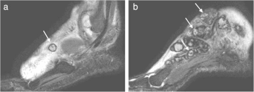
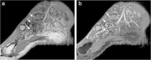
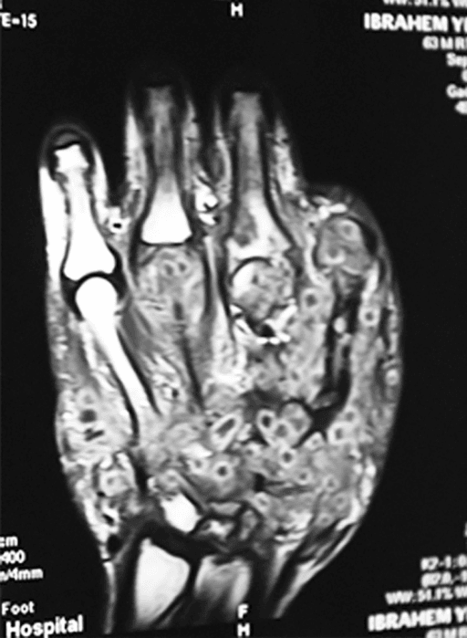
- The dot-in-circle sign on MRI (or ultrasound) is highly SUGGESTIVE: a low-signal grain within a high-signal granuloma, set in a low-signal fibrous matrix. Say suggestive, not specific - the sign was described in TWO cases and the authors themselves only speculated it was 'likely to be highly specific'. Crucially it does NOT separate eumycetoma from actinomycetoma, which is the distinction the whole management pathway turns on.
- Bone lesions are multiple small cavities with sclerotic margins and periosteal reaction — unlike the single large sequestrum-containing cavity of pyogenic osteomyelitis.
- Excision must be planned as a wide tumour resection through uninvolved tissue, not a curettage through a sinus tract.
- “Painless mass with sinuses in a barefoot farmer from the mycetoma belt is mycetoma until proven otherwise.
- “Grain colour is a useful bedside guide but is not definitive — black grains are almost always fungal, pale grains may be either.
- “Actinomycetoma responds to combination therapy usually built on co-trimoxazole with amikacin cycles for severe disease.
- “Eumycetoma medical therapy is typically itraconazole (or newer azoles) for many months, used to shrink and encapsulate lesions before excision.
- “Amputation is a treatment failure endpoint, not a first-line option — but it is definitive for extensive bone destruction.
- “Prevention is footwear and prompt wound care — a public health intervention with orthopaedic consequences.
Definition, Epidemiology and Global Burden
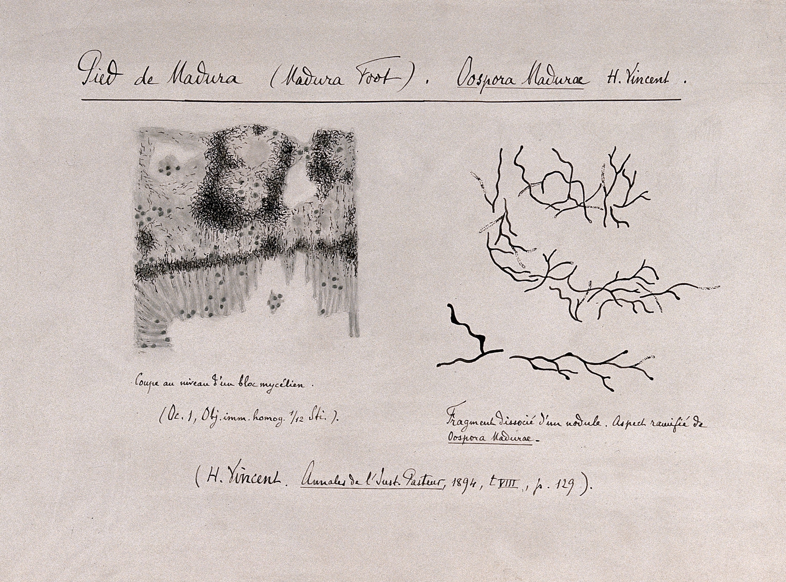
What it is. Mycetoma is a chronic, progressive, granulomatous implantation infection of the subcutaneous tissue that spreads to fascia, muscle and bone. Its signature is the grain, a compact colony of the causative organism, discharged through sinus tracts.
Two diseases under one name. Eumycetoma is caused by true fungi, with Madurella mycetomatis predominant in the African belt; actinomycetoma is caused by aerobic filamentous bacteria (actinomycetes) of the genera Nocardia, Actinomadura and Streptomyces. The two are clinically near-identical and therapeutically opposite, which is why the organism group has to be established before anything is decided.
Where. The "mycetoma belt" spans roughly 15 degrees south to 30 degrees north latitude: Sudan, Chad, Ethiopia, Somalia, Senegal, Mauritania, Niger, Mexico, Venezuela, Brazil, India, Yemen and Thailand. Sudan and Mexico are the classic reference cohorts, and they differ: Mexico is actinomycetoma-predominant, Sudan eumycetoma-predominant.
Who, and how. Young adult males aged 20 to 40, barefoot agricultural and pastoral workers, herders and labourers; the male predominance is occupational rather than biological. Inoculation is traumatic, classically a thorn prick (Acacia thorns in the African belt) or a splinter, or injury from soil-contaminated tools, and patients rarely recall the injury.
Why it matters beyond the belt. The World Health Organization recognises mycetoma as a neglected tropical disease, which drives diagnostic and drug-development programmes and regional referral centres. Cases present anywhere in the world in migrants and returning travellers, often years after leaving the endemic area, so a chronic painless foot mass with sinuses in such a patient must raise mycetoma.
Pathogenesis and Natural History
Natural history of mycetoma: inoculation to disabling deformity
A thorn, splinter or sharp stubble drives soil-dwelling organisms (actinomycetoma bacteria or eumycetoma fungi) into the subcutaneous tissue of a bare foot. Local defences contain the inoculum but do not eradicate it, and there is no acute cellulitic phase to bring the patient in.
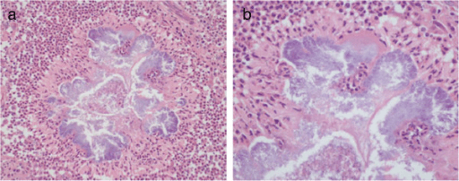
The organism aggregates into a grain: a cemented colony ringed by a neutrophil-rich zone, then granulomatous tissue, then dense fibrosis. The grain matrix impedes both drug penetration and phagocytosis, which is the structural reason antimicrobial courses run for months to years rather than weeks.
Firm, woody swelling, initially mobile on deep tissue and later fixed. Suppurating granulomas track to the surface as multiple sinuses discharging serosanguinous fluid containing grains; sinuses close and reopen elsewhere, so the skin carries scars of every generation. The triad of painless swelling, multiple sinuses and grain discharge is diagnostic.
Extension along fascial planes and tendon sheaths into intrinsic muscle, then to periosteum. Bone is invaded from outside in: periosteal reaction first, then cortical erosion, then multiple intramedullary cavities. Radiographic bone change therefore lags well behind clinical disease.
Grotesque enlargement of the foot with loss of normal architecture, fixed deformity, secondary pyogenic infection and inability to walk or work. Lymphatic spread to regional nodes occurs; haematogenous dissemination is uncommon.
Why it stays painless, and why patients present late. The granulomatous mass expands slowly without acute inflammatory pressure, so there is no pain to prompt review. Pain appears only with bone involvement, secondary bacterial infection or nerve compression. Painlessness is the diagnostic hallmark and the single greatest reason for presentation years after inoculation, often with established osseous disease.
Clinical Assessment
The history. Start with exposure: country of origin, rural residence, barefoot occupation, animal husbandry and thorn exposure. Duration is typically years, and the useful question is about the first swelling, not the first sinus. Ask directly about pain, because pain suggests bone involvement or superadded pyogenic infection.
Prior treatment. Repeated incision and drainage, curettage, and herbal or traditional remedies are common, and any previous antifungal or antibacterial course and its response matters: a response to co-trimoxazole retrospectively supports actinomycetoma. Function belongs in the history too, meaning the ability to walk, wear footwear and work, and school attendance for adolescents. Social stigma and the risk of treatment abandonment are part of the history in this disease.
Inspection. Examine the patient supine with both lower limbs fully exposed to the knee, then standing and walking. The forefoot dorsum and sole are the commonest sites; the ankle, leg, knee and hand are also affected, and the back or shoulder in head-load carriers. Look for a firm swelling with multiple sinus openings at different stages, some actively discharging, some granulating, some healed as puckered scars; grains may be visible at the sinus mouths or expressible. The skin is hyperpigmented and thickened, sometimes hypopigmented over old scars, with no overlying erythema unless secondarily infected.
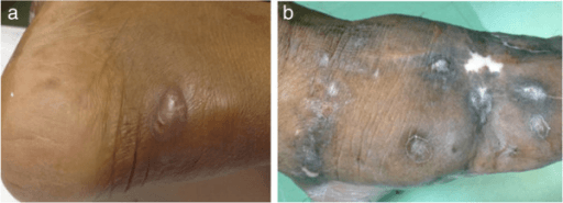
Palpation. Pinch the mass between thumb and index finger. The consistency is woody-hard and non-tender, and that is the key positive finding; tenderness is a red flag for secondary infection or bone destruction. Assess mobility over the deep fascia and bone: early lesions are mobile in the subcutaneous plane, late lesions are fixed, which indicates deep extension.
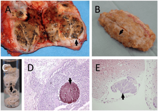
The grain at the bedside. Squeeze a sinus onto a slide to express grains and record their colour, size and consistency. Black, firm and gritty suggests eumycetoma; red, yellow, white or pale and soft suggests actinomycetoma. Colour guides but does not diagnose.
Regional and systemic. Regional lymph nodes are usually not enlarged; enlargement suggests secondary bacterial infection, lymphatic spread or an alternative diagnosis. Examine sensation, pulses and tendon function, since late disease and any planned resection depend on these. Systemic upset is absent in uncomplicated mycetoma, so fever and malaise mean secondary sepsis.
Traps. Chronic pyogenic osteomyelitis with multiple sinuses is tender and febrile at flare. Botryomycosis, a bacterial pseudomycetoma, produces grains and is a genuine mimic that requires culture. A neuropathic Charcot foot in an endemic country can coexist.
The differential. A painless woody swelling invites biopsy as a sarcoma, and bone cavities invite a tuberculosis label; grains on histology or ultrasound and the dot-in-circle sign settle it. Conversely, do not label every foot mass mycetoma, because synovial sarcoma and chronic osteomyelitis both mimic it.
- Discriminating Features
- Painless woody mass, multiple sinuses, grains expressed, years' duration
- Confirmatory Test
- Deep biopsy: grains on histology, culture, molecular identification
- Discriminating Features
- Painful flares, single large cavity with sequestrum, systemic upset, prior open fracture or acute illness
- Confirmatory Test
- Radiograph sequestrum, deep culture of pyogenic organism
- Discriminating Features
- Cold abscess, regional nodes, constitutional symptoms, juxta-articular osteopenia and erosion
- Confirmatory Test
- Tissue acid-fast bacilli, nucleic acid amplification testing, caseating granulomas
- Discriminating Features
- Grains present but bacterial (staphylococcal or Gram-negative) — a true mimic
- Confirmatory Test
- Culture and histology of grain (no filaments)
- Discriminating Features
- Painless mass but no sinuses or grains, may calcify, deep to fascia
- Confirmatory Test
- MRI plus core biopsy along a planned resection axis
- Discriminating Features
- Sensory loss, deformity, plantar ulceration rather than sinus-studded mass
- Confirmatory Test
- Sensory testing, glycaemic assessment, nerve thickening
Diagnosis: Getting the Species Right
The sample. The entire management plan hinges on identifying the causative organism group and, ideally, the species. The standard is a deep surgical biopsy under local or regional anaesthesia: a wedge of tissue from within the mass, taken through intact skin adjacent to, not through, a sinus. Ultrasound-guided fine-needle aspiration is a validated, lower-morbidity alternative for grain retrieval and cytology.
What not to send. A sinus swab or discharge from the skin surface is not a diagnostic sample. It grows skin and environmental contaminants, and the wrong drug class is then prescribed for years.
Splitting the specimen. Fresh saline-moistened tissue goes to microbiology (never formalin for culture), formalin-fixed tissue to histopathology, and a portion frozen or in a molecular transport medium for nucleic acid testing where available.
- What It Shows
- Fungal grains: broad septate hyphae 2 to 5 microns with chlamydospores in cement matrix. Actinomycetotic grains: fine filaments less than 1 micron
- Limitation
- Requires a grain; filament width is the key discriminator
- What It Shows
- Grain within neutrophil zone, granulomatous rim and fibrosis; Grocott stains fungal grains, Gram stains actinomycete filaments
- Limitation
- Species not determined; adequate deep tissue essential
- What It Shows
- Definitive species identification and drug susceptibility
- Limitation
- Slow (weeks for Madurella), contamination-prone, often negative after prior therapy
- What It Shows
- Rapid, species-level identification directly from grain or tissue
- Limitation
- Availability limited in many endemic settings; increasingly the reference standard
- What It Shows
- Supportive, and useful for treatment monitoring in research settings
- Limitation
- Not sufficiently specific for standalone diagnosis
- What It Shows
- Grains and inflammatory pattern with minimal morbidity
- Limitation
- Species identification still requires culture or molecular testing
Plan the diagnostic incision as though the definitive operation will follow. A transverse or badly placed biopsy scar over the dorsum of the foot can convert an excisable eumycetoma into an amputation, exactly as it can in soft-tissue sarcoma. Mark the tract, keep it short, obtain haemostasis without drains through separate stab wounds, and do not raise flaps.
Imaging
Views. Weight-bearing AP, lateral and oblique views of the foot, with dedicated views of any other involved region.
Early films. The bone is normal and the film shows only a soft-tissue mass with obliteration of the fat planes. Radiographs are insensitive early, so a normal film never excludes mycetoma.
The evolving bone lesion. As the lesion reaches the periosteum it raises smooth or lamellar new bone and erodes the cortex by pressure from outside. The characteristic pattern that follows is multiple small, round or oval lucent cavities with sclerotic margins in the medullary bone, classically in the metatarsals, tarsals, calcaneus and tibia. Late films show coalescence into larger defects, cortical destruction, pathological fracture, joint destruction and complete loss of tarsal architecture.
Density. Bone density is usually preserved between lesions; generalised osteopenia is unusual and suggests another process.
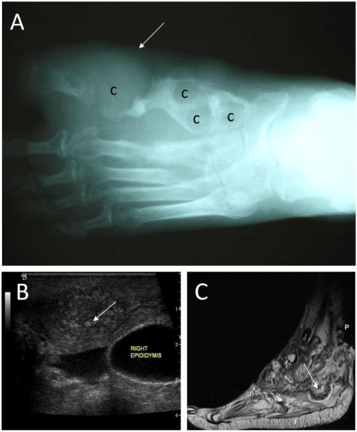
Mycetoma: multiple small cavities, sclerotic margins, periosteal reaction, bone invaded from outside in. Pyogenic osteomyelitis: single large cavity, sequestrum with involucrum, medulla outward. Tuberculosis: juxta-articular erosion, marked osteopenia, minimal periosteal reaction, little sclerosis.
Management: Two Diseases, Two Pathways
The rule. Actinomycetoma is a medically curable disease; eumycetoma is a surgical disease with medical adjuvant. Operating on actinomycetoma, or treating eumycetoma with antibiotics alone, are the two classic errors.
- Eumycetoma (fungal)
- Very indolent, well-encapsulated, later bone invasion
- Actinomycetoma (bacterial)
- Often more rapid, more inflammatory, earlier and more destructive bone invasion
- Eumycetoma (fungal)
- Prolonged azole antifungal PLUS wide surgical excision
- Actinomycetoma (bacterial)
- Prolonged combination antibacterial therapy alone
- Eumycetoma (fungal)
- Essential for localised disease; margins determine recurrence
- Actinomycetoma (bacterial)
- Rarely required; reserved for debulking massive disease or drainage of secondary abscess
- Eumycetoma (fungal)
- Many months to years; often continued after excision
- Actinomycetoma (bacterial)
- Months to over a year, continued until clinical and imaging resolution
- Eumycetoma (fungal)
- 75 per cent complete cure at 12 months on itraconazole PLUS surgery in the only randomised trial (Fahal 2024) — and that cohort EXCLUDED bone involvement, lymphatic extension, prior treatment and lesions over 16 cm. So a quarter of the most favourable patients were not cured; expect worse in advanced disease
- Actinomycetoma (bacterial)
- 28 of 31 (90.3 per cent) completely cured with antibiotics alone, despite bone involvement in about half (Cardenas-de la Garza 2020). Welsh regimen 90 per cent; TMP-SMX plus amoxicillin-clavulanate 100 per cent in a small subgroup
Principle. This is a medically curable infection, so do not operate first. Combination therapy is standard, both to improve efficacy and to prevent resistance, and the classic backbone is co-trimoxazole (trimethoprim-sulfamethoxazole) given continuously for many months.
Escalation. For extensive or bone-involving disease an intensive phase adds a second agent, historically amikacin in cycles (with audiometry and renal monitoring) or rifampicin. Alternative and adjunct agents include amoxicillin-clavulanate, dapsone and linezolid depending on species and susceptibility; species identification steers the choice, because Nocardia and Actinomadura differ in susceptibility.
Endpoint and monitoring. Continue until the sinuses close, the mass regresses, and ultrasound or MRI shows no residual grains, typically several months beyond apparent clinical cure. Monitor the full blood count, renal and liver function, audiometry if aminoglycoside cycles are used, and consider folate supplementation with long-term co-trimoxazole.
Where surgery still fits. Drainage of a secondary pyogenic abscess, debulking of a massive lesion impairing function, and rare sequestrectomy.
- 1Step 1
Painless, slowly enlarging foot mass with multiple discharging sinuses extruding grains
Species and extent both established before any operation
- 2Branch A
Grain identified as an actinomycete
Medical cure expected; no primary surgery
- 3Branch B
Grain identified as fungal
Lesion becomes resectable en bloc with less tissue sacrifice
- 4Branch B1
Localised subcutaneous mass, no bone involvement on MRI
Cure expected; margins determine recurrence
- 5Branch B2
Limited bone cavitation confined to one ray or one tarsal bone
A local operation, not a below-knee amputation
- 6Branch B3
Multi-compartment disease, extensive tarsal destruction, fixed deformity, non-functional foot
Function restored where limb preservation cannot succeed
- 7Failure route
Failed prolonged medical therapy with progressive destruction, uncontrolled secondary sepsis, or intractable pain
Applies to actinomycetoma and eumycetoma alike
- 8Monitoring
Response judged by sinus closure, mass regression and absence of residual grains on ultrasound or MRI
Treat several months beyond apparent clinical cure
Operative Technique
Indication. Localised eumycetoma, ideally after a pre-operative antifungal course, with MRI-defined extent within a resectable compartment. It is contraindicated in multi-compartment or extensive bone disease where margins are unachievable, in actinomycetoma (a medical disease), and in uncontrolled secondary sepsis, which is drained and treated first.
Why wide, not debulking. Grains lie beyond the visible mass, and intralesional surgery reliably recurs. Curettage or incisional debulking through a sinus track leaves grains in fascia and muscle and guarantees recurrence, so localised eumycetoma is excised like a soft-tissue tumour: through normal tissue, en bloc, with the sinus-bearing skin included.
Set-up. Supine, tourniquet on the thigh, exsanguinating by elevation rather than by Esmarch over the lesion, the foot on a sandbag and the whole limb prepped. Have the MRI on the screen and an image intensifier available if bone resection is planned, mark the MRI-defined extent sterilely, and lay out an oscillating saw and osteotomes, soft-tissue instruments, and containers for microbiology and histology. Antifungal therapy continues perioperatively; any secondary bacterial infection is treated first, nutrition and anaemia are optimised, and consent covers a possible ray amputation or conversion to a higher-level amputation.
Approach. A longitudinal incision in a line extensible proximally, encompassing an ellipse of all sinus-bearing skin, because the sinuses are part of the specimen. Do not raise wide flaps over uninvolved tissue.
Dissection. Work in normal tissue outside the fibrous pseudocapsule and do not enter the mass. Take a cuff of normal fat, fascia and, where necessary, muscle; divide involved tendon proximal and distal to the disease; send margins for histology.
Bone and reconstruction. Include involved bone en bloc, as a ray resection or partial tarsectomy. Fixation is usually unnecessary, but if a bone resection destabilises the medial column or hindfoot, plan arthrodesis or external fixation in the same setting. Reconstruct with local flaps, a split-skin graft over a healthy bed, or free tissue transfer where available; plantar defects need durable, sensate cover.
Structures at risk.
- Posterior tibial neurovascular bundle in the medial hindfoot
- Dorsalis pedis and the deep peroneal nerve dorsally
- Sural nerve laterally
- Flexor and extensor tendons
- The plantar weight-bearing skin envelope
Closure and aftercare. Close without tension, considering delayed primary closure or a negative-pressure dressing if contamination is a concern, and avoid drains through the resection bed where possible. Elevate and keep the patient non-weight-bearing until the soft tissues are secure, then progress to graduated weight-bearing in protective footwear. Continue antifungal therapy for months, with clinical and ultrasound or MRI surveillance for at least 2 years.
Pitfalls.
- Curetting through a sinus
- Failing to excise all sinus tracts
- Underestimating proximal fascial-plane spread on MRI
- Stopping antifungal therapy at wound healing
Treating a mycetoma like an abscess. Incision and drainage or curettage through a sinus disseminates grains along fascial planes, converts a localised excisable lesion into diffuse disease, and is the commonest reason a limb ultimately requires amputation. Biopsy first, identify the organism, then plan a tumour-style operation only if it is fungal.
MARGINPlanning an Excision
Hook:Treat black-grain disease like a sarcoma of the foot.
Complications
Recurrence. The dominant complication of eumycetoma surgery. Risk rises with inadequate margins, extensive or multi-compartment disease, bone involvement, poor adherence and interrupted drug therapy. Detect it with clinical review plus ultrasound or MRI. A discrete, resectable recurrence is re-excised widely; the antifungal class is switched with susceptibility guidance; extensive or repeated recurrence is the amputation group.
Bone destruction and pathological fracture. Progressive coalescent cavities weaken the metatarsals, tarsals and distal tibia. Manage with protected weight-bearing, resection or amputation; internal fixation into infected bone is generally avoided.
Deformity, contracture and disability. Fibrosis and tendon involvement produce fixed deformity and loss of gait. This is preventable only by early diagnosis; late cases need reconstruction or amputation with rehabilitation.
The social consequences. Loss of livelihood, school dropout, stigma and treatment abandonment follow the disease, and non-adherence over years is a social, not merely medical, problem.
- Where it arises
- Outpatient clinic, first presentation
- Mechanism
- Surface swab grows skin and secondary bacterial colonisers, not the causative eumycete or actinomycete, so the wrong organism drives therapy
- Avoid by
- Express or excise deep grains for direct microscopy, culture, histology and PCR; take a deep wedge or excisional biopsy rather than a surface swab
- If it happens
- Discard the misleading result, re-sample deep tissue or grains and re-classify eumycetoma versus actinomycetoma before committing to years of drugs
- Where it arises
- District hospital theatre
- Mechanism
- Opening infected planes seeds adjacent compartments and leaves grain-bearing tissue behind; margins are never defined
- Avoid by
- Image first with ultrasound or MRI to map compartments and bone, then plan a wide excision with defined margins under medical cover
- If it happens
- Expect early recurrence; restage with ultrasound or MRI, re-excise widely if disease is discrete, escalate to amputation if multi-compartment
- Where it arises
- Initiation of medical therapy
- Mechanism
- Eumycetoma needs prolonged azole therapy; actinomycetoma needs co-trimoxazole with or without amikacin. Treating one as the other wastes years and allows bone destruction
- Avoid by
- Confirm grain colour, microscopy, histology and culture or PCR before the first prescription
- If it happens
- Stop the ineffective agent, re-confirm the organism, restart the correct class and re-image for interval bone involvement
- Where it arises
- Radiology and surgical oncology referral
- Mechanism
- A painless firm mass with an MRI dot-in-circle sign can be read as tumour; equally a true sarcoma can be dismissed as mycetoma in an endemic area
- Avoid by
- Insist on tissue diagnosis before radical surgery; the dot-in-circle sign and multiple sinuses favour mycetoma, absence of grains does not exclude sarcoma
- If it happens
- Review histology with an experienced pathologist before any ablative procedure; unplanned excision of a sarcoma mandates oncological re-staging
- Where it arises
- Chronically discharging sinuses
- Mechanism
- Staphylococcal or Gram-negative colonisation of sinus tracts produces pain, erythema, fever and osteomyelitis that masquerade as disease progression
- Avoid by
- Culture-directed antibiotics and drainage first; operate on a quiet limb, not an acutely inflamed one
- If it happens
- Treat the acute infection and drain collections before definitive excision; defer wide excision until erythema, fever and discharge have settled
- Where it arises
- Months to years into medical therapy
- Mechanism
- Azole hepatotoxicity, interactions and cost; co-trimoxazole cytopenias, rash and renal effects; amikacin ototoxicity and nephrotoxicity. Distance to the referral centre and drug cost predict outcome as strongly as any clinical variable
- Avoid by
- Scheduled liver function, full blood count and renal monitoring with audiometry on amikacin; address travel, cost and school or work disruption as part of the treatment plan
- If it happens
- Non-adherence over years is the leading cause of failure: re-engage the patient, substitute or dose-adjust the toxic agent, and restage bone with imaging before assuming drug failure
Prevention and the Global-Health Dimension
- Footwear: closed protective shoes for agricultural and pastoral workers is the single most effective primary prevention, but adoption is limited by cost, climate and custom. Community footwear distribution programmes are the pragmatic intervention.
- Wound care: prompt cleansing and dressing of thorn and splinter injuries; removal of retained thorns.
- Early case detection: active community case-finding with ultrasound and grain microscopy at village and district level detects disease at the resectable, curable stage; late presentation is the strongest predictor of limb loss.
- Access to drugs: months to years of azole or co-trimoxazole therapy must be affordable and uninterrupted. Supply failure, not biology, causes many treatment failures.
- Stigma and education: sinus-discharging deformity leads to social exclusion, school dropout and treatment abandonment; community education is part of the treatment plan.
- Health-system framing: mycetoma is a disease of poverty and barefoot labour. Orthopaedic surgeons contribute the surgical arm of a fundamentally public-health disease, and should be advocates for decentralised diagnosis and drug access rather than only operators of last resort.
Guidelines, Registries & Global Practice
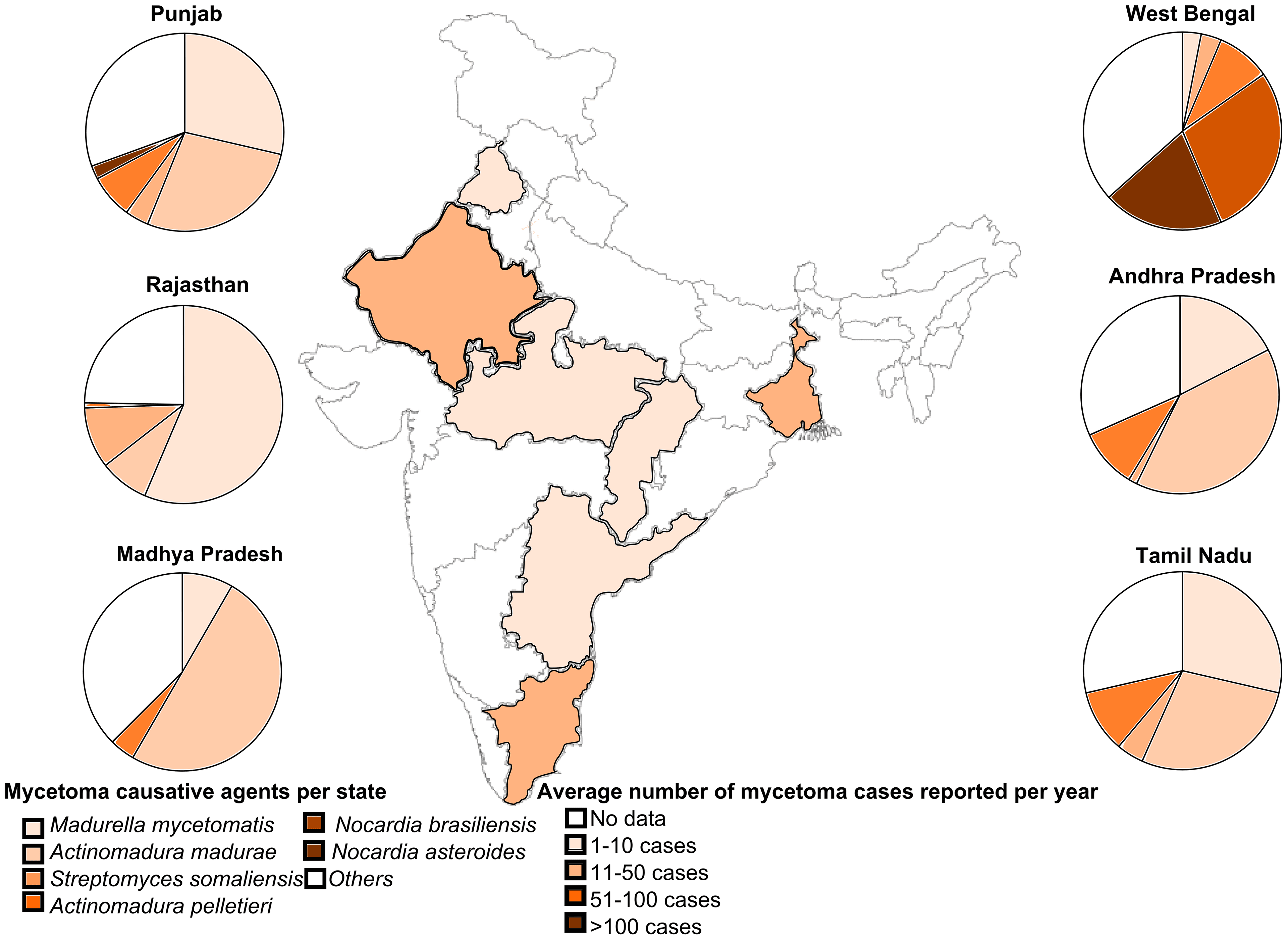
Global epidemiology
- Endemic across the tropical and subtropical belt; Sudan carries one of the highest reported burdens and hosts the principal dedicated referral centre. Mexico, India, Senegal, Mauritania, Chad, Ethiopia, Yemen, Venezuela and Brazil report substantial case series.
- Aetiological mix is geographically determined: eumycetoma predominates in the African belt and India; actinomycetoma predominates in Mexico and much of Latin America. Knowing the local mix informs empirical thinking while awaiting identification.
- True incidence is unknown; mycetoma is not notifiable in most countries and mapping is incomplete — one of the reasons for its neglected status.
Society and organisational guidance
- World Health Organization: mycetoma is on the NTD roadmap, with emphasis on early case detection, decentralised diagnosis (including ultrasound and grain microscopy at district level), access to affordable long-course therapy, and footwear-based prevention. WHO frames it as a disease of poverty requiring health-system rather than purely surgical solutions.
- National mycetoma programmes (notably Sudan, and dermatology-led services in Mexico and India) provide the operative protocols in widest use: species identification before therapy, medical therapy for actinomycetoma, and combined azole-plus-wide-excision for eumycetoma.
- General orthopaedic and musculoskeletal infection guidance (for example bone and joint infection principles endorsed by orthopaedic and infectious disease societies internationally) contributes the transferable rules: deep tissue sampling before antimicrobials, avoidance of sinus swabs, and multidisciplinary infection-team decision making. No major orthopaedic society publishes a mycetoma-specific guideline, and candidates should say so rather than invent one.
- Musculoskeletal tumour principles (biopsy along the line of definitive resection, en bloc excision, margin assessment) are explicitly borrowed for eumycetoma surgery and are the safest framework to quote in a viva.
Registries
- There is no implant registry relevance here. Disease registries are limited: dedicated national mycetoma registries exist in a small number of endemic countries and international case-collection efforts are developing under the NTD programme. This registry sparsity is itself an exam-worthy point about the neglected status of the disease.
Practice variation by resource setting
- Well-resourced centre with migrant patient: MRI, molecular species identification, susceptibility testing, multidisciplinary infection and tumour-service input, microvascular reconstruction available.
- Endemic referral centre: ultrasound and grain microscopy as frontline diagnostics, itraconazole or co-trimoxazole per protocol, wide excision with local flaps or skin grafting.
- District hospital: recognise the triad, resist the temptation to incise and drain, refer for biopsy and species identification. The single most valuable intervention at this level is not operating.
- Prevention: closed protective footwear, prompt cleansing of thorn injuries, community education, and case-finding outreach — cheap interventions with the largest population effect.
Controversies & Areas of Uncertainty
- Optimal antifungal agent and duration for eumycetoma: itraconazole remains the workhorse, but newer azoles and investigational agents from endemic-country trials may change practice; ideal duration before and after excision is not firmly established.
- How wide is wide?: no evidence-based margin has been defined for eumycetoma excision. Practice extrapolates from soft-tissue tumour principles, but the fascial-plane, skip-lesion behaviour of mycetoma may demand compartment-based rather than centimetre-based thinking.
- Role of pre-operative medical therapy: widely practised on the rationale of shrinkage and encapsulation, but the magnitude of benefit and the optimal pre-operative interval are not settled by trial data.
- Debulking in extensive disease: whether staged debulking plus long-term antifungal therapy is superior to early amputation for extensive fungal disease is unresolved, and depends heavily on prosthetic access and livelihood considerations.
- Should any actinomycetoma be operated? Some centres debulk very large lesions to accelerate response; others argue any surgery is unnecessary and risks dissemination.
- Diagnostic decentralisation: how far molecular identification can and should be pushed to district level in endemic countries, versus reliance on grain morphology and ultrasound.
- Amputation as primary treatment: ethically contested. In settings with no reliable drug supply or follow-up, an early definitive amputation may restore function faster — but it risks normalising limb loss in a curable disease.
MCQ Practice Points
Q: Which finding best distinguishes eumycetoma from actinomycetoma at the bedside?
A: Grain colour and consistency. Black, firm, gritty grains are almost always fungal; red, pink, yellow or white softer grains suggest actinomycetes. It is a guide only — definitive separation requires filament width on microscopy (broad septate hyphae versus filaments less than 1 micron), histological staining, culture and molecular identification. Note that species cannot be read off the lesion either: reviewing 12,379 eumycetoma cases, van de Sande and Fahal found no difference in lesion appearance between the 69 causative species.
Q: What is the correct diagnostic sample, and what is wrong with the obvious one?
A: Deep tissue biopsy or ultrasound-guided fine-needle aspiration through intact skin. A sinus swab yields colonising skin flora and misdirects therapy. Split the sample: fresh tissue for culture and grain microscopy, formalin tissue for histology, and material for molecular identification. The last of those matters more than it once did — culture and histology are prone to misidentification, and species affects treatment response.
Q: Which MRI sign points to mycetoma, and how far can you push it?
A: The dot-in-circle sign — small low-signal grains each surrounded by a high-signal rim of granulation tissue within a low-signal fibrous matrix on T2 or STIR, with an analogous grain-within-granuloma appearance on high-resolution ultrasound. Answer the second half too. It is widely called highly specific, but that word traces to a two-patient report in which the authors wrote only that the sign was "likely to be a highly specific sign" — a speculation never since given a denominator. And it identifies mycetoma, not which mycetoma: eumycetoma and actinomycetoma look identical, so the sign cannot make the decision the whole pathway turns on.
Q: What is the radiographic bone pattern of mycetoma, and what does it distinguish it from?
A: Multiple small round cavities with sclerotic margins plus periosteal reaction, with bone invaded from the outside in. This contrasts with the single large cavity and sequestrum of pyogenic osteomyelitis, and with the juxta-articular erosion and osteopenia of tuberculosis.
Q: Which form is usually cured by drugs alone, and what are the actual cure rates?
A: Actinomycetoma — cured medically in roughly nine of ten. In a Mexican reference series of 31 patients, 28 of 31 (90.3 per cent) were completely cured with antibiotics alone, despite bone involvement in about half; the Welsh regimen (co-trimoxazole plus amikacin) cured 90 per cent and co-trimoxazole plus amoxicillin-clavulanate 100 per cent of a small subgroup. No primary resection was needed. Set that against eumycetoma: in the only randomised trial ever conducted, twelve months of itraconazole plus surgery produced complete cure in 75 per cent (27 of 36) — in a cohort that excluded bone involvement, lymphatic extension, prior treatment and lesions over 16 cm. A quarter of the most favourable patients were not cured. This asymmetry is the reason grain identification is the single most consequential step on the page.
Q: What is the principal determinant of recurrence after eumycetoma excision?
A: Inadequate margins — intralesional or marginal surgery — compounded by extensive or bone-involving disease and interrupted antifungal therapy. The operation must be planned like a wide tumour resection, including all sinus-bearing skin, with antifungal therapy continued for months afterwards. Note also that the standard regimen is standard by default: the fosravuconazole trial that tested an alternative was terminated for futility, so itraconazole plus surgery holds the field without ever having been shown to be the best available option.
Exam Viva Scenarios
Practise clinical reasoning and management decisions out loud
“A 28-year-old male agricultural worker from Sudan, now living abroad, presents with a 4-year history of a painless enlarging swelling of the right forefoot with several discharging sinuses. He is systemically well. How do you approach him?”
“Biopsy confirms eumycetoma with black grains and broad septate hyphae; molecular testing identifies Madurella mycetomatis. MRI shows disease confined to the dorsum of the second ray with a small cavity in the second metatarsal. Plan your management.”
“A patient is referred after two previous incision and drainage procedures and a curettage through a sinus for a 'chronic foot abscess'. He now has a diffuse swollen foot with eight sinuses, and radiographs show multiple cavities across the tarsus. Culture from a previous swab grew Staphylococcus aureus.”
“A 35-year-old presents with a painless firm mass over the dorsum of the foot. There are no sinuses. Ultrasound shows a heterogeneous mass. How do you avoid a diagnostic disaster?”
Definition and Triad
- Chronic implantation infection of subcutaneous tissue spreading to fascia, muscle and bone
- Triad: painless woody swelling + multiple discharging sinuses + expressed grains
- Inoculation by thorn prick in barefoot agricultural workers of the mycetoma belt
- WHO-recognised neglected tropical disease
The Two Diseases
- Eumycetoma (fungal, typically black grains, broad septate hyphae) — azole therapy PLUS wide excision
- Actinomycetoma (filamentous bacteria, red/pale grains, filaments less than 1 micron) — prolonged combination antibiotics alone, usually curative
- Eumycetoma predominates in the African belt and India; actinomycetoma in Mexico and Latin America
- Species identification by culture and molecular testing is the pivot of management
Imaging
- Radiographs: multiple small cavities with sclerotic margins plus periosteal reaction
- Ultrasound: hyperechoic grains with acoustic shadowing within thick-walled cavities (sharper in eumycetoma)
- MRI: dot-in-circle sign — low-signal grain with high-signal rim in a low-signal matrix; highly suggestive, not specific
- MRI stages compartment and bone involvement and determines resectability
Diagnosis
- Deep biopsy or ultrasound-guided FNA through intact skin — NEVER a sinus swab
- Split sample: fresh tissue for culture and grain microscopy, formalin for histology, portion for molecular testing
- Histology: grain in neutrophil zone with granulomatous rim; Grocott for fungi, Gram for actinomycetes
- Plan the biopsy incision along the line of any future definitive resection
Surgical Principles
- Operate only for eumycetoma (or complications) — actinomycetoma is a medical disease
- Pre-operative antifungal course to shrink and encapsulate
- Wide en bloc excision including all sinus-bearing skin; work outside the pseudocapsule
- Include involved bone (ray resection, partial tarsectomy) rather than curetting cavities
- Continue antifungal therapy for months post-operatively; surveillance for at least 2 years
Amputation Thresholds
- Extensive multi-compartment disease with tarsal destruction and non-functional foot
- Failed prolonged medical and surgical therapy with progression
- Uncontrolled secondary sepsis or intractable pain
- Amputate through uninvolved tissue; continue antimicrobials if margins uncertain; plan prosthetic rehabilitation first
Classic Errors
- Incision and drainage as for an abscess
- Curettage through a sinus track
- Treating a sinus-swab coloniser as the pathogen
- Antibiotics alone for eumycetoma, or surgery for actinomycetoma
- Stopping drug therapy when the wound heals
Differential Diagnosis
- Chronic pyogenic osteomyelitis — painful, single large cavity with sequestrum
- Tuberculosis — cold abscess, osteopenia, juxta-articular erosion, caseating granulomas
- Botryomycosis — bacterial grains without filaments
- Soft-tissue sarcoma — painless mass without sinuses or grains; biopsy correctly
Prevention and Global Health
- Protective closed footwear and prompt thorn-wound care
- Community case-finding with ultrasound and grain microscopy for early, resectable disease
- Uninterrupted affordable drug supply is the main determinant of cure
- Stigma, livelihood loss and distance to care drive treatment abandonment
Evidence
An Updated List of Eumycetoma Causative Agents and Their Differences in Grain Formation and Treatment Response
- 12,379 eumycetoma cases reviewed - the largest synthesis available - identifying 69 different fungal species as causative agents
- MADURELLA MYCETOMATIS is by far the most commonly reported agent
- In most studies identification was by CULTURE OR HISTOLOGY, which the authors state is prone to MISIDENTIFICATION; molecular tools have since identified new agents
- Clinically, NO DIFFERENCE in the appearance of the lesion between species - but grain formation and antifungal susceptibility do vary
- Limited case series suggest FUSARIUM eumycetoma responds LESS WELL to treatment than Madurella mycetomatis
- In 2023 the WHO designated eumycetoma causative agents HIGH-PRIORITY PATHOGENS on its fungal priority pathogens list
MRI of Mycetoma of the Foot: Two Cases Demonstrating the Dot-in-Circle Sign
- MRI showed multiple 2-5 mm high-signal lesions interspersed within a LOW-signal matrix, each frequently containing a minute low-intensity central focus
- Ultrasound showed the same architecture - distinct hyperechoic foci within a hypoechoic mass
- The proposed correlation: low-signal matrix equals fibrous tissue, high-signal lesions equal granulomata, and the central low-signal dot equals the organised fungal GRAIN
- The authors' own wording is careful - they 'speculate' on the correlation and conclude the sign 'is LIKELY to be a highly specific sign for this lesion'