A firm swelling in the palm — the discriminators that get you from 'lump' to diagnosis
- Any solid palmar lump that is enlarging, greater than 2 cm, deep to fascia or ulcerating is a sarcoma until proven otherwise — image before you excise.
- Never shell out an undiagnosed solid hand mass: an unplanned excision of epithelioid sarcoma converts a limb-sparing operation into a wider resection.
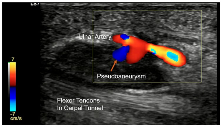
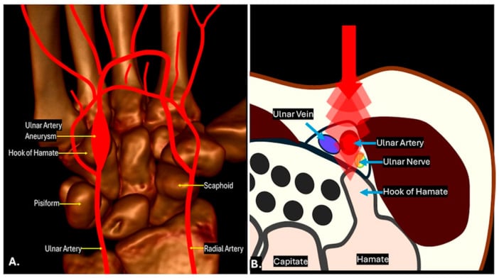
- A pulsatile or thrill-bearing hypothenar mass in a manual worker or hammer user is an ulnar artery aneurysm — do not biopsy it.
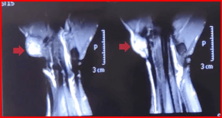
- Giant cell tumour of tendon sheath is the commonest solid hand tumour after ganglion and characteristically shows blooming on gradient-echo MRI from haemosiderin.
- A Dupuytren nodule is often tender early and precedes any cord or contracture — tenderness does not make it something sinister.
- “Ask the patient to flex the digit: a lesion arising from the flexor sheath travels proximally/distally with the tendon; a Dupuytren nodule does not.
- “Transillumination is unreliable in the thick palmar skin — use ultrasound rather than a torch.
- “Radiographs are still worth taking: pressure erosion (GCTTS), phleboliths (venous malformation), tophi with overhanging edges (gout), radiodense foreign body.
Dupuytren nodules are skin-tethered, ulnar-sided, at or distal to the distal palmar crease, and occur in a patient with the diathesis (family history, Ledderhose, Garrod pads). A midline or radial firm nodule in a 25-year-old with no diathesis is not Dupuytren.
The commonest disaster in hand oncology. Small size is falsely reassuring — epithelioid sarcoma is typically less than 3 cm at presentation. Note how often the bedside impression is simply wrong: in 134 consecutive excised finger masses, clinical and pathological diagnoses agreed only 76.1 per cent of the time (Gok 2025). Image, then core or plan a formal excision with margins.
A hypothenar mass with a positive Allen test on the ulnar side is hypothenar hammer syndrome with ulnar artery thrombosis or aneurysm. Incising it in clinic is a bleeding disaster and misses the digital embolic ischaemia.
Cystic-feeling does not exclude malignancy: myxoid sarcomas and synovial sarcoma can feel fluctuant, and a "ganglion" that recurs twice after aspiration deserves imaging, not a third needle.
Recognising the Pattern
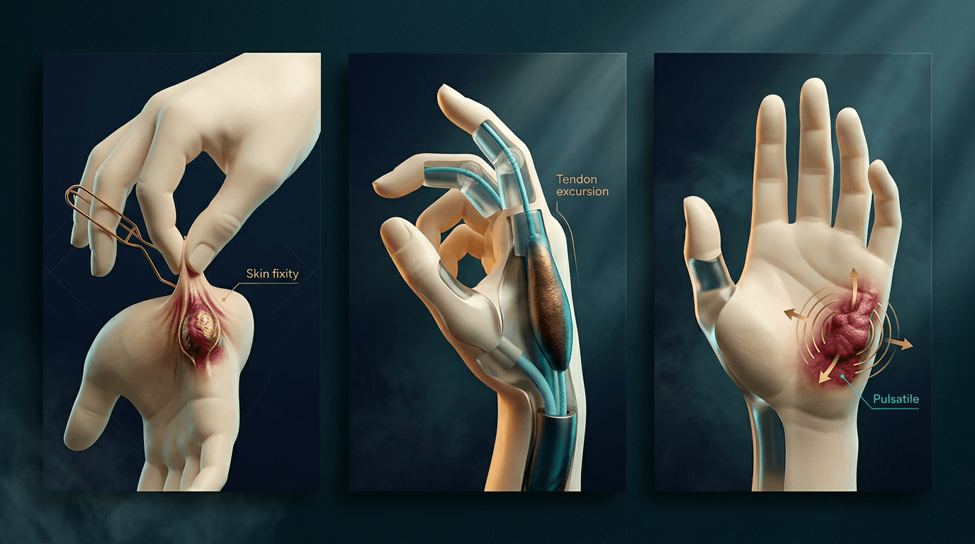
a discrete swelling within the palm, bounded superficially by glabrous skin and deep by the palmar aponeurosis, flexor sheaths, neurovascular bundles and interossei. The anatomical layer in which the lump sits does most of the diagnostic work.
- Palpate with the wrist and digits relaxed; the palmar fascia tenses in extension and can create a pseudo-mass over a normal fascial band.
- Compare with the opposite hand — normal hypothenar muscle bulk, a prominent hook of hamate or a bifid palmaris longus are frequently mistaken for tumours.
- Fix the skin between finger and thumb: skin fixity implies dermal or fascial origin (Dupuytren, epidermal inclusion cyst, epithelioid sarcoma).
- Actively flex and extend the finger: excursion of the lump with tendon movement implies flexor sheath origin (giant cell tumour of tendon sheath, retinacular cyst, fibroma of tendon sheath).
- Check for pulsatility, thrill and Allen test before any needle.
"There is a firm, non-tender, 1.5 cm mass in the ulnar palm at the level of the distal palmar crease. It is fixed to the overlying skin but not to the flexor tendon, there is no pulsation, no digital neurological deficit, and no contracture. The overlying skin is intact with no puckering elsewhere. My differential is led by a Dupuytren nodule, but I must exclude a soft tissue sarcoma."
- Prominent hook of hamate or an anomalous muscle belly (accessory abductor digiti minimi) — symmetrical, non-progressive, moves with muscle contraction.
- Callus over a metacarpal head in a manual worker — pure dermal thickening, no deep component.
- A flexor tendon nodule of trigger finger — palpable at the A1 pulley, moves with the tendon, and clicks.
- Retained foreign body granuloma — the history of a thorn or glass injury is the whole diagnosis.
- Reactive lymphadenopathy is not a palmar finding; there are no palmar nodes, so do not offer it.
Next Investigation
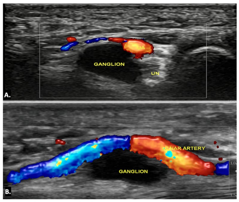
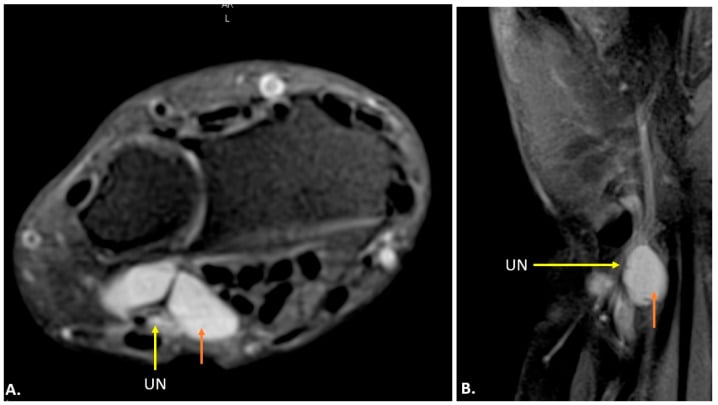
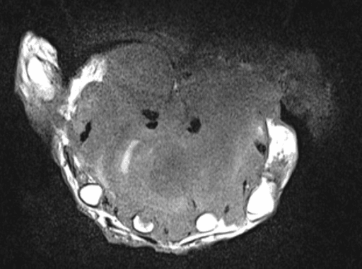
The single sentence that keeps you safe in a viva: "Because this is a solid soft tissue mass in the hand, I would obtain an MRI with contrast before any tissue is removed, and if the imaging is not clearly benign I would refer to the sarcoma multidisciplinary team for a planned biopsy rather than excise it myself."
The Differential
- Typical age / setting
- Young adult 20-40, male predominance, volar hand and forearm
- Discriminating feature
- Small (often less than 3 cm), painless, firm, multinodular along fascial planes; may ulcerate and mimic a non-healing wound or granuloma
- What confirms it
- MRI with contrast then core biopsy through a planned incision; INI1 (SMARCB1) loss on immunohistochemistry
- Typical age / setting
- 15-40 years, deep volar hand/wrist
- Discriminating feature
- Long history of a slowly growing painful mass; calcification on radiograph in up to a third
- What confirms it
- MRI showing heterogeneous 'triple sign'; biopsy with SS18 (SYT) translocation
- Typical age / setting
- Manual worker, hammer user, cyclist; 30-60 years
- Discriminating feature
- Pulsatile or expansile hypothenar mass with cold, pale ulnar-sided digits and abnormal Allen test
- What confirms it
- Duplex ultrasound or CT/MR angiography — never a diagnostic incision
- Typical age / setting
- Any age; penetrating injury, diabetes, immunosuppression
- Discriminating feature
- Hot, exquisitely tender, digit held flexed with pain on passive extension; systemically unwell
- What confirms it
- Clinical Kanavel signs, inflammatory markers, ultrasound showing fluid in the sheath; urgent theatre
- Typical age / setting
- Northern European, 45-70, male, family history, diabetes, alcohol
- Discriminating feature
- Firm nodule fixed to skin with dimpling, ulnar-sided, at or distal to the distal palmar crease; painless once mature; no tendon excursion
- What confirms it
- Clinical diagnosis — no imaging needed if typical; look for Garrod pads and plantar fibromatosis
- Typical age / setting
- 30-50 years, volar digit or palm, female predominance
- Discriminating feature
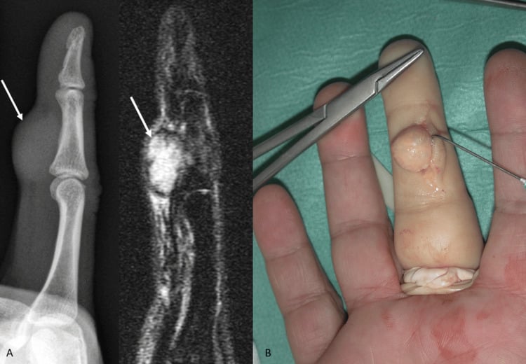
- Firm, lobulated, painless mass that moves with the flexor tendon; commonest solid hand tumour after ganglion
- What confirms it
- MRI: low signal on T1 and T2 with blooming on gradient-echo from haemosiderin; excision biopsy confirms
- Typical age / setting
- 20-40 years, at the A1/A2 pulley
- Discriminating feature
- Pea-sized, very firm, tender on gripping, does NOT move with the tendon (it is fixed to the sheath)
- What confirms it
- Ultrasound shows an anechoic cyst adjacent to the sheath; aspiration yields clear gel
- Typical age / setting
- Manual worker or previous penetrating injury; any age
- Discriminating feature
- Skin-fixed, sometimes with a punctum; may discharge keratin; can cause a lytic phalangeal lesion if intraosseous
- What confirms it
- Ultrasound shows a well-defined subdermal lesion with internal echoes; excision with the cyst wall intact
- Typical age / setting
- 40-60 years, thenar or deep palmar space
- Discriminating feature
- Soft, mobile, slowly growing, often large before presentation; may cause median nerve compression if deep
- What confirms it
- MRI: signal identical to subcutaneous fat on all sequences with complete suppression on fat-saturated imaging
- Typical age / setting
- Middle-aged onward, known hyperuricaemia, often multiple joints
- Discriminating feature
- Chalky yellow-white material visible through thin skin; may discharge toothpaste-like material
- What confirms it
- Radiograph: juxta-articular erosion with overhanging edge and preserved joint space; polarised microscopy of aspirate
- Typical age / setting
- Any age; thorn, glass or wood splinter weeks to months earlier
- Discriminating feature
- Point tenderness exactly at a healed puncture scar, with recurrent low-grade discharge
- What confirms it
- Ultrasound detects radiolucent wood/thorn that radiographs miss; glass and metal show on plain film
- Typical age / setting
- 30-60 years, along the common digital nerve line
- Discriminating feature
- Mobile transversely but not longitudinally; percussion gives paraesthesiae in the nerve distribution (Tinel over the mass)
- What confirms it
- MRI: fusiform lesion in continuity with the nerve, target sign; schwannoma is eccentric and enucleable
- Typical age / setting
- Childhood to young adult, present for years
- Discriminating feature
- Compressible, bluish, enlarges on dependency or Valsalva
- What confirms it
- Radiograph shows phleboliths; MRI shows lobulated T2-hyperintense channels with slow flow on duplex
- Typical age / setting
- 20-50 years, volar hand, male predominance
- Discriminating feature
- Small, very hard, well-circumscribed nodule attached to the sheath, mimics GCTTS but no haemosiderin
- What confirms it
- MRI low signal on T1 and T2 without blooming; histology of dense collagen with bland fibroblasts
Narrowing It Down
- 1Step 1 — Is it hot, painful and acute?
Ask how many days, not how many months. Look for erythema, fusiform digital swelling, a flexed resting posture and pain on passive extension, and take the diabetes and immunosuppression history.
Any of these makes it pyogenic flexor tenosynovitis or a deep palmar space collection — a theatre problem, not a tumour problem. Antibiotics and surgical exploration, not an MRI request.
- 2Step 2 — Is it pulsatile, compressible or bluish?
Palpate for expansile pulsation or thrill in the hypothenar eminence, perform an Allen test, take an occupational history for repetitive palmar impact, and look for splinter haemorrhages and cold digits. Test whether the lump empties on elevation and refills on dependency.
Pulsatile plus abnormal Allen test is ulnar artery aneurysm — duplex or angiography. Compressible with dependency and phleboliths on film is venous malformation. In both, needling is contraindicated, so this step must come before any aspiration.
- 3Step 3 — Which layer is it in? Test skin fixity and tendon excursion.
Fix the skin between finger and thumb, then ask for active digit flexion and extension while holding the lump.
Skin-tethered with no excursion: Dupuytren nodule, epidermal inclusion cyst — or epithelioid sarcoma, which is also fascial and skin-involving, so this finding is not by itself reassuring. Travels with the tendon: flexor sheath origin (GCTTS, fibroma of tendon sheath). Fixed to the sheath, does not travel, pea-hard: retinacular ganglion. Deep, soft, mobile: lipoma.
- 4Step 4 — Does the patient have the Dupuytren diathesis?
Ask for family history and ancestry, examine both hands and the soles, and look for Garrod knuckle pads, plantar fibromatosis, early onset, diabetes and antiepileptic use.
Diathesis present with a skin-fixed ulnar palmar nodule: this is Dupuytren and needs no imaging. Diathesis ABSENT in a young patient with a firm fascial nodule: this raises rather than lowers the suspicion of sarcoma — the same physical sign means opposite things depending on the answer here.
- 5Step 5 — Apply the red flag filter to solid lesions.
Measure it. Then check each red flag explicitly: greater than 2 cm, deep to the palmar aponeurosis, progressive growth, ill-defined margins, rest or night pain, skin ulceration, or a previous 'unplanned excision' that recurred.
Any single one takes the lesion out of clinic and into an MRI scanner and a sarcoma multidisciplinary discussion before any tissue is removed.
- 6Step 6 — Neurological and vascular mapping.
Percuss the mass for a Tinel, map any paraesthesiae to digital fields, test median and ulnar sensation, and document the deficit in the notes before operating.
Tinel with paraesthesiae into two adjacent digital fields localises a common digital nerve schwannoma. Median territory numbness with a deep thenar mass suggests a space-occupying lipoma or ganglion extending distally out of the carpal tunnel.
- 7Step 7 — Is it solitary or one of many?
Examine the whole hand and the other hand, and look at the skin for cafe-au-lait patches and at other joints for tophi.
Multiple firm nodules over joints with tophaceous discharge is gout. Multiple soft nodules with cafe-au-lait patches is neurofibromatosis. Multiple fascial nodules in the SAME ray along a fascial plane is the multinodular pattern of epithelioid sarcoma and demands biopsy.
MCQ Practice Points
Q: Which imaging feature is most characteristic of giant cell tumour of tendon sheath, and what else does it tell you?
A: Low signal on both T1 and T2-weighted sequences with marked blooming artefact on gradient-echo imaging, reflecting haemosiderin deposition. Pressure erosion of the adjacent phalanx may be seen on radiographs — and that erosion does two jobs, because it is also one of the three measured predictors of recurrence. In Reilly's 70 excisions, 27 per cent recurred, and the significant risk factors were adjacent degenerative joint disease, a distal joint location (finger DIP or thumb IP), and radiographic osseous pressure erosion. Size was not one of them.
Q: A firm pea-sized tender nodule at the A1 pulley that does not move with the flexor tendon — diagnosis?
A: Volar retinacular (flexor sheath) ganglion. It arises from the sheath itself, so it stays put while the tendon glides beneath it, and it hurts on gripping a handlebar or steering wheel. Aspiration yields clear gel and cures a proportion; excision is definitive. Contrast this with the lesion it is most often confused for: a giant cell tumour of tendon sheath arises from the sheath lining and TRAVELS with the tendon.
Q: Which hand sarcoma classically shows loss of INI1 (SMARCB1) on immunohistochemistry?
A: Epithelioid sarcoma. It presents in young adults as a small, firm, painless, often ulcerating volar hand lesion, spreads along fascia and tendon sheaths, and unusually for a sarcoma also metastasises to regional lymph nodes. In Kawai's 13 hand and wrist sarcomas the only two local recurrences were both epithelioid sarcoma, and it was the single histology the authors would not extend their otherwise reassuring conclusion to.
Q: Which lesion is most reliably diagnosed by MRI alone with no biopsy required?
A: Lipoma — signal follows subcutaneous fat on every sequence and suppresses completely with fat saturation, with no thick septa, no nodular enhancement and no non-adipose component. Any of those features raises the possibility of an atypical lipomatous tumour and warrants biopsy.
Q: A palm lump with phleboliths on plain radiograph — what is it and what test next?
A: A venous malformation. It is compressible, enlarges with dependency and Valsalva, and phleboliths are effectively pathognomonic. MRI shows lobulated T2-hyperintense channels; duplex confirms slow flow. Management is usually conservative with compression, with sclerotherapy for symptomatic lesions.
Exam Viva Scenarios
Practise clinical reasoning and management decisions out loud
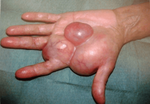
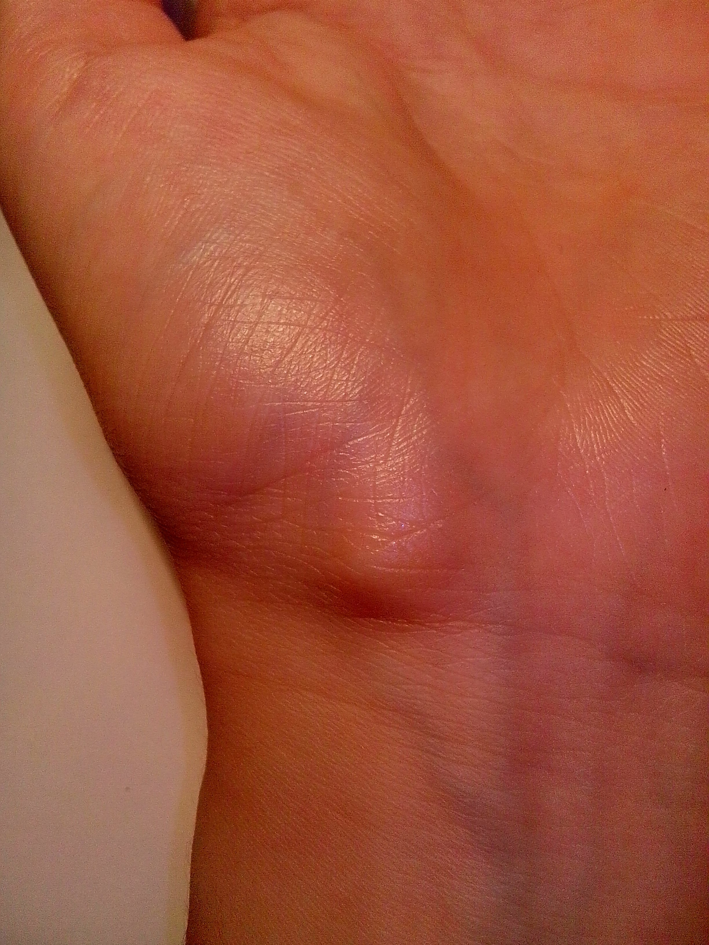
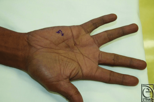
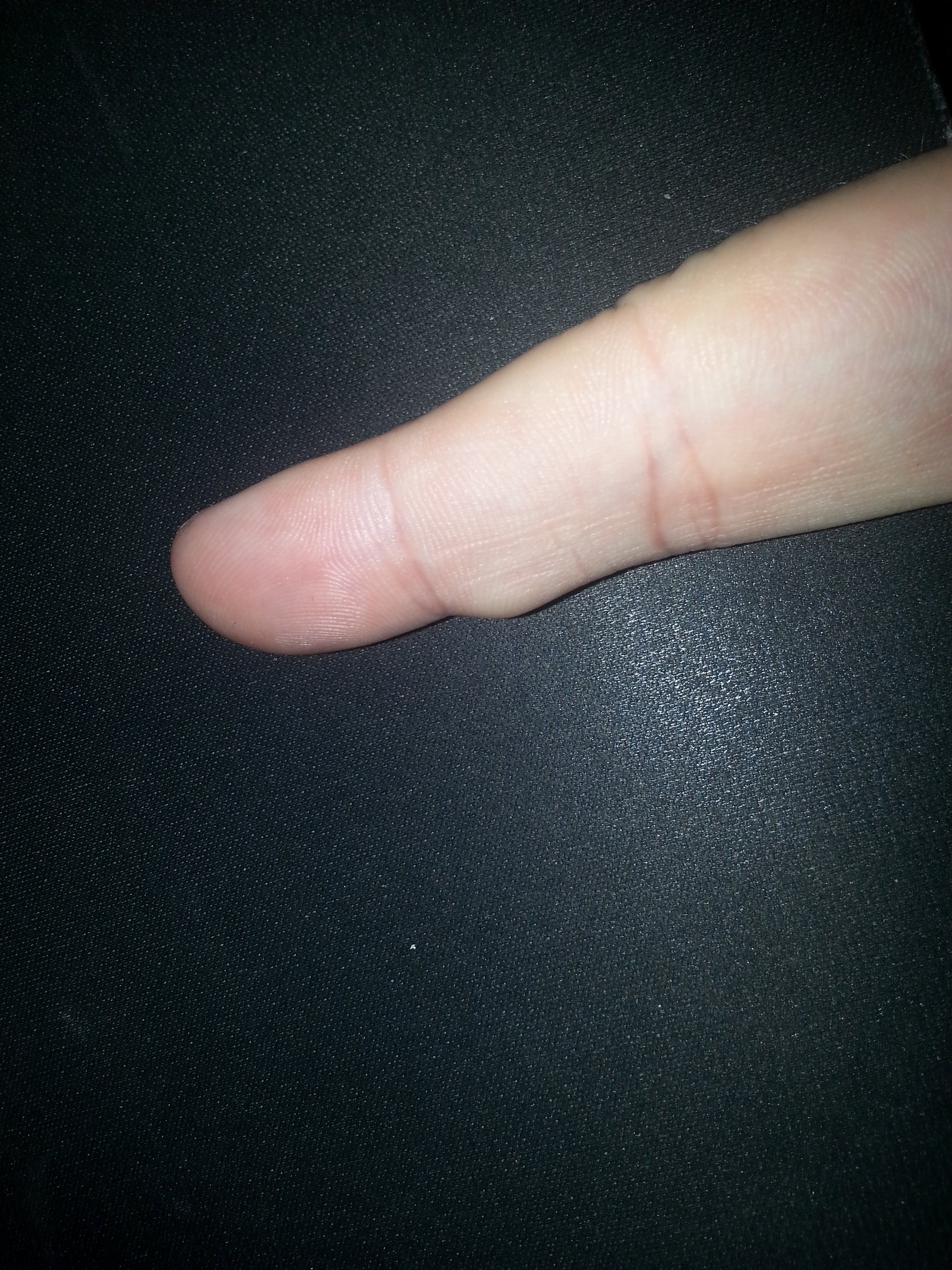
“You are shown a clinical photograph of the palm of a 58-year-old diabetic joiner. There is a firm nodule at the ulnar side of the distal palmar crease with skin puckering. The little finger is straight. He asks whether it is cancer.”
“You are shown a photograph of a 1.8 cm firm nodule on the volar aspect of the palm of a 27-year-old man, present for eight months, painless, with a small area of superficial skin breakdown that has been treated as an infected wart. There is a second smaller nodule 2 cm proximally.”
“You are shown a photograph of the hand of a 45-year-old panel beater with a 2 cm swelling over the hypothenar eminence. He reports cold, painful ring and little fingers, and there is a small ulcer at the tip of the little finger.”
Bedside discriminators
- Skin-fixed with puckering: Dupuytren, epidermal inclusion cyst, epithelioid sarcoma
- Moves with the flexor tendon: GCTTS, fibroma of tendon sheath
- Fixed to the sheath, does not move with tendon, pea-hard: retinacular ganglion
- Pulsatile hypothenar plus abnormal Allen test: ulnar artery aneurysm
- Tinel over the mass with digital paraesthesiae: nerve sheath tumour
Red flags for sarcoma
- Greater than 2 cm, deep to palmar aponeurosis, progressive growth
- Ill-defined margins, rest or night pain, skin ulceration
- Recurrence after previous unplanned excision
- Multinodular spread along a fascial plane in a young adult
Investigation ladder
- Classic Dupuytren or trigger nodule: no imaging
- Radiograph: phleboliths, foreign body, erosions, calcification
- Ultrasound first line: cystic vs solid vs vascular, dynamic and cheap
- MRI with contrast: all solid lesions greater than 2 cm or with red flags
- Angiography for any pulsatile mass; never aspirate it
Do not do
- Do not excise an undiagnosed solid hand mass
- Do not needle a pulsatile or compressible lesion
- Do not re-aspirate a 'ganglion' that has recurred twice without imaging
- Do not rely on transillumination in glabrous palmar skin
Signature findings
- Gradient-echo blooming: giant cell tumour of tendon sheath
- Complete fat suppression on all sequences: lipoma
- Phleboliths: venous malformation
- Overhanging-edge juxta-articular erosion: gouty tophus
- INI1 loss: epithelioid sarcoma
Evidence Base
Recurrent Giant Cell Tumors of the Tendon Sheath
- Seventy excised giant cell tumours of the tendon sheath, followed for a mean of 3 years 4 months: 19 of 70 (27 per cent) had a surgically and histologically documented recurrence
- Recurrence came at a mean of 2 years 3 months, but the range was 3 months to 10 years - so a clinic discharge at one year proves nothing
- Statistically significant risk factors were adjacent degenerative joint disease, location at the DIP joint of a finger or the IP joint of the thumb, and radiographic osseous pressure erosion
- Age, gender, SIZE, and volar versus dorsal location within the digit were NOT risk factors
- Eight of the 19 recurrences (42 per cent) were in patients who had already recurred once