Expendable, Absent, and Indispensable to the Surgeon
- Origin from the medial epicondyle via the common flexor origin; insertion into the flexor retinaculum and the apex of the palmar aponeurosis, with which it is continuous.
- It is the ONLY long flexor tendon that passes SUPERFICIAL to the flexor retinaculum - it is not a carpal tunnel structure.
- The median nerve lies DEEP and slightly RADIAL to the palmaris longus tendon at the wrist - the single most important surgical relation of this muscle.
- Agenesis is common: in a 300-subject population study, unilateral absence in 16 per cent and bilateral absence in 9 per cent, with the right side more often affected in unilateral cases.
- Loss of the muscle produces no measurable functional deficit, which is precisely why it is the default tendon graft donor.
- “The carpal tunnel incision is placed in line with the radial border of the ring finger, ULNAR to the palmaris longus axis - this keeps the knife away from both the median nerve and its palmar cutaneous branch.
- “The Schaeffer test - oppose the thumb to the little finger and flex the wrist - is the classic bedside method of demonstrating the tendon.
- “A surgeon who mistakes the median nerve for palmaris longus at harvest can excise the nerve - always confirm that the structure passes SUPERFICIAL to the retinaculum before dividing anything.
- “Absence must be established BEFORE consenting a patient for a reconstruction that depends on the graft; have plantaris, a superficialis slip or extensor retinaculum planned as alternatives.
Overview
Palmaris longus (PL) is a slender, entirely expendable muscle of the superficial flexor compartment. In evolutionary terms it is a regressing muscle - absent in a substantial minority of people, with no measurable functional cost when it is missing. In surgical terms it is one of the most useful structures in the upper limb, for two entirely separate reasons.
First, it is the landmark. It sits in the midline of the volar wrist, superficial and easily palpable, and the median nerve lies immediately deep and slightly radial to it. Every safe volar wrist incision - carpal tunnel release, median nerve block, tendon graft harvest - is planned in relation to the palmaris longus axis. Second, it is the graft. It provides 12 to 15 cm of strong, thin, round tendon that can be taken through two small incisions with essentially no donor deficit, and it is the default free tendon graft for flexor reconstruction, collateral ligament reconstruction at the thumb and elbow, and pulley reconstruction.
The muscle that is not there is as important as the one that is. Consenting a patient for a reconstruction that depends on a palmaris longus graft without first confirming the tendon is present is a well-recognised way to be embarrassed in theatre.
The relationship that governs volar wrist surgery is simple and must be stated precisely: at the wrist, the median nerve lies DEEP to palmaris longus and slightly RADIAL to it. From that single fact three surgical rules follow:
- Carpal tunnel release incision. Place it in line with the radial border of the ring finger, that is, ULNAR to the palmaris longus axis. Going ulnar achieves two things at once: it stays away from the median nerve (which is radial and deep), and it stays away from the palmar cutaneous branch of the median nerve (which arises 5 to 8 cm proximal to the crease and runs in the interval between palmaris longus and flexor carpi radialis, that is, radial to the palmaris axis).
- Dividing the transverse carpal ligament. Divide it at its ULNAR margin, immediately radial to the hook of hamate. This keeps the knife away from the median nerve and from the recurrent motor branch, which - depending on the Lanz variant - may be extraligamentous, subligamentous or transligamentous.
- Median nerve block at the wrist. The needle is placed between palmaris longus and flexor carpi radialis at the proximal wrist crease. Where palmaris longus is absent, the landmark becomes approximately 1 cm ulnar to the flexor carpi radialis tendon.
And the corollary that catches people out: in some individuals the median nerve lies directly beneath the palmaris longus tendon rather than slightly radial to it. That is the anatomy behind the reported catastrophe of a surgeon harvesting the median nerve believing it to be palmaris longus. Never divide a structure at graft harvest until you have confirmed it passes SUPERFICIAL to the flexor retinaculum - palmaris longus is the only long flexor tendon that does.
Attachments, Innervation and Relations
Origin
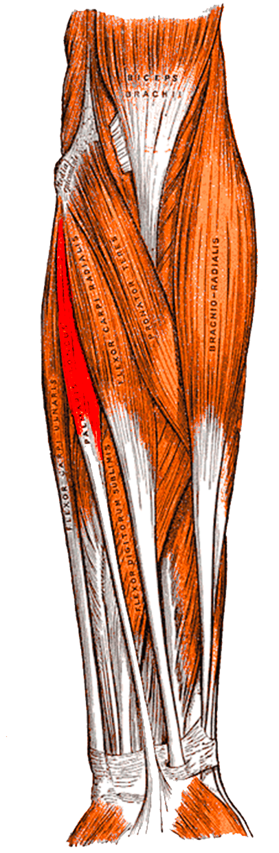
- The medial epicondyle of the humerus via the common flexor origin, lying between flexor carpi radialis (radially) and flexor carpi ulnaris (ulnarly).
- Additional fibres from the adjacent intermuscular septa and the deep antebrachial fascia.
Muscle belly and tendon
- A short, fusiform belly in the proximal third of the forearm giving way to a long, thin, round tendon that occupies the distal two-thirds.
- The tendon is typically 12 to 15 cm in usable length and 3 to 5 mm in diameter - dimensions that make it ideal as a free graft.
Insertion
- The flexor retinaculum (transverse carpal ligament) and, in direct continuity, the apex of the palmar aponeurosis.
- Some fibres continue into the thenar fascia and the abductor pollicis brevis region - the anatomical continuity exploited in the Camitz opponensplasty, where a strip of palmar aponeurosis is harvested in continuity with the tendon to gain length.
The defining anatomical fact
- Palmaris longus passes SUPERFICIAL to the flexor retinaculum. It is the only long flexor tendon of the forearm that is not a carpal tunnel structure.
- Practical consequences: a palmaris longus tendon is not decompressed by carpal tunnel release; a structure identified at wrist harvest that dives deep to the retinaculum is not palmaris longus; and a volar wrist mass superficial to the retinaculum may be an anomalous palmaris belly.
A reported and preventable disaster.
- The median nerve lies deep and slightly radial to palmaris longus, and in some people directly beneath it.
- Rule: before dividing anything, confirm the structure passes SUPERFICIAL to the flexor retinaculum. Palmaris longus is the only long flexor tendon that does.
- A nerve is soft, has visible longitudinal fascicular striations and vasa nervorum, and does not glide with wrist flexion the way a tendon does. Traction on it produces paraesthesiae in an awake patient.
Radial to the palmaris axis.
- Arises 5 to 8 cm proximal to the crease and runs between palmaris longus and flexor carpi radialis.
- Injury: painful thenar neuroma; the patient cannot bear weight through the heel of the hand.
- Avoidance: keep incisions ULNAR to the palmaris axis; cross the wrist crease obliquely if at all.
Action and Biomechanics
Actions
- Weak wrist flexion - a minor contributor alongside flexor carpi radialis and flexor carpi ulnaris.
- Tensing the palmar aponeurosis - its most plausible functional role, stiffening the palmar skin for grip and possibly contributing to cupping of the palm.
- Anchoring the palmar skin against shear during grasp.
- Weak thumb abduction through its fascial continuity with the thenar region - the anatomical basis of the Camitz transfer, although the native contribution is negligible.
The honest answer about function
- No measurable functional deficit follows its absence. Grip strength, pinch strength and wrist flexion power are not significantly different between people with and without palmaris longus. This is the single fact that justifies its universal use as a graft donor.
- It is best described as a phylogenetically regressing muscle - substantial in some primates, vestigial in humans.
Numbers examiners ask for
- Palmaris Longus
- 16 per cent in a 300-subject study
- Comparator
- -
- Significance
- One in six people lack it on one side
- Palmaris Longus
- 9 per cent in the same study
- Comparator
- -
- Significance
- Roughly one in eleven have no palmaris longus at all
- Palmaris Longus
- 25 per cent - 75 of the same 300 subjects
- Comparator
- -
- Significance
- Derived by adding the two rows above (49 unilateral + 26 bilateral). ONE PATIENT IN FOUR is missing a palmaris longus somewhere - this is the figure that should shape your expectation in clinic, and the study does not print it
- Palmaris Longus
- About 17 per cent - 101 of 600 limbs
- Comparator
- -
- Significance
- Derived: (49 x 1) + (26 x 2) = 101 absent tendons in 600 limbs. This is the rate that applies when you need a graft from ONE nominated arm; it is close to the unilateral figure by coincidence, not because they measure the same thing
- Palmaris Longus
- Right
- Comparator
- Left less often
- Significance
- Not statistically significant in the original study - state it as a trend
- Palmaris Longus
- 12 to 15 cm
- Comparator
- Plantaris up to 30 cm
- Significance
- Adequate for most upper limb reconstructions
- Palmaris Longus
- 3 to 5 mm
- Comparator
- -
- Significance
- Ideal for collateral ligament and pulley reconstruction
- Palmaris Longus
- SUPERFICIAL
- Comparator
- All other long flexors pass deep
- Significance
- The identification test at harvest
- Palmaris Longus
- Median, C7-C8
- Comparator
- -
- Significance
- Denervated and unavailable in a HIGH median palsy
Graft properties compared
- Length
- 12 to 15 cm
- Advantages
- Same limb, same field, thin and round, no functional deficit, quick harvest
- Limitations
- Absent in a substantial minority; limited length for long reconstructions
- Length
- Up to 30 cm
- Advantages
- Long, thin, ideal where great length is needed
- Limitations
- Absent in a proportion of legs; requires a second surgical field and a leg incision
- Length
- Long
- Advantages
- Reliable presence, good length
- Limitations
- Second field; donor site morbidity at the foot; flat rather than round
- Length
- Variable
- Advantages
- Same field, strong, always present
- Limitations
- Donor swan neck risk; loss of independent PIP flexion; unavailable in high median palsy
- Length
- Short
- Advantages
- Excellent for pulley reconstruction, gliding surface on one side
- Limitations
- Limited length; dorsal incision required
Surface Anatomy and Examination
Where to look
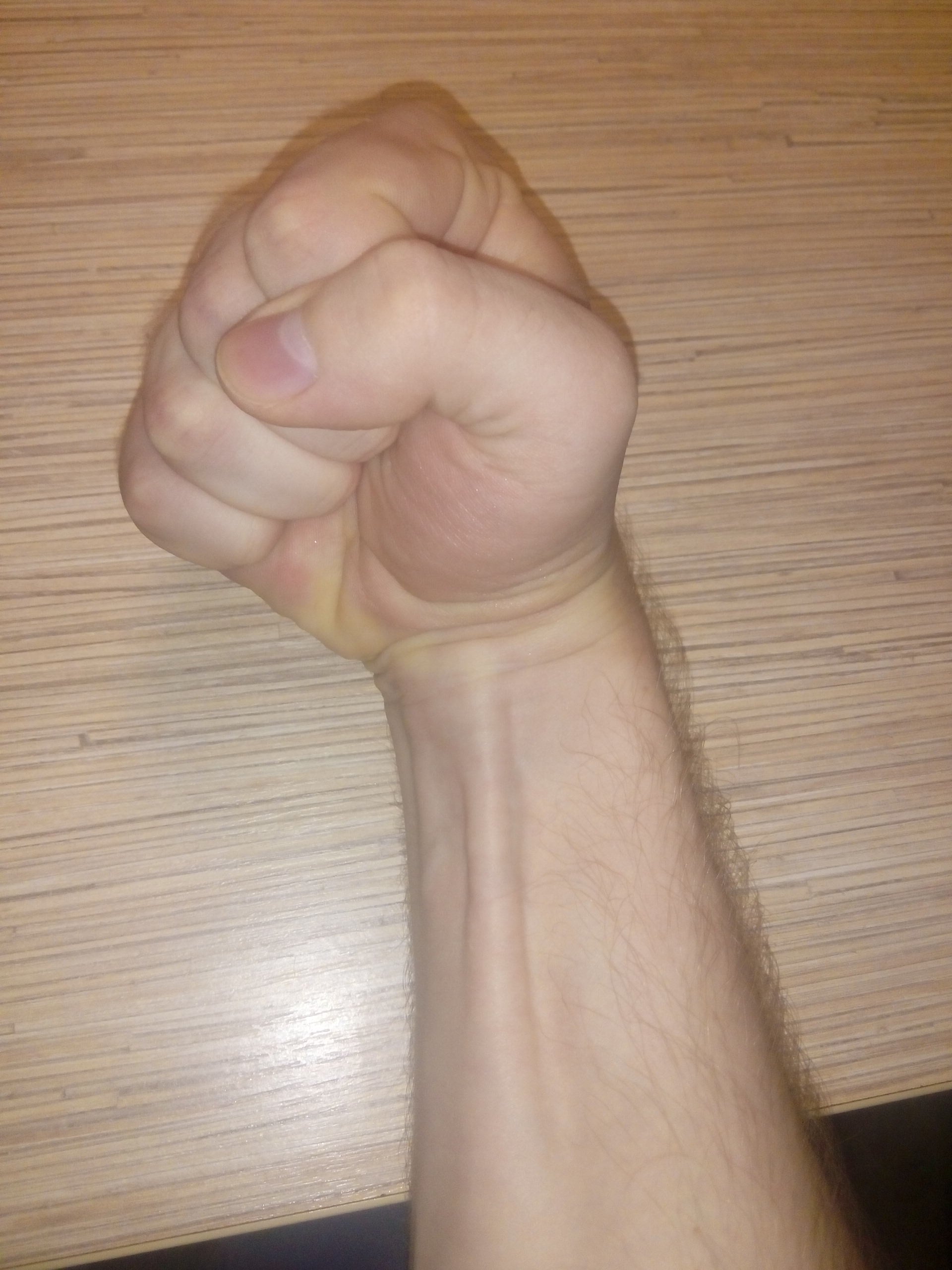
- The palmaris longus tendon appears as the thin, superficial cord in the MIDLINE of the volar wrist, between the more prominent flexor carpi radialis (radially) and flexor carpi ulnaris (ulnarly).
- It is thinner and more superficial than flexor carpi radialis, and it disappears distally by blending into the palmar aponeurosis rather than by diving under the retinaculum.
- In an obese or oedematous wrist it may be impalpable even when present - a false negative that must be considered before declaring agenesis.
Detection tests
- How to perform
- Oppose the thumb to the little finger and flex the wrist
- Positive finding
- The tendon stands out in the midline of the volar wrist
- Notes
- The classical and most widely used test
- How to perform
- Make a fist, flex the wrist, and place the thumb OVER the flexed fingers
- Positive finding
- Tendon becomes prominent
- Notes
- Useful when the Schaeffer test is equivocal
- How to perform
- Passively hyperextend the metacarpophalangeal joints, then ask for active wrist flexion
- Positive finding
- Tendon becomes prominent
- Notes
- Recruits the muscle by pre-tensioning the palmar aponeurosis
- How to perform
- Abduct the thumb fully against resistance with the wrist slightly flexed
- Positive finding
- Tendon becomes prominent
- Notes
- Exploits the fascial continuity with the thenar region
- How to perform
- Fully extend the index and middle fingers, flex the ring, little and thumb, then flex the wrist
- Positive finding
- Tendon becomes prominent
- Notes
- Reported as more sensitive than the standard test in equivocal wrists
- How to perform
- Scan the volar wrist in the transverse plane
- Positive finding
- A discrete tendon superficial to the retinaculum
- Notes
- Definitive when clinical tests are equivocal, and confirms the median nerve relationship
Use more than one test before declaring the tendon absent. No single clinical test is perfect, and the practical consequence of a false negative is that a patient is offered a plantaris or superficialis graft they did not need; the consequence of a false positive is a surgeon opening a wrist expecting a tendon that is not there.
What the tendon must NOT be mistaken for
- Flexor carpi radialis - larger, more radial, and it dives deep into its own tunnel over the trapezium.
- The median nerve - deep and slightly radial to palmaris longus, occasionally directly beneath it. Soft, with fascicular striations and vasa nervorum, and it does not glide with wrist motion. In an awake patient, traction produces paraesthesiae.
- A superficial vein or a persistent median artery - both are compressible and do not tension with wrist flexion.
The relevant preoperative examination
Before any operation that depends on a palmaris longus graft:
- Test both wrists with at least two detection tests.
- Document the finding in the notes and in the consent discussion.
- Plan and consent the alternative - plantaris, a superficialis slip, extensor retinaculum, or a long toe extensor - so that a missing tendon is a change of plan, not a crisis.
- If planning to use plantaris as the alternative, remember it too is absent in a proportion of legs, so consent for a second option there as well.
Complications
Donor site complications after harvest
- Median nerve injury - the catastrophic and entirely preventable complication. Prevented by the retinaculum test, by not using a blind stripper before identification, and by operating with the anatomy explicitly in mind rather than by feel.
- Palmar cutaneous branch neuroma from an incision placed radially or crossing the crease transversely - produces a hypersensitive scar and inability to bear weight through the heel of the hand.
- Haematoma from an unligated superficial vein.
- Scar tenderness and hypertrophic scarring at the wrist crease - minimised by short incisions in line with, or oblique to, the crease.
- No functional deficit: this is the expected outcome and should be stated at consent.
Complications of the Camitz transfer
- Bowstringing of the transferred tendon across the wrist - reduced by passing the transfer beneath the abductor pollicis brevis fascia or by using a formal pulley.
- Painful wrist scar from the conventional long incision straddling the wrist crease - the specific problem addressed by the minimally invasive modification.
- Inadequate pronation: the transfer restores palmar abduction rather than true opposition, which must be explained preoperatively so the patient's expectation matches the operation.
- Loss of the graft option: taking palmaris longus for a Camitz removes it as a future graft donor in that limb.
Failure to plan for absence
- Intraoperative change of plan without consent, requiring either an unconsented second donor site or abandonment of the reconstruction.
- Prolonged tourniquet and anaesthetic time while an alternative is found.
- A plantaris harvest that also fails, because plantaris is likewise absent in a proportion of legs and was not checked either.
Graft-related complications
- Adhesion of the graft to a scarred bed - the reason a two-stage reconstruction with a silicone rod is preferred where the bed is unfavourable.
- Graft rupture or elongation from over-tensioning or premature loading.
- Incorporation failure in an avascular or irradiated bed.
Clinical Relevance
The prevalence figures
- In a population study of 300 Caucasian subjects aged 18 to 40 (150 men and 150 women) assessed by clinical inspection:
- Unilateral absence in 49 subjects (16 per cent)
- Bilateral absence in 26 subjects (9 per cent)
- Unilateral and bilateral absence were more common in males, but not statistically significantly so.
- In those with unilateral absence, the right side was more commonly affected, again without reaching statistical significance.
- Roughly one quarter of people therefore lack a palmaris longus on at least one side.
Ethnic and population variation
- Reported agenesis rates vary widely between populations. Rates in European-descended populations are generally the highest, in the region reported above, whereas published series from East Asian and African populations report substantially lower rates of absence.
- The practical message is not the precise figure for any one group but the principle: the prevalence of absence differs meaningfully between populations, so a surgeon should quote local or study-specific data rather than assume a universal figure, and should always examine the individual patient rather than rely on a population rate.
Inheritance and associations
- Absence appears to be inherited, with reported patterns most consistent with autosomal dominant transmission with variable penetrance, and it is not associated with any functional deficit or with any specific syndrome in the general population.
- It is not associated with reduced grip strength, pinch strength or wrist flexion power in published comparisons - the finding that underpins its status as a free graft donor.
Why the number matters clinically
- A patient consented for a thumb ulnar collateral ligament reconstruction, an elbow medial collateral ligament reconstruction, a flexor tendon graft or a pulley reconstruction on the assumption of an available palmaris longus has roughly a one in four chance of the tendon being absent on the operative side if it is never checked.
- The examination takes ten seconds. Not doing it is indefensible.
Roughly one in four people lack a palmaris longus on at least one side. Before consenting a patient for a thumb ulnar collateral ligament reconstruction, an elbow medial collateral reconstruction, a flexor tendon graft or a pulley reconstruction, examine both wrists with two detection tests, document the finding, and consent the alternative graft as well. This is a ten-second examination that prevents an intraoperative change of plan for which the patient has not consented.
Surgical Relevance
Preoperative steps
- Confirm the tendon is present on the intended donor side with at least two clinical tests; use ultrasound if equivocal.
- Consent for the alternative graft (plantaris, superficialis slip, extensor retinaculum, long toe extensor).
- Mark the tendon with the patient awake, using the Schaeffer test.
Technique
- Distal incision: a short transverse incision at the distal wrist crease, directly over the tendon. Incise skin only and spread bluntly - the tendon is immediately subcutaneous.
- Confirm identity before dividing. The three checks:
- It is superficial to the flexor retinaculum - the definitive test. Any structure diving deep to the retinaculum is not palmaris longus.
- Traction on it tensions the palmar aponeurosis and does not move a finger.
- It is a tendon: firm, glistening white, without fascicular striations or longitudinal vasa nervorum. In an awake patient, traction produces no paraesthesiae.
- Divide distally, place a holding suture, and deliver the tendon proximally either:
- through one or two additional small transverse counter-incisions at intervals up the forearm, or
- with a closed tendon stripper advanced proximally along the tendon to the musculotendinous junction.
- Preserve the paratenon on the graft where possible; it aids incorporation.
- Take 12 to 15 cm and confirm the graft length against the requirement before closing.
Hazards and how to avoid them
- Harvesting the median nerve. The reported catastrophe. Prevented absolutely by the retinaculum test and by never using a blind stripper before positive identification.
- Palmar cutaneous branch injury from a transverse incision crossing the crease radially - keep the incision small, over the tendon, and cross the crease obliquely if extension is needed.
- Injury to a superficial vein producing a haematoma - ligate rather than avulse.
- Blind stripper injury to the median nerve or to muscle if the stripper deviates from the tendon; advance it gently, in line, without force.
- Incomplete harvest where the tendon avulses at the musculotendinous junction - use a counter-incision if resistance is felt.
What the graft is used for
- Flexor tendon reconstruction: single-stage grafting and the second stage of a two-stage silicone rod reconstruction.
- Thumb ulnar collateral ligament reconstruction for chronic gamekeeper thumb.
- Elbow medial collateral ligament reconstruction - palmaris longus is the classic donor for the docking and figure-of-eight techniques.
- Pulley reconstruction (Kleinert-Weilby, Bunnell loop, Karev belt-loop) for A2 or A4 loss.
- Scapholunate and other carpal ligament reconstructions, interposition arthroplasty and static suspension procedures.
- Outside orthopaedics: eyelid slings for facial palsy, lip augmentation, tympanoplasty grafts and temporomandibular joint reconstruction - all exploiting the same expendability.
Before dividing any structure during palmaris longus harvest, confirm that it passes SUPERFICIAL to the flexor retinaculum. Palmaris longus is the only long flexor tendon that does. A structure that dives deep to the retinaculum is a flexor tendon, an anomalous palmaris profundus, or - the reported catastrophe - the median nerve. Never advance a blind tendon stripper before this check.
Rich Frenchmen Prefer Perfect ManicuresVolar Wrist, Radial to Ulnar
Hook:The median nerve hides behind palmaris longus - so the incision goes ULNAR to it.
Guidelines, Registries & Global Practice
Anatomical variation across populations
- The most-cited population figures come from a study of 300 Caucasian subjects: unilateral absence 16 per cent, bilateral absence 9 per cent, with a non-significant male predominance and a non-significant right-sided predominance in unilateral cases.
- Reported agenesis rates differ substantially between populations. Series from European-descended populations generally report the highest rates, while series from East Asian and African populations report substantially lower rates. The safest examination answer quotes the study-specific figure, acknowledges the population variation, and states that the individual patient must be examined regardless.
- Absence appears heritable, with reported patterns most consistent with autosomal dominant transmission with variable penetrance, and it has no association with any functional deficit.
- Variants - reversed, digastric, duplicated and palmaris profundus - occur at low frequency and are principally relevant as causes of a volar wrist mass or of nerve compression.
Differences in described technique between sources
- Position on palmaris longus practice
- Places the carpal tunnel incision in line with the radial border of the ring finger and emphasises protection of the palmar cutaneous branch and the recurrent motor branch.
- Position on palmaris longus practice
- Recommend documenting palmaris longus presence preoperatively where a graft is planned, and consenting for the alternative donor.
- Position on palmaris longus practice
- Uses palmaris longus and flexor carpi radialis as the surface landmarks defining the safe volar wrist corridors.
- Position on palmaris longus practice
- Palmaris longus remains the preferred autograft for medial collateral ligament reconstruction of the elbow, with gracilis or a toe extensor as the standard alternative where it is absent.
Evidence positions worth stating
- No functional deficit follows harvest. Comparative studies of grip strength, pinch strength and wrist flexion power in people with and without palmaris longus have not demonstrated meaningful differences. This is the evidential basis for its use.
- Camitz modifications: comparative data show that adding a flexor retinaculum pulley improves thumb flexion and pronation over the classical transfer, and that a minimally invasive technique avoiding an incision across the wrist crease substantially reduces painful wrist scars and bowstringing. Neither modification is supported by randomised evidence, but the comparative series are consistent.
- No high-level evidence favours palmaris longus over other autografts for any specific reconstruction; the choice rests on availability, required length and the acceptability of a second surgical field.
High- versus limited-resource practice
- Well-resourced settings: preoperative ultrasound confirmation of palmaris longus, closed tendon strippers, allograft as a fallback where no autograft is available, and formal hand therapy after transfers.
- Limited-resource settings: palmaris longus is arguably the single most valuable structure in reconstructive hand surgery in a low-resource environment - it is free, in the same operative field, requires no implant and needs only a knife and a holding suture. Where allograft and ultrasound are unavailable, the discipline of clinical detection with two tests and a planned alternative donor is what makes reconstruction reliable. The Camitz opponensplasty is likewise attractive in these settings because it needs no implant and can be combined with the carpal tunnel release in a single, short operation.
MCQ Practice Points
Q: How commonly is palmaris longus absent? A: In a 300-subject population study, unilaterally in 16 per cent and bilaterally in 9 per cent - roughly a quarter of people lack it on at least one side.
Q: In unilateral absence, which side is more often affected? A: The right, though the difference did not reach statistical significance in the original study.
Q: Where does the median nerve lie in relation to palmaris longus at the wrist? A: DEEP and slightly RADIAL to it - and in some individuals directly beneath it.
Q: Where is the open carpal tunnel incision placed and why? A: In line with the radial border of the ring finger, ULNAR to the palmaris longus axis - keeping away from both the median nerve and its palmar cutaneous branch.
Q: How do you positively identify palmaris longus at harvest? A: It passes SUPERFICIAL to the flexor retinaculum - the only long flexor tendon that does.
Q: Describe the Schaeffer test. A: Oppose the thumb to the little finger and flex the wrist; the tendon stands out in the midline of the volar wrist.
Q: Where does palmaris longus insert? A: Into the flexor retinaculum and the apex of the palmar aponeurosis, with which it is continuous.
Q: How much graft does it provide? A: Approximately 12 to 15 cm in length and 3 to 5 mm in diameter.
Q: What does the Camitz transfer restore? A: Palmar abduction, not true opposition. It provides little thumb pronation unless a pulley modification is added.
Q: How does palmaris profundus differ, and why does it matter? A: It lies DEEP to the retinaculum, within the carpal tunnel, and is therefore a genuine cause of median nerve compression - and it is not palmaris longus at harvest.
Q: Is palmaris longus available as a transfer in a high median nerve palsy? A: No - it is median-innervated and therefore denervated. Use extensor indicis proprius or an abductor digiti minimi (Huber) transfer.
Exam Viva Scenarios
Practise clinical reasoning and management decisions out loud
“You are about to reconstruct a chronic thumb ulnar collateral ligament rupture in a 32-year-old skier using a palmaris longus graft. In theatre, with the tourniquet up, you cannot find the tendon. What went wrong and what do you do now?”
“Show me where you would place your incision for an open carpal tunnel release, and justify every part of it in terms of anatomy.”
“A 74-year-old man has had numbness in his hand for four years and now has profound thenar wasting with no palmar abduction. Nerve conduction studies show a severe median neuropathy at the wrist with no motor response. He wants his thumb to work. What do you offer, and what will and will not improve?”
Anatomy
- Origin: medial epicondyle via common flexor origin
- Insert: flexor retinaculum and apex of the palmar aponeurosis
- Nerve: median, C7-C8
- SUPERFICIAL to the flexor retinaculum - not a carpal tunnel structure
- Short proximal belly, long thin tendon
The Landmark
- Median nerve lies DEEP and slightly RADIAL to it
- Palmar cutaneous branch runs between PL and FCR
- Carpal tunnel incision ULNAR to the PL axis
- Median nerve block between PL and FCR; if absent, 1 cm ulnar to FCR
Agenesis
- Unilateral 16%, bilateral 9% in a 300-subject study
- Right side more often absent in unilateral cases
- Rates vary substantially between populations
- No functional deficit when absent
Detection Tests
- Schaeffer: thumb to little finger, flex the wrist
- Thompson: fist, flex wrist, thumb over the fingers
- Mishra I and II; Pushpakumar two-finger sign
- Ultrasound if equivocal - use two tests before declaring absence
Uses
- Default free tendon graft: 12-15 cm, 3-5 mm
- Thumb UCL, elbow MCL, pulley and flexor reconstruction
- Camitz opponensplasty - abduction, not opposition
- PL to rerouted EPL in radial nerve palsy
- Unavailable in HIGH median nerve palsy
Evidence Base
Absence of the Palmaris Longus Muscle: A Population Study
- 300 Caucasian subjects (150 male, 150 female) aged 18 to 40 assessed by clinical inspection
- Forty-nine subjects had unilateral absence of palmaris longus (16 per cent)
- The tendon was absent bilaterally in 26 subjects (9 per cent)
- Unilateral and bilateral absence were more common in males but not significantly so
- In those with unilateral absence the right side was more commonly affected, again without statistical significance
Anatomical Variations of the Median Nerve in the Carpal Tunnel
- 246 operatively explored carpal tunnels with 29 variations in the course of the median nerve identified
- Four groups: variation in the thenar branch course, accessory distal branches, high divisions, and accessory branches proximal to the canal
- The famous thenar branch figures are NOT Lanz's own data: he quotes POISEL's 100 cadaver hands - extraligamentous 46 per cent, subligamentous 31 per cent, transligamentous 23 per cent
- The findings emphasise approaching the median nerve from the ULNAR side when opening the carpal tunnel
Opponensplasty Using Palmaris Longus Tendon and Flexor Retinaculum Pulley in Patients with Severe Carpal Tunnel Syndrome
- Twelve cases of long-standing severe carpal tunnel syndrome with severe thenar atrophy treated by modified Camitz opponensplasty with a pulley at the ulnar remnant of the flexor retinaculum
- Mean grade of thenar atrophy improved from 3 preoperatively to 0.83 at final follow-up
- Maximal palmar abduction improved from 3.6 to 6.8 cm and spatial rotation from 54 to 83 degrees
- Kapandji tip opposition improved from 65 to 85 per cent symmetry
- All patients were satisfied or very satisfied
Minimally Invasive Modified Camitz Opponensplasty for Severe Carpal Tunnel Syndrome
- Twenty-eight hands treated by a minimally invasive modified Camitz passing the palmaris longus beneath the abductor pollicis brevis fascia through palm and thumb incisions only, compared with 10 conventional Camitz procedures
- Thumb palmar abduction, pinch power and grip strength improved significantly in both groups
- The minimally invasive group had no bowstringing, no painful wrist scars and no palmar cutaneous branch injuries
- The conventional group had four painful wrist scars and nine cases of transferred tendon bowstringing
Flexor Carpi Radialis Tendinitis. Part I: Operative Anatomy
- Twenty-five cadaveric specimens studied with sections, tenograms and histology of the volar radial wrist
- Defines the relationship of the flexor carpi radialis tendon to the trapezium, carpal canal septum and adjacent structures
- The tendon enters a fibro-osseous tunnel at the proximal border of the trapezium and is separated from the carpal canal by a thick septum
- The tendon lies within a few millimetres of the distal radius, the scaphoid tubercle, the scaphoid-trapezium-trapezoid joint and the thumb carpometacarpal joint
Proximal Median Nerve Compression: Pronator Syndrome
- Review of pronator syndrome, a compressive neuropathy of the median nerve in the proximal forearm
- Symptoms often overlap with those of carpal tunnel syndrome, so making the correct diagnosis is challenging
- Electrodiagnostic studies are often negative in pronator syndrome, so the diagnosis rests on clinical assessment
- The review concludes that a thorough understanding of median nerve anatomy, the possible sites of compression and the characteristic clinical findings is essential to a reliable diagnosis