The Small Muscle That Organises the Axilla
- Origin: outer surfaces and upper borders of the third, fourth and fifth ribs near their costal cartilages, with the intervening fascia.
- Insertion: the medial border and superior surface of the coracoid process.
- Innervation: medial pectoral nerve (C8, T1), which pierces the muscle, with a contribution via the ansa pectoralis.
- Actions: protraction, depression, downward rotation and anterior tilt (tipping) of the scapula; accessory inspiration with the scapula fixed.
- It is enclosed by the clavipectoral fascia, with the costocoracoid membrane above and the suspensory ligament of the axilla below.
- “Pectoralis minor divides the axillary artery into three parts: first medial to, second behind, third lateral to the muscle.
- “Axillary node levels follow the same rule: level I lateral and inferior, level II posterior, level III medial and superior to pectoralis minor.
- “A shortened pectoralis minor tilts the scapula anteriorly and internally rotates it, narrowing the subacromial space — the mechanical link to impingement.
- “In the Latarjet, incomplete release of pectoralis minor from the medial coracoid limits graft mobilisation and tethers the plexus.
Overview
The pectoralis minor is a small triangular muscle running from the third, fourth and fifth ribs to the medial border of the coracoid process, lying deep to the pectoralis major and enclosed within the clavipectoral fascia. It contributes little to gross strength and its loss produces almost no measurable functional deficit — Poland syndrome patients, in whom it is often absent alongside the sternocostal head of pectoralis major, function essentially normally.
Its importance is positional. It is the muscle by which surgeons and radiologists orient themselves in the axilla: it divides the axillary artery into three parts and the axillary lymph nodes into three levels. It tips the scapula forward when it shortens, which is the mechanical bridge between soft-tissue tightness and subacromial narrowing. It forms the roof of a space in which the brachial plexus cords and axillary vessels can be compressed. And it is the structure that must be released before the coracoid can be moved in a Latarjet procedure.
Understand what pectoralis minor does to the scapula and every clinical association follows.
- Its line of pull runs from the coracoid, which is the anterosuperior corner of the scapula, downward and medially to the anterior chest wall. Contraction therefore pulls the coracoid downward and forward.
- Result on the scapula: anterior tilt (the inferior angle lifts off the chest wall as the coracoid drops forward), internal rotation (the medial border lifts), protraction and downward rotation.
- Normal elevation of the arm requires the opposite — posterior tilt, external rotation and upward rotation of the scapula, driven by serratus anterior and lower trapezius.
- Therefore a shortened pectoralis minor directly opposes the scapular motion required for elevation. It holds the acromion tipped forward and down over the greater tuberosity, narrowing the subacromial space at exactly the moment it needs to open.
- The clinical chain: tight pectoralis minor leads to anterior tilt and internal rotation, leads to reduced subacromial clearance and altered glenohumeral kinematics, leads to impingement-type pain and scapular dyskinesis. This is the mechanism examiners want articulated, not merely the observation that "pec minor is tight".
One Two Three, Medial Behind LateralPectoralis Minor Divides the Axilla into Thirds
Hook:The number of branches from each part of the axillary artery equals the part number: one, two, three.
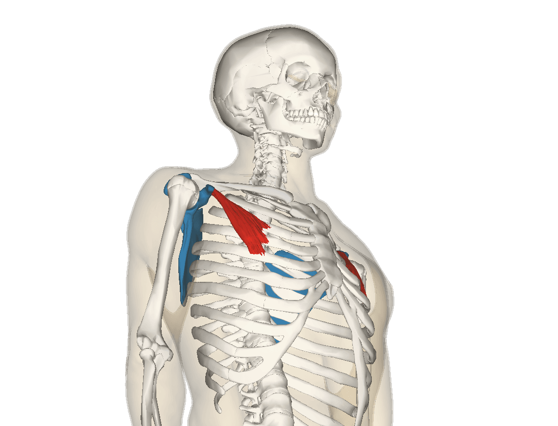
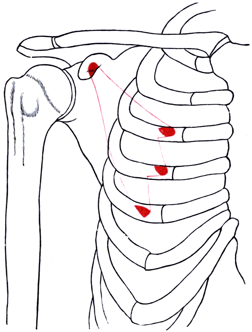
Attachments, Innervation and Relations
Origin
- The outer surfaces and superior borders of the third, fourth and fifth ribs, close to their costal cartilages, together with the fascia over the intervening external intercostal muscles.
- A common variant takes the origin from the second to fourth ribs rather than the third to fifth, and additional slips from the second or sixth rib are described.
Insertion
- The medial border and superior surface of the coracoid process, over a footprint of approximately 1.5 to 2 cm.
- Important insertional variants: in a minority of shoulders the tendon does not stop at the coracoid. Fibres continue over the coracoid to blend with the coracoacromial ligament, the coracohumeral ligament, or to pass into the rotator interval and glenohumeral joint capsule. Reported prevalence depends on the modality: 1.5 per cent of 335 MR arthrograms in Lee and colleagues' series, where the aberrant tendon reached the joint capsule and the coracohumeral ligament was absent in every case, against 9.6 per cent of 606 shoulders on dynamic ultrasound in Homsi's series, with a left-sided and female predominance and no correlation with symptoms. The variant matters in three ways: it can contribute to subcoracoid impingement, it complicates the plane of dissection during coracoid osteotomy, and it means that releasing the tendon at the coracoid may not fully release the muscle.
Fascial envelope
- Pectoralis minor is enclosed within the clavipectoral fascia, a structure the examiners like because its named parts are precise.
- Above the muscle the fascia extends to the clavicle and subclavius as the costocoracoid membrane, which is pierced by the lateral pectoral nerve, the thoracoacromial artery, the cephalic vein and lymphatics.
- Below the muscle the fascia continues to the axillary fascia as the suspensory ligament of the axilla, which pulls the axillary fascia upward when the arm is abducted, producing the hollow of the axilla.
1 to 2 cm deep.
- The axillary artery and vein and all three cords of the brachial plexus lie immediately deep to pectoralis minor as it inserts on the coracoid.
- Release the tendon on the coracoid, from lateral to medial, hugging the bone.
- Never plunge medially or deep. Diathermy in this plane without direct vision is how the axillary vein is opened.
Inside the muscle.
- The medial pectoral nerve pierces the muscle belly, so dividing the belly rather than the tendon risks denervating both pectoralis minor and the sternocostal head of pectoralis major.
- Tenotomy at the coracoid avoids the nerve entirely.
- A visible loss of the inferior chest contour after an axillary procedure is the clinical signature of this injury.
Action and Biomechanics
Actions on the scapula
- Protraction (abduction) of the scapula — drawing it forward around the chest wall.
- Depression of the shoulder girdle.
- Downward rotation of the scapula, opposing the upward rotation produced by trapezius and serratus anterior.
- Anterior tilt (tipping) in the sagittal plane — the coracoid moves anteroinferiorly and the inferior angle lifts away from the chest wall.
- Internal rotation of the scapula in the transverse plane — the medial border lifts away from the chest wall.
- Accessory inspiration: with the scapula fixed by trapezius and levator scapulae, pectoralis minor elevates the third to fifth ribs. This is why a breathless patient braces the arms and why the muscle is described as an accessory respiratory muscle.
Stabilising role
- Pectoralis minor is a scapular stabiliser working in the opposite direction to the serratus anterior and lower trapezius. In balance it contributes to controlled scapular positioning; out of balance it dominates.
- Its short resting length and its position deep to pectoralis major make it prone to adaptive shortening in sustained protracted, forward-shouldered postures.
Length-tension and adaptive shortening
- The muscle is short and has a limited excursion, so a relatively small absolute loss of length produces a substantial change in scapular resting position.
- Measured length is expressed as the pectoralis minor index: the distance from the medial coracoid to the fourth rib at the sternocostal junction, normalised to the subject's height. A shorter index indicates a shorter resting length. This is a research measure, but it underpins the clinical tests.
Synergists and antagonists
- Synergists: serratus anterior (protraction), levator scapulae and rhomboids (downward rotation), latissimus dorsi and lower pectoralis major (girdle depression), subclavius (girdle depression).
- Antagonists: trapezius and serratus anterior together (upward rotation and posterior tilt), lower trapezius specifically (posterior tilt and external rotation), rhomboids (retraction).
The link is subacromial clearance, and it is mechanical rather than inflammatory.
- Normal arm elevation requires the scapula to posteriorly tilt, externally rotate and upwardly rotate, all of which lift the anterior acromion away from the greater tuberosity.
- A shortened pectoralis minor holds the scapula anteriorly tilted and internally rotated, so the anterior acromion sits lower and further forward throughout the arc.
- The consequence is reduced subacromial and subcoracoid clearance and altered glenohumeral kinematics, producing impingement-type symptoms in a shoulder with a structurally normal acromion and an intact cuff.
- Therapeutic implication: the treatment is a stretching and motor control programme addressing pectoralis minor length and lower trapezius and serratus anterior activation, not an acromioplasty. Resecting bone from an acromion that is being tipped forward by soft tissue treats the wrong structure.
- The examiner's follow-up is usually to ask how you would demonstrate this at the bedside — with the pectoralis minor length test and by observing scapular motion from behind during elevation and lowering.
Surface Anatomy and Examination
Palpation and positioning
- The muscle is deep to pectoralis major and is not directly palpable over most of its length. It is palpated indirectly at two points.
- At the coracoid: the coracoid process is found about 2 cm inferior and slightly medial to the anterolateral clavicle in the deltopectoral groove — the "lighthouse of the shoulder". The pectoralis minor insertion is on its medial border, and firm palpation there in a relaxed patient reproduces the tenderness characteristic of pectoralis minor syndrome.
- At the ribs: with the patient supine and the pectoralis major relaxed, the tendinous origins can be appreciated over the third to fifth ribs lateral to the sternal border.
- Observe the scapula from behind during active elevation and, importantly, during lowering, when dyskinesis is often most obvious. Look for medial border prominence and inferior angle lift-off.
Named tests and clinical assessment
- How to perform
- Patient supine, arms at the sides, shoulders relaxed; measure the distance from the couch to the posterior aspect of the acromion
- Positive finding
- A distance greater than approximately 2.6 cm, or a clear side-to-side difference
- What it means
- Shortened pectoralis minor holding the scapula in anterior tilt
- False positives and pitfalls
- Thoracic kyphosis and a large posterior chest wall produce the same measurement; compare sides and assess posture
- How to perform
- Firm palpation of the medial border of the coracoid
- Positive finding
- Marked focal tenderness compared with the other side
- What it means
- Pectoralis minor insertional tenderness; a component of the SICK scapula and of pectoralis minor syndrome
- False positives and pitfalls
- Subcoracoid bursitis and biceps pathology are tender nearby
- How to perform
- Observe both scapulae from behind during three repetitions of elevation and lowering, with weights if needed
- Positive finding
- Medial border or inferior angle prominence, early or excessive elevation, dysrhythmia
- What it means
- Altered scapular control; classify as obvious or subtle
- False positives and pitfalls
- Fatigue reveals dyskinesis that is absent at rest; a single repetition is not enough
- How to perform
- Manually assist scapular upward rotation and posterior tilt, or manually retract the scapula, and reassess symptoms and strength
- Positive finding
- Symptoms reduce or strength improves
- What it means
- The scapular position is contributing to the symptoms; supports a rehabilitation-first approach
- False positives and pitfalls
- A negative test does not exclude dyskinesis
- How to perform
- Abduct and externally rotate the arm to 180 degrees; assess symptoms and radial pulse
- Positive finding
- Reproduction of neurological symptoms, with or without pulse diminution
- What it means
- Compression in the retropectoralis minor space beneath the coracoid
- False positives and pitfalls
- Pulse diminution occurs in a substantial proportion of asymptomatic people; symptom reproduction matters more than the pulse
- How to perform
- Image-guided local anaesthetic infiltration at the pectoralis minor insertion
- Positive finding
- Substantial temporary relief of symptoms
- What it means
- Supports pectoralis minor syndrome and predicts response to tenotomy
- False positives and pitfalls
- Diffusion to adjacent structures reduces specificity
Scapular dyskinesis is far more obvious during the eccentric, lowering phase of elevation and after three to five repetitions, when the scapular stabilisers fatigue. A single slow elevation with the patient concentrating will look normal in most patients with genuine dyskinesis. Examine from directly behind, with the patient's back exposed, and ask for repeated repetitions with a light weight.
Complications
Vascular injury
- Mechanism: dissection deep or medial to the pectoralis minor tendon at the coracoid, or blind diathermy in a bloody field. The axillary vein is the most commonly injured structure because it is the most superficial and the most fragile.
- Consequence: major haemorrhage that is difficult to control through a small anterior or transaxillary incision, potentially requiring extension of the exposure and vascular repair.
- Avoidance: stay on bone, work lateral to medial, and never coagulate without direct vision.
Brachial plexus injury
- Mechanism: traction on the cords during retraction, or direct injury during a release carried too deep.
- Consequence: a plexopathy in a patient operated on for a compressive neurological syndrome — the worst possible outcome of a decompression.
- Avoidance: the same discipline of plane and depth, and intermittent rather than sustained retraction.
Medial pectoral nerve injury
- Mechanism: dividing the muscle belly rather than the tendon at the coracoid, or dissection in the axilla during node clearance.
- Consequence: denervation of pectoralis minor, which is silent, and of the sternocostal head of pectoralis major, which is not — a visible loss of the inferior chest contour and measurable weakness of adduction.
- Avoidance: release the tendon at the coracoid, where the nerve is not present.
Musculocutaneous nerve injury
- Mechanism: sustained medial retraction of the conjoined tendon during coracoid work, or transfer of the coracoid without recognising the tethering.
- Consequence: loss of elbow flexion power and lateral forearm sensation.
- Avoidance: gentle intermittent retraction; identify the nerve when the conjoined tendon is mobilised.
Incomplete or excessive release
- Incomplete release in the Latarjet limits graft mobilisation, prevents flush apposition on the glenoid neck, and tethers the plexus. Look specifically for fibres continuing over the coracoid into the coracoacromial or coracohumeral ligament.
- Incomplete release in a tenotomy for pectoralis minor syndrome produces persistent symptoms and an apparently failed operation.
- Excessive dissection medially in either setting is what injures the vessels and the plexus.
Failure of a tenotomy for pectoralis minor syndrome
- The dominant cause of failure is diagnostic, not technical. Patients whose symptoms arise principally at the interscalene triangle will not be relieved by a distal release.
- Avoidance: insist on focal coracoid tenderness, symptom reproduction with hyperabduction, and substantial temporary relief from an image-guided diagnostic block before offering surgery.
- Recognise that the two frequently coexist, and that coexistence is the single best predictor of failure: in Sanders and Rao's series tenotomy was good or excellent in 90 per cent when the pectoralis minor was the sole site, but in only 35 per cent when neurogenic thoracic outlet syndrome coexisted, with 46 per cent outright failures. Counsel accordingly that a further scalene or first rib procedure may be needed.
Persistent dyskinesis after rehabilitation
- Scapular dyskinesis is a motor control problem and requires sustained rehabilitation rather than a course of a few weeks. Recurrence with return to overhead sport is common if load management is not addressed.
- Operating on the acromion for what is a scapular positioning problem produces a patient with the same symptoms and a resected acromion.
Clinical Relevance
Pectoralis minor syndrome and thoracic outlet compression
Thoracic outlet compression is conventionally described at three sites. Pectoralis minor defines the third and most distal.
- Interscalene triangle — between the anterior and middle scalene muscles and the first rib. Classical neurogenic thoracic outlet syndrome, and the site of a cervical rib or fibrous band.
- Costoclavicular space — between the clavicle and the first rib. The usual site of venous compression and effort thrombosis (Paget-Schroetter).
- Retropectoralis minor (subcoracoid) space — behind the pectoralis minor as it passes to the coracoid. The site of pectoralis minor syndrome.
- Anatomy of the space: the axillary artery and vein and all three cords of the brachial plexus pass immediately deep to the pectoralis minor tendon. Abduction and external rotation of the arm draws the neurovascular bundle tightly against the taut tendon and the coracoid, which is exactly why the hyperabduction manoeuvre is provocative.
- Presentation: arm pain, paraesthesia and heaviness, worse with overhead and sustained abducted activity, often with anterior chest wall pain and focal tenderness at the coracoid. Symptoms overlap almost completely with classical neurogenic thoracic outlet syndrome.
- Coexistence matters: in Sanders and Rao's series, more than 75 per cent of patients diagnosed with neurogenic thoracic outlet syndrome also had neurogenic pectoralis minor syndrome, and about 30 per cent had pectoralis minor syndrome alone. That distinction is prognostic, not academic: after tenotomy, 90 per cent of the pectoralis-minor-alone group were good or excellent at one to three years, against only 35 per cent of the combined group, of whom 46 per cent failed and most went on to thoracic outlet decompression.
- Diagnosis: clinical, supported by focal coracoid tenderness, a positive hyperabduction test with symptom reproduction, and substantial temporary relief after an image-guided pectoralis minor block. There is no confirmatory imaging test.
- Management: physiotherapy addressing posture, pectoralis minor length and scapular control first. Pectoralis minor tenotomy for refractory cases with a positive block — a considerably smaller operation than first rib resection, and a reasonable first surgical step.
Scapular dyskinesis and the SICK scapula
- Typical Patient
- Overhead athletes, desk workers with protracted posture
- Presentation
- Impingement-type pain with a structurally normal shoulder
- Key finding
- Positive pectoralis minor length test, dyskinesis on repeated elevation
- Management
- Stretching, postural and scapular motor control programme
- Typical Patient
- Throwing athletes late in a season
- Presentation
- Anterior shoulder and coracoid pain, loss of velocity
- Key finding
- Coracoid tenderness, inferior medial border prominence
- Management
- Rest and structured scapular rehabilitation; surgery is rarely indicated
- Typical Patient
- Any age, often coexisting with neurogenic thoracic outlet syndrome
- Presentation
- Arm pain and paraesthesia worse overhead, coracoid tenderness
- Key finding
- Positive hyperabduction test with symptom reproduction; relief after a pectoralis minor block
- Management
- Physiotherapy first; pectoralis minor tenotomy for refractory cases
- Typical Patient
- Middle-aged, or after a coracoid-altering procedure
- Presentation
- Anterior pain on flexion, adduction and internal rotation
- Key finding
- Coracohumeral distance less than 6 mm on MRI; subscapularis fraying
- Management
- Physiotherapy, then coracoplasty with subscapularis assessment
- Typical Patient
- Congenital
- Presentation
- Absent anterior axillary fold with ipsilateral hand hypoplasia
- Key finding
- Absent sternocostal head of pectoralis major, often with absent pectoralis minor
- Management
- Hand reconstruction as indicated; chest wall reconstruction for contour
- Typical Patient
- Anatomical variant
- Presentation
- May contribute to subcoracoid impingement; complicates coracoid osteotomy
- Key finding
- Tendon fibres continuing over the coracoid into the coracoacromial or coracohumeral ligament or the rotator interval
- Management
- Recognition; complete release when the coracoid must be mobilised
Imaging
- No routine imaging establishes pectoralis minor tightness; it is a clinical measurement.
- MRI demonstrates the muscle and its insertion, identifies aberrant coracoid insertions, and measures the coracohumeral distance when subcoracoid impingement is suspected. A coracohumeral distance of less than 6 mm supports subcoracoid narrowing.
- Dynamic ultrasound can demonstrate the neurovascular bundle being compressed against the coracoid on hyperabduction, and guides the diagnostic pectoralis minor block.
- Vascular imaging with duplex or angiography in provocative positions is reserved for suspected vascular thoracic outlet compression, remembering that positional pulse diminution occurs in a substantial proportion of asymptomatic people.
- Chest radiograph and cervical spine imaging are obtained in suspected thoracic outlet syndrome to identify a cervical rib, an elongated C7 transverse process, or a healed clavicular malunion.
Surgical Relevance
Pectoralis minor tenotomy
- Detail
- Refractory pectoralis minor syndrome with a positive diagnostic block; adjunct to thoracic outlet decompression; release of a tethered coracoid
- Rationale and risk
- A far smaller operation than first rib resection and a reasonable first surgical step
- Detail
- Transaxillary, or an infraclavicular deltopectoral-style approach to the coracoid
- Rationale and risk
- The transaxillary approach reaches the tendon from below; the anterior approach gives direct vision of the coracoid
- Detail
- Release the TENDON at the coracoid, hugging bone, working from lateral to medial
- Rationale and risk
- Avoids the medial pectoral nerve, which pierces the muscle belly rather than the tendon
- Detail
- The axillary artery and vein and all three cords lie 1 to 2 cm deep to the tendon
- Rationale and risk
- Never plunge medially or deep; no diathermy without direct vision
- Detail
- Check for fibres continuing over the coracoid into the coracoacromial or coracohumeral ligament
- Rationale and risk
- An aberrant insertion means a coracoid tenotomy alone may leave the muscle tethered
- Detail
- Symptom relief in appropriately selected patients with a positive block; poorer results when the diagnosis is uncertain
- Rationale and risk
- Selection, not technique, determines the result
- Detail
- Negligible; the muscle's actions are duplicated and its loss is well tolerated
- Rationale and risk
- Poland syndrome patients function normally without it
Pectoralis minor in the Latarjet procedure
The Latarjet transfers the coracoid with the conjoined tendon to the anterior glenoid. Pectoralis minor is the structure that must be dealt with first, and it is a favourite viva point.
- The attachments differ: the conjoined tendon (coracobrachialis and the short head of biceps) attaches to the tip of the coracoid; the pectoralis minor attaches to its medial border; the coracoacromial ligament attaches laterally.
- The osteotomy is made at the "knee" of the coracoid, and the graft cannot be mobilised until pectoralis minor has been released from the medial border, working from lateral to medial and staying on bone.
- Incomplete release has two consequences. It limits mobilisation of the graft so that it will not sit flush on the glenoid neck, and it tethers the graft medially, which in turn tensions the brachial plexus lying immediately deep.
- Over-enthusiastic release in the wrong plane is worse: the axillary vessels and the cords are only a centimetre or two deep to the medial coracoid, and the musculocutaneous nerve runs in the conjoined tendon that is about to be pulled laterally.
- The related rule: retract the conjoined tendon gently and intermittently after transfer, because the musculocutaneous nerve is tethered and traction, not laceration, is the usual mechanism of palsy in this operation.
Other surgical contexts
- Axillary lymph node dissection: pectoralis minor stages the clearance. Level I is lateral and inferior to the muscle, level II posterior to it, level III medial and superior to it, with the interpectoral (Rotter's) nodes between pectoralis major and minor. A Patey modified radical mastectomy divides pectoralis minor to access level III; the Auchincloss modification preserves it and accepts a level I and II clearance. The medial pectoral nerve is at risk during this dissection.
- Axillary vascular exposure: the muscle divides the artery into three parts, and dividing the tendon is the standard manoeuvre for exposing the second part.
- Free functional pectoralis minor transfer for facial reanimation, particularly in children, exploiting its small flat shape and dedicated motor nerve.
- Pectoralis minor transfer to the lesser tuberosity for subscapularis insufficiency has been described but is limited by short excursion and is largely superseded.
- Coracoplasty for subcoracoid impingement requires recognition of the pectoralis minor insertion on the medial coracoid and of any aberrant continuation over the coracoid.
Whether releasing pectoralis minor for a tenotomy, mobilising the coracoid for a Latarjet, or performing a coracoplasty, the discipline is identical.
- Stay on bone. Work from lateral to medial along the coracoid, keeping the instrument in contact with the periosteum.
- The axillary artery, axillary vein and all three cords lie 1 to 2 cm deep. There is no margin for a plunge, and no role for blind diathermy.
- Identify the musculocutaneous nerve if the conjoined tendon is to be mobilised or retracted. The main trunk enters coracobrachialis a mean of roughly 5 cm distal to the coracoid but the range runs from 17 to 72 mm, and the most proximal motor twigs enter as close as 22 mm — so work within about 3 cm and identify the nerve rather than trusting the traditional 5 cm safe zone.
- Retract gently and intermittently. Sustained medial retraction of the conjoined tendon is the classic mechanism of musculocutaneous palsy.
- If bleeding obscures the field, pack and obtain direct vision before doing anything else. An axillary vein injury at this depth is difficult to control through a small anterior incision.
- 1Confirm the structure is normalIntact cuff on imaging, no significant acromial pathology, coracohumeral distance not reduced. If the structure is abnormal, treat the structure.
- 2Assess the scapulaObserve from behind during repeated elevation and lowering. Look for medial border and inferior angle prominence, and use the scapular assistance and retraction tests.
- 3Measure pectoralis minor lengthSupine, measure couch to posterior acromion; greater than about 2.6 cm or a clear side-to-side difference indicates shortening. Palpate the medial coracoid for focal tenderness.
- 4Treat the soft tissue, not the boneA structured programme of pectoralis minor stretching, thoracic extension mobility, and lower trapezius and serratus anterior activation. Reserve surgery for pectoralis minor syndrome with a positive diagnostic block.
Guidelines, Registries & Global Practice
Anatomical variation across populations
- The rib origin varies, commonly from the second to fourth rather than the third to fifth, with additional slips described. This has no functional consequence but explains discrepancies between textbooks.
- Aberrant coracoid insertion, in which tendon fibres continue over the coracoid to blend with the coracoacromial ligament, the coracohumeral ligament or the glenohumeral capsule, is reported in 1.5 per cent of shoulders on MR arthrography and 9.6 per cent on dynamic ultrasound, the latter with a left-sided and female predominance. It is associated with rotator interval and labral pathology and means a coracoid tenotomy may be incomplete.
- Absence of pectoralis minor occurs as part of Poland syndrome, alongside absence of the sternocostal head of pectoralis major, and is functionally silent.
- The ansa pectoralis and the branching pattern of the pectoral nerves vary, which affects how much of the muscle is denervated by a given injury.
Side-by-side guidance
- Position Relevant to Pectoralis Minor
- Guidance on subacromial pain emphasises structured non-operative management including scapular rehabilitation, and does not support isolated acromioplasty for impingement pain without a structural lesion.
- Position Relevant to Pectoralis Minor
- Recommend a structured physiotherapy programme addressing scapular control and soft-tissue length as first-line management of subacromial pain, and discourage isolated subacromial decompression.
- Position Relevant to Pectoralis Minor
- Reporting standards for thoracic outlet syndrome define the neurogenic, venous and arterial subtypes and recognise the retropectoralis minor space as a distinct compression site requiring a diagnostic block before surgery.
- Position Relevant to Pectoralis Minor
- Define axillary clearance by node level relative to pectoralis minor, with the Patey modification dividing the muscle for level III access and the Auchincloss modification preserving it.
Registry and outcome signals
- Randomised trials of arthroscopic subacromial decompression against placebo surgery for subacromial pain without a full-thickness cuff tear have shifted international practice decisively toward rehabilitation. Pectoralis minor length and scapular control are part of what that rehabilitation addresses, and this is one of the clearest examples in shoulder practice of a mechanical soft-tissue explanation displacing a bony one.
- Thoracic outlet surgery is performed in low volume and is not captured by registries. Published series are institutional and consistently emphasise that patient selection, particularly the response to a diagnostic block, determines the outcome far more than the technique.
- Axillary node dissection practice has changed substantially with sentinel node biopsy and with trials showing that completion axillary clearance can be omitted in selected patients. Where clearance is still performed, the level definitions relative to pectoralis minor remain the standard of description.
- There are no registry data specific to pectoralis minor tenotomy.
High-resource versus limited-resource practice
- Well-resourced settings: image-guided diagnostic pectoralis minor blocks, dynamic ultrasound of the neurovascular bundle in provocation, MRI for coracohumeral distance, and supervised physiotherapy programmes.
- Limited-resource settings: every essential element of this assessment is clinical and free. Observing the scapula from behind during repeated elevation and lowering, measuring couch-to-acromion distance supine, and palpating the medial coracoid together identify the patient whose problem is scapular position rather than subacromial structure. The treatment, a stretching and motor control programme, requires no equipment.
- The universally applicable principle is the same in both settings: do not resect bone from an acromion that is being tipped forward by soft tissue.
MCQ Practice Points
Q: Where does pectoralis minor arise and insert? A: From the outer surfaces and superior borders of ribs three, four and five near their costal cartilages, to the medial border and superior surface of the coracoid process.
Q: How does pectoralis minor relate to the axillary artery? A: It divides it into three parts: first part medial to the muscle (one branch), second part behind it (two branches), third part lateral to it (three branches).
Q: How are axillary lymph node levels defined relative to pectoralis minor? A: Level I lateral and inferior, level II posterior, level III medial and superior to the muscle. The interpectoral (Rotter's) nodes lie between pectoralis major and minor.
Q: What supplies pectoralis minor, and what is anatomically distinctive about it? A: The medial pectoral nerve (C8, T1) from the medial cord, which pierces the muscle on its way to supply the sternocostal head of pectoralis major.
Q: What are the parts of the clavipectoral fascia above and below pectoralis minor? A: The costocoracoid membrane above and the suspensory ligament of the axilla below. The costocoracoid membrane is pierced by the lateral pectoral nerve, thoracoacromial artery, cephalic vein and lymphatics.
Q: What does pectoralis minor do to the scapula, and why does that matter clinically? A: Protraction, depression, downward rotation and anterior tilt with internal rotation. Shortening therefore opposes the posterior tilt and upward rotation needed for elevation, narrowing the subacromial space.
Q: How is pectoralis minor length assessed clinically? A: Supine with the arms at the sides, measure the distance from the couch to the posterior aspect of the acromion. A distance greater than approximately 2.6 cm, or a clear side-to-side difference, suggests shortening.
Q: Name the three sites of thoracic outlet compression and which one involves pectoralis minor. A: The interscalene triangle, the costoclavicular space, and the retropectoralis minor (subcoracoid) space — the last is pectoralis minor syndrome.
Q: Which structure attaches to the medial border of the coracoid and must be released in a Latarjet? A: Pectoralis minor. The conjoined tendon attaches to the tip and the coracoacromial ligament laterally. Incomplete release limits mobilisation and tethers the plexus.
Q: How deep to the pectoralis minor tendon at the coracoid do the axillary vessels and brachial plexus cords lie? A: Only 1 to 2 cm. Release the tendon staying on bone, working lateral to medial, and never coagulate without direct vision.
Exam Viva Scenarios
Practise clinical reasoning and management decisions out loud
“A 34-year-old office worker and recreational swimmer has six months of anterolateral shoulder pain on overhead activity. The cuff is intact on ultrasound and the acromion is type I. He has been offered a subacromial decompression. What is your assessment?”
“You are performing an open Latarjet for recurrent anterior instability with glenoid bone loss. You have exposed the coracoid. What attaches where, what do you release, and what is one to two centimetres away from your instruments?”
“A 42-year-old hairdresser has two years of right arm pain, heaviness and paraesthesia in all fingers, worse with sustained overhead work. Nerve conduction studies are normal. Cervical spine MRI is unremarkable. She has marked tenderness at the right coracoid. How do you proceed?”
Attachments
- Origin: outer surfaces and upper borders of ribs 3, 4 and 5 near the cartilages
- Variant origin from ribs 2 to 4
- Insertion: medial border and superior surface of the coracoid
- Aberrant fibres may continue into the coracoacromial or coracohumeral ligament
- Enclosed by the clavipectoral fascia
Nerve and Vessels
- Medial pectoral nerve C8-T1, which PIERCES the muscle
- Contribution via the ansa pectoralis
- Pectoral branch of the thoracoacromial trunk is the dominant supply
- Lateral thoracic and superior thoracic arteries contribute
- Free functional transfer used in facial reanimation
The Three-Part Rule
- Axillary artery: 1st medial, 2nd behind, 3rd lateral to the muscle
- Branches per part: one, two, three
- Nodes: level I lateral and inferior, II posterior, III medial and superior
- Rotter's interpectoral nodes lie between pectoralis major and minor
- Costocoracoid membrane above, suspensory ligament of the axilla below
Function and Dysfunction
- Protraction, depression, downward rotation, anterior tilt, internal rotation
- Accessory inspiration with the scapula fixed
- Shortening opposes the posterior tilt needed for elevation
- Length test: couch to posterior acromion greater than about 2.6 cm
- SICK scapula: Scapular malposition, Inferior medial border, Coracoid pain, dysKinesis
Surgical Rules
- Vessels and all three cords 1 to 2 cm deep to the tendon
- Release the tendon at the coracoid, lateral to medial, on bone
- Tenotomy at the coracoid avoids the medial pectoral nerve
- Latarjet: release pectoralis minor before mobilising the coracoid
- Retropectoralis minor space is the third thoracic outlet compression site
Evidence Base
The Forgotten Pectoralis Minor Syndrome: 100 Operations for Pectoralis Minor Syndrome Alone or Accompanied by Neurogenic Thoracic Outlet Syndrome
- More than 75 per cent of patients diagnosed with neurogenic thoracic outlet syndrome also had neurogenic pectoralis minor syndrome, and about 30 per cent had pectoralis minor syndrome alone
- Diagnosis rested on history, tenderness over the pectoralis minor tendon, and pectoralis minor and scalene muscle blocks with lidocaine
- 100 tenotomies in 76 patients through a 5 to 7 cm transaxillary incision as a day-case procedure
- Pectoralis minor alone: 90 per cent good or excellent at 1 to 3 years. Combined with thoracic outlet syndrome: only 35 per cent good or excellent, 46 per cent failed
Pectoralis Minor Syndrome: Subclavicular Brachial Plexus Compression
- Review article: diagnosis of brachial plexus compression, whether neurogenic thoracic outlet syndrome or neurogenic pectoralis minor syndrome, rests on history and physical examination
- Scalene muscle and pectoralis minor muscle blocks are used to confirm a diagnosis suspected on clinical grounds
- Electrodiagnostic studies can confirm nerve compression but cannot establish the diagnosis
- This is explicitly not a diagnosis of exclusion; the differential and associated causes of upper limb pain must always be considered
The Disabled Throwing Shoulder: Spectrum of Pathology Part III — The SICK Scapula, Scapular Dyskinesis, the Kinetic Chain, and Rehabilitation
- Note: this article has no abstract indexed on PubMed, so the summary below reflects the content for which Part III is universally cited rather than a quotable abstract
- Introduced the SICK scapula: Scapular malposition, Inferior medial border prominence, Coracoid pain and malposition, and dysKinesis of scapular movement
- Described the syndrome in throwing athletes as an overuse, fatigue-related alteration of scapular position rather than a structural lesion
- Presented the kinetic chain framework and a rehabilitation-led approach to scapular position and control
The Effect of Long Versus Short Pectoralis Minor Resting Length on Scapular Kinematics in Healthy Individuals
- Fifty volunteers without shoulder pain divided into long and short groups by pectoralis minor resting length normalised to height
- Three-dimensional electromagnetic motion capture measured scapular orientation during arm elevation in three planes
- The short group's scapula remained anteriorly tipped at higher elevation angles in every plane of elevation
- The short group also showed greater scapular internal rotation at lower elevation angles in the coronal plane
Clinical Implications of Scapular Dyskinesis in Shoulder Injury: The 2013 Consensus Statement from the Scapular Summit
- Second international consensus conference on the scapula, covering scapular involvement in shoulder injury, sports participation, clinical evaluation, interventions and outcomes
- Scapular dyskinesis is present in a high percentage of most shoulder injuries
- The exact role of the dyskinesis in creating or exacerbating shoulder dysfunction is not clearly defined
- Shoulder impingement symptoms are particularly affected by the presence of dyskinesis
Unusual Variation of the Rotator Interval: Insertional Abnormality of the Pectoralis Minor Tendon and Absence of the Coracohumeral Ligament
- Retrospective review of 335 shoulder MR arthrograms for anomalous pectoralis minor insertion
- Anomalous insertion was found in 5 of 335 shoulders (1.5 per cent); in every case the tendon crossed over the coracoid to attach to the glenohumeral joint capsule
- The coracohumeral ligament was absent in all five
- Three of the five (60 per cent) had a superior labrum anterior to posterior lesion
Anomalous Insertion of the Pectoralis Minor Muscle: Ultrasound Findings
- Prospective ultrasound study of 606 shoulders in 303 individuals
- Anomalous insertion, with distal fibres passing over the coracoid rather than inserting on it, was demonstrated in 9.6 per cent of shoulders
- Significant left-sided predominance (12.2 versus 6.9 per cent) and female predominance (12.2 versus 4.7 per cent)
- No significant correlation with symptoms
2014 Neer Award Paper: Neuromonitoring the Latarjet Procedure
- Continuous intra-operative neuromonitoring during 34 Latarjet procedures divided into nine defined stages
- 26 of 34 patients (76.5 per cent) had 45 nerve alert episodes; glenoid exposure and graft insertion were the highest-risk stages
- The axillary nerve was involved in 35 alerts and the musculocutaneous nerve in 22
- 7 patients (20.6 per cent) had a clinical deficit post-operatively, all axillary, and all resolved between 28 and 165 days