Hallucal Sesamoid Pathology | Tibial and Fibular Sesamoids | Sesamoidectomy Complications
- Tibial (medial) sesamoid bears more weight and is most commonly affected (25:1 ratio)
- Bipartite sesamoid has smooth corticated margins, bilateral in 85%, larger total volume
- MRI is gold standard for differentiating sesamoiditis, fracture, and AVN
- Conservative management successful in over 90% of cases - trial 3-6 months minimum
- Sesamoidectomy risks: hallux valgus (tibial excision), hallux varus (fibular excision), cock-up toe
- “Passive dorsiflexion test (Windlass mechanism) reproduces pain by loading sesamoid complex
- “Axial (skyline) radiograph view is critical for visualizing sesamoid-metatarsal articulation
- “FHB tendon repair during sesamoidectomy is mandatory to prevent cock-up deformity
- “Blood supply enters proximally and plantarly (Sobel) - distal pole and mid-substance fractures have high AVN and nonunion risk
Overview and Epidemiology
Hallucal sesamoid pathology runs from a benign anatomical variant, the bipartite sesamoid, to conditions disabling enough to need surgery, such as AVN and nonunion. Exam scenarios turn on the differential diagnosis and the imaging that separates it.
Who. Athletes, dancers and runners are most affected, typically aged 20-40 for acute injuries, and men and women are affected equally. The activities that bring them in:
- Ballet - the relevé position
- Running - the push-off phase
- Basketball - jumping
High heels, cleats or minimalist shoes increase forefoot loading.
Risk factors.
- Cavus foot - rigid, with a peaked MTPJ angle
- Plantarflexed first ray
- Gastrocnemius tightness - the equinus prolongs forefoot loading
- Training errors - a sudden increase in volume, hard surfaces
- Previous sesamoid pathology, which increases the risk of reinjury
Anatomy and Biomechanics
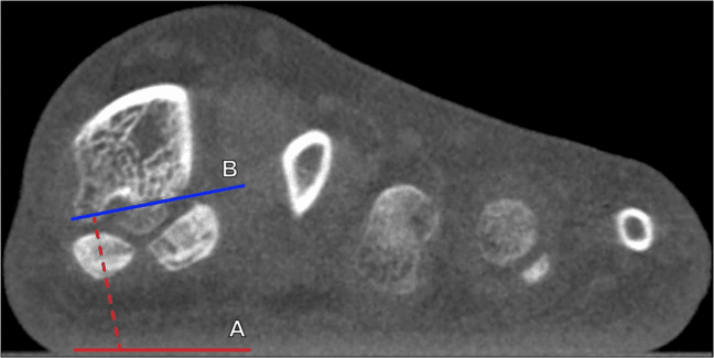
The complex. The hallucal sesamoids are two small ovoid bones embedded within the tendons of flexor hallucis brevis (FHB), plantar to the first metatarsal head. The dorsal surface of each articulates with the crista on the plantar metatarsal head; the plantar, non-articular surface gives attachment to the plantar plate and FHB. Six soft-tissue structures relate to them, and their attachments explain both the injuries and the deformities that follow excision.
- Relationship to Sesamoids
- Medial head encases tibial sesamoid; lateral head encases fibular
- Clinical Significance
- Main stabiliser - repair mandatory during sesamoidectomy
- Relationship to Sesamoids
- Strong fibrous band connecting the two sesamoids
- Clinical Significance
- Prevents separation; rupture occurs in severe turf toe
- Relationship to Sesamoids
- Passes in groove between sesamoids plantarly
- Clinical Significance
- Protected from ground contact; can develop tendinitis
- Relationship to Sesamoids
- Inserts partially on tibial sesamoid medially
- Clinical Significance
- Tibial excision allows valgus drift from loss of medial pull
- Relationship to Sesamoids
- Inserts on fibular sesamoid laterally
- Clinical Significance
- Fibular excision causes varus from unopposed abductor
- Relationship to Sesamoids
- Thick fibrocartilaginous structure attaching to sesamoids
- Clinical Significance
- Turf toe involves plantar plate rupture with sesamoid retraction
Tibial and fibular. The tibial sesamoid carries more of the load and is far more often the one that fractures. Which of the two is removed decides the direction of any drift that follows.
- Tibial (Medial) Sesamoid
- Typically more elongated
- Fibular (Lateral) Sesamoid
- More rounded
- Tibial (Medial) Sesamoid
- Medial, more prominent plantarly
- Fibular (Lateral) Sesamoid
- Lateral, less prominent
- Tibial (Medial) Sesamoid
- Approximately 60% of the sesamoid complex load
- Fibular (Lateral) Sesamoid
- Approximately 40%
- Tibial (Medial) Sesamoid
- 25 times more likely to fracture than the fibular
- Fibular (Lateral) Sesamoid
- Fractures and AVN less common
- Tibial (Medial) Sesamoid
- Hallux valgus risk
- Fibular (Lateral) Sesamoid
- Hallux varus risk
- Tibial (Medial) Sesamoid
- Equal to the fibular sesamoid on injection studies (Sobel 1992)
- Fibular (Lateral) Sesamoid
- Equal to the tibial sesamoid (Sobel 1992)
Blood supply. The sesamoids are supplied from the first plantar metatarsal artery (Rath 2009). Sobel's injection study found two major entry sources, proximal at the FHB attachment and plantar near the midline, with a minor distal contribution, and both sesamoids equally well vascularised. The watershed pattern means distal pole and mid-substance fractures can compromise perfusion and predispose to avascular necrosis and nonunion; at surgery, the proximal and plantar surfaces should be respected to preserve it.
Function. The sesamoids do four jobs in the first ray:
- Mechanical advantage - they lift the FHB tendon away from the joint centre and increase its moment arm
- Load absorption - 2-3 times body weight in walking, up to 300% during running
- FHL protection - they elevate the metatarsal head and shield the FHL tendon from compressive forces
- Weight distribution - they spread force across the metatarsal head during gait
The windlass. At toe-off, hallux dorsiflexion tightens the plantar fascia, raising the arch and depressing the first metatarsal head, which greatly increases the force transmitted through the sesamoid complex. Any pathology that makes the sesamoids painful disrupts this mechanism, and the compensatory gait changes can lead to transfer metatarsalgia.
Pathophysiology
Overuse. Sesamoiditis is an inflammatory response to chronic repetitive loading. In athletes who repeat toe-off, in ballet, running and jumping, cumulative microtrauma exceeds the tissue's healing capacity, and the result is:
- Bone marrow oedema visible on MRI
- Periosteal inflammation
- Soft-tissue swelling in the FHB tendons and plantar plate
- Bursitis between sesamoid and metatarsal head
Stress fracture. When cyclical loading exceeds the bone's capacity to remodel, a stress fracture develops through a predictable sequence:
- Stress reaction - increased osteoclastic activity creates microscopic trabecular damage
- Cortical disruption - a fracture line appears as remodelling fails to keep pace
- Propagation - continued loading extends the fracture through the cortex
- Displacement - in severe cases, a complete cortical break with fragment separation
Acute fracture. High-energy axial loading (forced dorsiflexion, direct impact) exceeds the bone's ultimate tensile strength and fractures it at once. How far it displaces depends on the magnitude of the force and the integrity of the surrounding soft-tissue envelope.
Why the tibial sesamoid. It is the more frequently injured of the two, and the reason is mechanical: it sits directly beneath the first metatarsal head in the line of weight transfer and carries the larger share of the load. Its larger size creates greater stress concentration, and first ray plantarflexion drives it into ground contact. Sobel found both sesamoids equally well vascularised, so the widely repeated claim that the tibial sesamoid has a poorer blood supply is not supported; what determines AVN risk is where the fracture line sits relative to the proximal and plantar vascular entry points, not which sesamoid it is.
Avascular necrosis. AVN develops when the blood supply is critically compromised, most commonly by:
- Fracture - distal pole fractures disrupt the proximal vascular pedicle
- Idiopathic - gradual vascular insufficiency from chronic repetitive trauma
- Surgery - excessive soft-tissue stripping
The sequence then runs from osteocyte death, through failed repair in persistently ischaemic bone, to subchondral collapse as necrotic bone gives way under load. Fragmentation and sclerosis follow as the body attempts to revascularise, and secondary degenerative change develops in the metatarsosesamoid joint.
Arthritis. Chronic overload or post-traumatic change degrades the cartilage of the metatarsosesamoid articulation. Progressive joint-space narrowing, osteophyte formation and subchondral sclerosis follow the usual osteoarthritic pattern.
Why they heal poorly. The blood supply is limited and the loading constant during walking. The bone is small, which limits revascularisation, it sits embedded in a tendon that creates shear forces across it, and its plantar soft-tissue coverage is poor.
Classification Systems
Classification is descriptive, by aetiology and imaging, along a continuum from normal variant to surgical condition.
- Mechanism
- Repetitive microtrauma, overuse inflammation
- Imaging Features
- Radiographs normal; MRI shows bone marrow oedema
- Treatment Approach
- Conservative: offloading, NSAIDs, activity modification
- Mechanism
- Cyclical loading exceeds bone remodelling capacity
- Imaging Features
- Fine cortical break; MRI high T2 signal with surrounding oedema
- Treatment Approach
- NWB immobilisation 6-8 weeks in walking boot
- Mechanism
- Single high-energy traumatic event
- Imaging Features
- Sharp irregular fracture line, possible displacement
- Treatment Approach
- Cast immobilisation vs ORIF if displaced over 2mm
- Mechanism
- Vascular compromise, often post-fracture
- Imaging Features
- Sclerosis, fragmentation, collapse, loss of height
- Treatment Approach
- Sesamoidectomy if symptomatic and failed conservative
- Mechanism
- Congenital variant (multipartite ossification)
- Imaging Features
- Smooth corticated margins, bilateral 85%, larger volume
- Treatment Approach
- Usually asymptomatic; conservative if symptomatic synchondrosis
- Mechanism
- Degenerative changes in metatarsosesamoid articulation
- Imaging Features
- Joint space narrowing, osteophytes, subchondral sclerosis
- Treatment Approach
- Conservative first; sesamoidectomy or shaving if severe
Clinical Assessment
History. Onset and mechanism usually point to the pathology. Insidious onset suggests sesamoiditis or a stress fracture, and acute onset a traumatic fracture or turf toe. Forced dorsiflexion points to turf toe, repetitive push-off to sesamoiditis and a single impact to an acute fracture. The pain is localised plantarly and worse in the toe-off phase of gait, and prior episodes suggest chronic overload or a bipartite variant.
- Sesamoiditis: 25-year-old ballet dancer, insidious onset over weeks, worsened by relevé
- Stress Fracture: 30-year-old marathon runner, progressive pain over 2-4 weeks, no specific event
- Acute Fracture: 22-year-old footballer, immediate pain after forced dorsiflexion tackle
- Bipartite: Incidental finding on radiographs for unrelated foot pain, bilateral
Look. Examination localises the pathology and rules out the differential.
- Swelling - mild plantar MTPJ oedema suggests active inflammation
- Erythema - may indicate gout, infection or acute injury
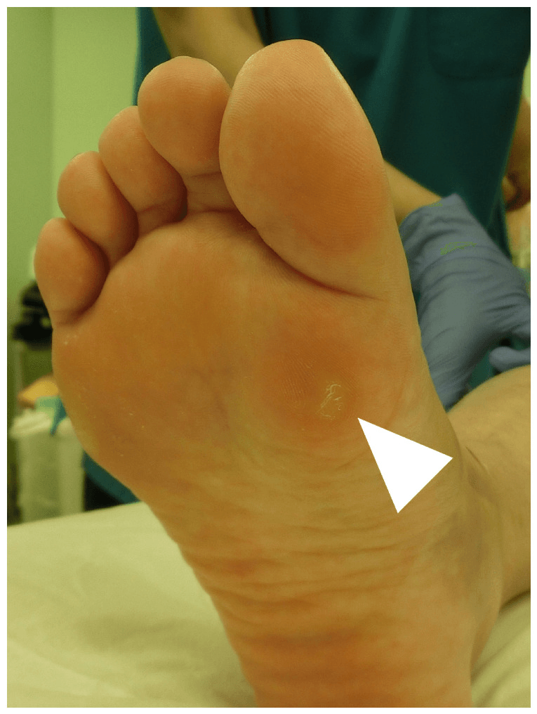
- Callosities - an intractable plantar keratosis (IPK) under a sesamoid indicates prominence
- Alignment - cavus foot, hallux valgus or first ray plantarflexion
- Gait - antalgic, with a shortened stride and early heel rise to avoid push-off
Feel.
- Point tenderness - direct pressure over the specific sesamoid reproduces the pain
- Dorsal or plantar - make sure the pain is plantar (sesamoid), not dorsal (arthritis, gout)
- Joint line - palpate the MTPJ for synovitis or effusion
- FHL excursion - pain with FHL gliding suggests tendinitis
- Neurovascular - sensation (medial plantar nerve) and pulses
Provoke.
- Passive dorsiflexion test - extending the hallux while palpating the sesamoid reproduces pain by loading the complex
- Resisted flexion - active flexion against resistance tests FHB and FHL integrity
- Push-off test - inability to perform a single-leg heel raise indicates significant pain
- Grind test - compression plus rotation reproduces arthritic pain
- Vertical Lachman - dorsal translation indicates plantar plate injury (turf toe)
- Erythema + Fever + Systemic Symptoms: Septic arthritis or osteomyelitis
- Gout Crystals: Aspirate and send for crystal analysis if suspicious
- Neurovascular Compromise: Rare but check pulses and sensation
- Unable to Weight Bear: Suggests displaced fracture or complete plantar plate rupture
Differential diagnosis of sesamoid pain. Turf toe aside (see Classification):
- Trauma - acute fracture, or stress fracture from repetitive loading
- Inflammation - sesamoiditis from overuse, bursitis, gout
- Symptomatic bipartite sesamoid - usually asymptomatic, but the synchondrosis can be painful
- Infection - osteomyelitis (rare) or septic arthritis
- Arthritis or AVN
Investigations
Radiographs first. Weight-bearing films in three views:
- AP foot - sesamoid alignment, fracture diastasis, bipartite margins
- Lateral foot - sesamoid height and proximal migration (turf toe)
- Axial (skyline) sesamoid view - critical for visualising the sesamoid-metatarsal articulation
Always obtain bilateral comparison radiographs to check a two-part sesamoid for symmetry. AVN shows as sclerosis, fragmentation, collapse and loss of height; arthritis as joint-space narrowing, osteophytes and subchondral cysts.
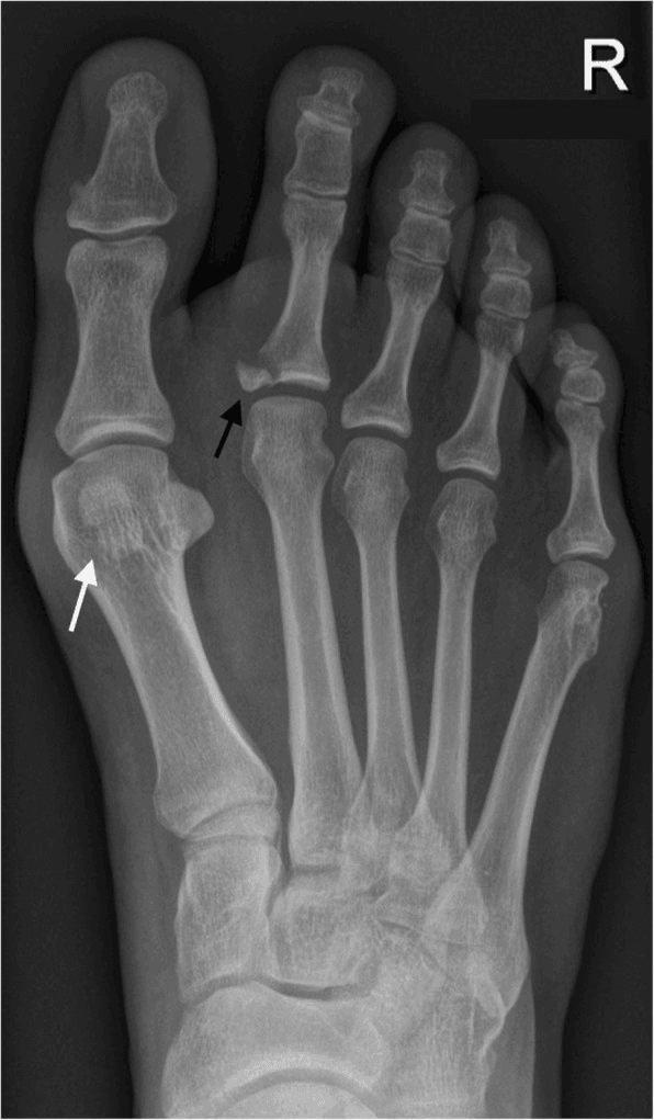
Bipartite or fractured. This is the distinction the films must make. A bipartite sesamoid is a congenital variant, present in 10-30% of the population, and the tibial sesamoid is 10 times more likely to be bipartite than the fibular. A fracture has a sharp, irregular line, is unilateral and shows bone marrow oedema on MRI. Five features point to a bipartite variant instead:
- Smooth, rounded corticated margins, against a sharp jagged fracture line
- Bilateral symmetry - present in both feet in 85% of cases
- Larger total volume - the fragments together exceed a normal single sesamoid
- Unequal fragment sizes - typically one large and one small piece
- Normal MRI signal - no bone marrow oedema unless the synchondrosis is symptomatic
If unsure, MRI is definitive: a fracture shows surrounding oedema, and a bipartite sesamoid does not unless it is symptomatic. An asymptomatic bipartite sesamoid needs no treatment.
MRI. The gold standard for sesamoid pathology, and the definitive test when radiographs are normal or equivocal.
- T1 - low signal indicates oedema, AVN or chronic change
- T2/STIR - high signal confirms acute bone marrow oedema
- Proton density - cartilage integrity and the plantar plate
Each diagnosis has its pattern:
- Sesamoiditis - bone marrow oedema, intact cortex, normal FHB and plantar plate
- Stress fracture - linear T2 hyperintensity, surrounding oedema, cortical disruption
- AVN - low T1 and T2 signal (sclerosis), fragmentation, subchondral collapse
- Symptomatic bipartite - oedema at the synchondrosis; an asymptomatic one shows normal signal
- Turf toe - plantar plate rupture, capsular tear, joint effusion, sesamoid retraction
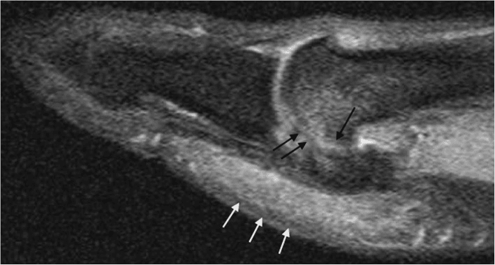
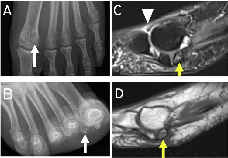
CT and bone scan. CT is best for assessing fracture healing (union or nonunion), bony architecture and surgical planning. A bone scan has high sensitivity for stress reactions and sesamoiditis but low specificity, and is the fallback if MRI is contraindicated.
Management Algorithm
Conservative first, for everything. Over 90% of sesamoid disorders resolve without surgery, and a minimum 3-6 month trial is mandatory before surgery is considered. It succeeds in the vast majority when compliance is good and offloading adequate.
Fractures. A stress fracture is immobilised non-weight-bearing for 6-8 weeks (see Classification). An acute fracture goes into a cast or boot for 6 weeks non-weight-bearing, and ORIF is considered if it is displaced. Phase 1 below gives a shorter non-weight-bearing period.
Stepwise Conservative Protocol
Stop the aggravating activity (running, dancing, jumping) completely and move to non-impact exercise such as swimming or cycling. A stiff-soled or rocker-bottom shoe limits MTPJ extension; a short leg walking (CAM) boot is for acute fractures or severe pain. An acute fracture is kept non-weight-bearing for 2 weeks, then heel weight-bearing.
- Dancer's pad - felt with a U-shaped cutout to float the painful sesamoid
- Sesamoid relief orthotic - a depression under the sesamoid with a metatarsal bar proximal
- Full-length custom orthotic - with first ray extension and arch support
- NSAIDs - ibuprofen 400mg TDS or naproxen 500mg BD for 2-4 weeks
- Ice - 15-20 minutes TDS to reduce inflammation
Physiotherapy addresses the biomechanical factors and rebuilds strength:
- Intrinsic strengthening - towel curls, marble pickups
- Gastrocnemius stretching for equinus contracture
- Arch strengthening - short foot exercises
- FHL and FHB strengthening with resistance bands
A corticosteroid injection is diagnostic and therapeutic for sesamoiditis, placed under fluoroscopic or ultrasound guidance to ensure accuracy. Give 1-2 at most, because of the risk of fat-pad atrophy and tendon weakening, and not in the presence of infection or an acute fracture. Adjuncts are a bone stimulator for stress fractures or delayed healing, contrast baths (vascular flushing) and iontophoresis for dexamethasone penetration.
- Weeks 1-2: walking tolerance without pain
- Weeks 3-4: brisk walking and light jogging
- Weeks 5-8: running progression (50%, 75%, 100% intensity)
- Weeks 9-12: sport-specific drills and return to full activity
The changes are permanent: orthotics indefinitely for impact activities, correction of biomechanical factors (cavus foot, equinus) and footwear that avoids minimalist shoes and high heels.
Surgical Technique
Total sesamoidectomy through a medial approach is described here for the tibial sesamoid, the one most commonly affected.
Before the operation.
- Confirm the diagnosis - MRI showing AVN, nonunion or arthritis
- Document a conservative trial of at least 6 months
- Counsel on hallux valgus (10-15%), cock-up toe and transfer metatarsalgia
- Review the imaging of the remaining sesamoid - avoid a double sesamoidectomy
Setup. Supine, with a sandbag under the ipsilateral hip to prevent external rotation. A thigh tourniquet is preferred, though an ankle tourniquet is acceptable. Anaesthesia is general or a popliteal regional block, the block giving excellent postoperative pain control, and a mini C-arm should be available to confirm complete excision.
Operative Steps
The medial approach is preferred: a longitudinal 3-4cm incision centred over the medial aspect of the MTPJ, deepened to the capsule in line with the skin. It avoids a direct plantar incision and its painful weight-bearing scar. Identify and protect the dorsomedial cutaneous nerve, retracting it dorsally.
The plantar approach is the alternative, reserved for plantar pathology needing direct access: a curvilinear incision plantar to the sesamoid, between the weight-bearing areas. It gives direct visualisation at a higher risk to the medial plantar nerve branches and a potentially painful scar. The choice depends on the location of the pathology and surgeon preference.
- Longitudinal capsulotomy along the sesamoid axis
- Identify the sesamoid within the FHB tendon substance
- Divide the metatarsosesamoid (suspensory) ligament
- Inspect the metatarsal head articular surface for arthritis
- Assess plantar plate integrity
The sesamoid is embedded in the tendon, not attached to it, so careful "shelling out" preserves the flexor mechanism. This is the most critical step for FHB function.
- Use a Beaver blade (#64 or #67) or a small #15 scalpel
- Dissect the sesamoid carefully from the surrounding FHB fibres
- Preserve as much tendon as possible - do not excise tendon with the bone
- Remove every bony fragment, checking with fluoroscopy
- Inspect the FHL tendon deep to the sesamoid for pathology
Tendon repair is the most important step in preventing complications. Close the longitudinal defect in the FHB with #1 Vicryl or PDS, figure-of-8 or horizontal mattress, restoring tension to prevent cock-up deformity, and make sure the hallux can plantarflex actively before closure.
Then balance the soft tissues and test alignment: the hallux should sit in neutral without drift.
- Tibial sesamoidectomy - consider abductor hallucis release if there is pre-existing valgus
- Fibular sesamoidectomy - consider adductor hallucis release to prevent varus
- Capsule with 2-0 Vicryl, to restore static stability
- Subcutaneous layer with 3-0 Monocryl
- Skin with 3-0 or 4-0 nylon, or Monocryl subcuticular
- Sterile dressing and a post-op shoe or walking boot
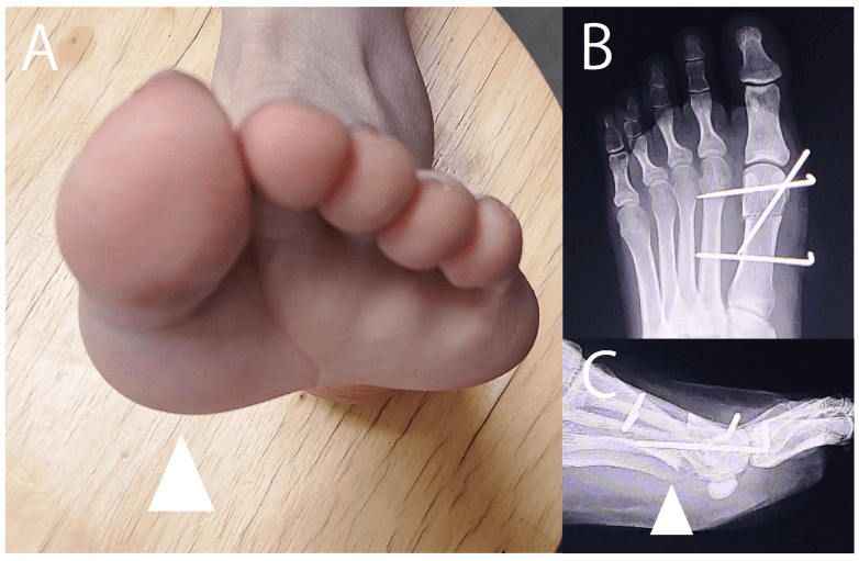
Failure to repair the FHB tendon defect is the single most common cause of cock-up toe deformity after sesamoidectomy. The intrinsic flexor power is lost, and unopposed extensor pull causes MTP hyperextension and IP flexion (claw toe).
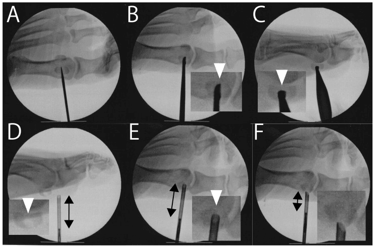
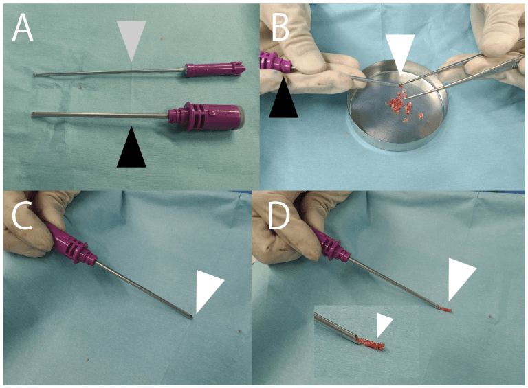
Sesamoid Shaving (Planing)
Indication. A painful plantar prominence or intractable plantar keratosis under the tibial sesamoid, with a preserved metatarsosesamoid joint and no fracture, AVN or arthritis: the problem is mechanical prominence, not intra-articular disease. It is the least destructive surgical option and preserves the sesamoid and the FHB.
Technique. Through a medial or plantar-medial approach the sesamoid is exposed within the FHB without shelling it out. A burr or osteotome shaves the plantar (non-articular) half flush, reducing the prominence while leaving the dorsal articular surface and the FHB attachment intact. Because the FHB is not divided, the flexor mechanism and the medial buttress are preserved, avoiding the hallux valgus and cock-up risks of total excision.
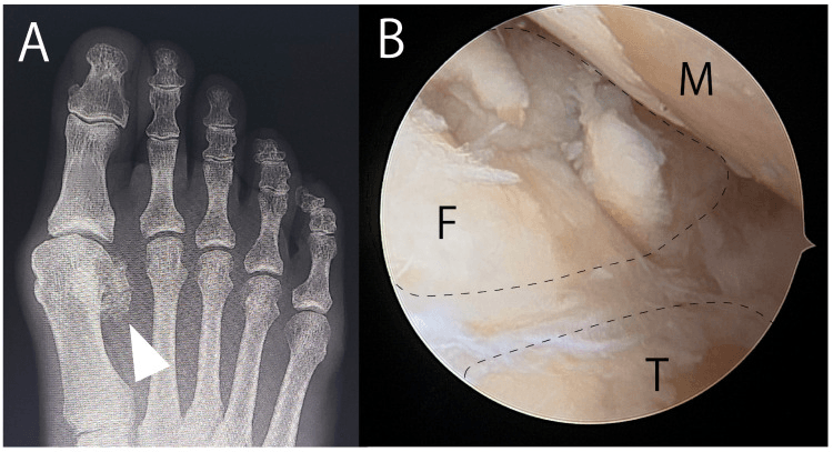
Endoscopic shaving. A medial portal and blunt plantar dissection establish access, fluoroscopy controls resection of the plantar half with an abrasion burr, and endoscopy confirms a smooth residual surface without FHL injury.
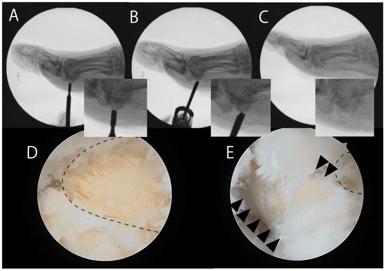
Aftercare and pitfalls. Offloading is similar to the non-operative protocol, with an earlier return than after excision. The main risks are inadequate debridement (persistent keratosis), fracture of the thinned sesamoid and violation of the articular surface. Shaving does not help if the pain is intra-articular (arthritis or AVN), where excision is required.
Unloading osteotomy. A plantar prominence of the first metatarsal head can instead be addressed with a dorsoproximal sliding osteotomy, which unloads the sesamoid complex without excising it; overcorrection risks transfer metatarsalgia.
Partial Sesamoidectomy
What it is. Removal of only the diseased fragment of a sesamoid, typically the smaller, avascular or ununited pole, while retaining the viable fragment and the FHB attachment. It is used for a fractured or symptomatic bipartite sesamoid where one pole is diseased, or for a stress-fracture nonunion of a bipartite sesamoid, most often as the proximal-fragment excision described for medial (tibial) sesamoid stress fractures.
Why it helps. Preserving the larger viable pole and the FHB maintains more of the flexor lever and the medial or lateral buttress than total excision. That reduces the risk of hallux valgus or varus and cock-up deformity while still removing the pain source, a middle option between planing and total sesamoidectomy.
When it is enough. Only when one pole is clearly the pain source and the retained pole is viable; if the whole sesamoid is necrotic or arthritic, total excision with FHB repair is required. Biedert and Hintermann excised the proximal fragment in young athletes with medial sesamoid stress fractures, and all were pain-free and back to full sport within six months.
Partial (Proximal-Fragment) Sesamoidectomy for Medial Sesamoid Stress Fracture
- 5 athletes (6 feet, mean age 16.8 years), mostly rhythmic gymnasts, with medial great-toe sesamoid stress fractures
- Bone scan and axial/sagittal CT were more reliable than plain films or MRI for confirming the fracture
- After failed conservative care, excision of the proximal fragment (partial sesamoidectomy) was performed in all
- All patients were pain-free and returned to full sport within six months with no complications (mean AOFAS 95.3)
Complications
- Mechanism
- Tibial sesamoidectomy removes medial buttress; unopposed lateral pull from adductor
- Prevention Strategy
- Meticulous FHB repair; consider abductor release if pre-existing valgus
- Management
- Early: orthotic and taping; Late: distal metatarsal osteotomy or MTP fusion
- Mechanism
- Fibular sesamoidectomy removes lateral stabiliser; unopposed medial pull
- Prevention Strategy
- FHB repair; consider adductor release at time of excision
- Management
- EHL split transfer (reverse Jones) or MTP fusion if rigid
- Mechanism
- Loss of FHB flexor function from inadequate tendon repair
- Prevention Strategy
- Robust tendon repair with #1 Vicryl; test active plantarflexion before closure
- Management
- Girdlestone-Taylor FDL transfer or MTP fusion
- Mechanism
- Loss of first ray weight-bearing; overload of lesser metatarsals
- Prevention Strategy
- Custom orthotic with met pad; avoid excessive first ray elevation
- Management
- Metatarsal osteotomies (Weil) to equalise metatarsal parabola
- Mechanism
- Injury to medial plantar nerve or dorsomedial cutaneous nerve
- Prevention Strategy
- Careful dissection; early identification and retraction of nerves
- Management
- Nerve excision proximal to weight-bearing area; bury in muscle
- Mechanism
- Incomplete excision of fragments; arthritis in remaining sesamoid
- Prevention Strategy
- Fluoroscopy to confirm complete removal; assess other sesamoid preop
- Management
- Revision excision or contralateral sesamoidectomy (if necessary)
- Mechanism
- Capsular fibrosis, prolonged immobilisation
- Prevention Strategy
- Early ROM exercises (2 weeks postop); aggressive PT
- Management
- Manipulation under anaesthesia; capsular release if severe
How often. Published series report:
- Overall satisfaction 85-95% at 2-year follow-up
- Hallux valgus or varus 5-15% (reduced with modern repair techniques)
- Cock-up toe 5-10% (preventable with FHB repair)
- Transfer metatarsalgia 10-20% (managed with orthotics)
- Neuroma 5% (nerve protection critical)
Shimozono's systematic review, in the Evidence Base, found an overall complication rate of 22.5%, although the mean hallux valgus angle did not change significantly. The key to minimising complications is meticulous technique, with emphasis on preserving and repairing the FHB tendon.
Postoperative Care and Rehabilitation
Rehabilitation Protocol After Sesamoidectomy
A post-op stiff-soled shoe or short leg walking boot, with heel weight-bearing only to avoid push-off, and strict elevation to prevent haemarthrosis and swelling. Keep the wound clean and dry, watch for infection, and remove sutures or staples at 14 days.
Early mobilisation prevents stiffness.
- Passive and active MTPJ range of motion from 2 weeks, avoiding aggressive dorsiflexion to protect the repair
- FHL excursion exercises (toe curls, towel grabs)
- Progress from heel weight-bearing to full forefoot loading in a stiff-soled shoe, weaning from the boot by week 4-6 according to pain
- Scar massage and desensitisation (especially plantar incisions) and silicone sheeting to minimise hypertrophic scarring
- Physiotherapy - intrinsic strengthening (short foot exercises), gastrocnemius and soleus stretching, proprioception (single-leg balance), progressive resistance
- Footwear - a supportive athletic shoe, a custom orthotic with a sesamoid cutout or dancer's pad, and a rocker-bottom sole to reduce MTPJ extension forces
- Month 3: unlimited walking, stationary cycling, swimming
- Month 4: light jogging, elliptical trainer
- Month 5: running progression (50%, 75%, 100% intensity)
- Month 6: sport-specific drills, return to full activity
Watch for hallux drift (valgus or varus), which early taping can correct; for transfer metatarsalgia, which calls for orthotic adjustment; and for lost push-off power, which calls for FHL and FHB strengthening. Orthotics continue indefinitely for impact sports, with no minimalist footwear or high heels and gastrocnemius flexibility maintained.
After bone grafting. Rehabilitation is stricter and return to sport slower than after sesamoidectomy:
- Strict non-weight-bearing for 8 weeks in a short leg cast or boot
- Radiographs at 6 and 12 weeks to confirm union
- A weight-bearing boot from week 8 to 12
- Full weight-bearing by 12 weeks if healed
- Return to sport at 6-9 months
Guidelines, Registries & Global Practice
Global Epidemiology
- Population at risk: Predominantly athletes and dancers worldwide. Sesamoid injuries account for roughly 9% of foot and ankle injuries in athletic populations and up to 12% of great-toe injuries.
- Sport distribution (international): Running and sprinting (push-off loading), ballet and dance (relevé), football/soccer and rugby (forced dorsiflexion), basketball and gymnastics (jump landings), and cricket fast bowling (repetitive forefoot impact).
- Bipartite prevalence: 10-30% of the general population; bilateral in around 85%; tibial sesamoid roughly 10 times more likely to be bipartite than fibular.
- Fracture distribution: Tibial-to-fibular fracture ratio approximately 25:1, reflecting greater medial load-bearing.
Side-by-Side Society Guidance
There is no dedicated AAOS or NICE clinical practice guideline specific to sesamoid disorders; recommendations are extrapolated from foot and ankle society consensus, AO Foundation principles, and sports-medicine bodies. Where guidance genuinely differs, it is on the threshold for surgery and choice of preservation vs excision.
- Emphasis
- Preservation-first; biomechanical balance
- Practical Recommendation
- Prolonged offloading trial; sesamoid-preserving fixation/grafting before excision; mandatory FHB repair if excising
- Emphasis
- Conservative trial then staged surgery
- Practical Recommendation
- Minimum 3-6 months non-operative care; MRI confirmation before resection; avoid double sesamoidectomy
- Emphasis
- Return-to-sport optimisation in elite athletes
- Practical Recommendation
- Internal fixation favoured for stress-fracture nonunion to preserve push-off; sport-specific counselling
- Emphasis
- Individualised, stepwise algorithm
- Practical Recommendation
- Tailored decision (Artioli 2025); excision reserved for refractory symptomatic nonunion or AVN
Registry and Evidence Notes
- No national joint registry captures sesamoid procedures (they are non-arthroplasty), so the evidence base is systematic reviews of case series rather than registry survivorship data.
- Best available syntheses: Shimozono 2018 (sesamoidectomy, 196 feet), Robertson 2017 (stress-fracture return to sport), and Artioli 2025 (nonunion algorithm).
High- vs Limited-Resource Practice Variation
- High-resource settings: Early MRI and CT for diagnosis; custom orthoses; access to arthroscopic and sesamoid-preserving fixation/grafting; bone stimulators for delayed union.
- Limited-resource settings: Diagnosis often radiograph-based with contralateral comparison films; conservative management with stiff-soled footwear and felt dancer's pads; open sesamoidectomy is the dominant surgical option where preservation techniques or implants are unavailable.
Universal Consent Points for Sesamoidectomy
- Hallux valgus (after tibial excision) or varus (after fibular excision)
- Cock-up toe deformity from inadequate FHB repair
- Transfer metatarsalgia to the lesser rays
- Neuroma (medial plantar or dorsomedial cutaneous nerve)
- Loss of push-off power, career-relevant for sprinters and dancers
- Possible revision surgery
- Always document a minimum 3-6 month conservative trial and MRI-confirmed pathology, and discuss preservation alternatives in athletes.
Related pages: Turf Toe is the acute plantar-plate injury that shares this anatomy and mechanism, and the injury in which a proximally migrated sesamoid on the lateral radiograph tells you the complex has ruptured; Plantar Plate Insufficiency for the same structure failing chronically at the lesser toes; Hallux Valgus for the deformity that both causes sesamoid pain through subluxation of the complex and follows tibial sesamoidectomy - Shimozono found the mean angle change non-significant, but the individual deformity is the reason both sesamoids are never excised; Hallux Rigidus for the other cause of a painful stiff first MTP joint that must be excluded before the pain is attributed to a sesamoid; Metatarsalgia and Metatarsal Stress Fractures for the neighbouring forefoot pain generators and the same overload physiology; Freiberg Disease for the osteonecrosis analogue at the lesser metatarsal head; and Stress Fractures Overview for the bone-stress framework this sits inside.
Controversies and Areas of Uncertainty
The evidence base for sesamoid disorders is composed almost entirely of small case series and systematic reviews of them; no randomised trials and no registry data exist. Several genuine areas of debate remain.
- Argument For
- Preservation maintains FHB power and avoids deformity; union 90-100% in series
- Argument Against / Caveat
- Technically demanding, prolonged NWB, persistent nonunion risk; excision more predictable for pain
- Argument For
- Older series quote low deformity rates with good FHB repair
- Argument Against / Caveat
- Shimozono 2018 found an overall complication rate of 22.5%, higher than commonly taught
- Argument For
- Classic teaching: blood enters the proximal pole only
- Argument Against / Caveat
- Sobel 1992 shows proximal AND plantar are both major sources; distal is minor
- Argument For
- Diagnostic and therapeutic for refractory sesamoiditis
- Argument Against / Caveat
- Risk of fat-pad atrophy and tendon weakening; no high-level evidence of durable benefit
- Argument For
- Internal fixation gives best return-to-sport in athletes (Robertson 2017)
- Argument Against / Caveat
- Optimal modality undefined; many resolve with 2-6 months conservative care
MCQ Practice Points
Q: Which sesamoid is most commonly affected by fracture and AVN?
A: Tibial (medial) sesamoid - 25:1 ratio compared to fibular sesamoid. This is due to increased weight-bearing forces (tibial bears 60% of load), more prominent plantar position, and more variable vascular supply with higher AVN risk.
Q: What percentage of the population has bipartite sesamoids?
A: 10-30% of the population has bipartite sesamoids, and they are bilateral in 85% of cases. The tibial sesamoid is 10 times more likely to be bipartite than the fibular sesamoid. This is why bilateral comparison radiographs are mandatory when evaluating suspected sesamoid fractures.
Q: Why are distal pole sesamoid fractures at higher risk for AVN?
A: The blood supply enters the sesamoid via the proximal pole (branches of medial plantar artery and plantar arch). Fractures through the distal pole disrupt this vascular pedicle, creating a distal fragment with compromised blood supply that is prone to avascular necrosis and nonunion.
Q: Which sesamoid excision leads to hallux valgus deformity?
A: Tibial (medial) sesamoidectomy leads to hallux valgus. The tibial sesamoid and medial FHB act as a medial stabilizer. Excision removes this buttress, allowing unopposed lateral pull from the adductor hallucis, causing valgus drift. Prevention: meticulous FHB repair and consider prophylactic abductor hallucis release.
Q: What percentage of sesamoid disorders resolve with conservative management?
A: The majority of sesamoid disorders resolve with adequate conservative management (offloading orthotics, activity modification, NSAIDs, stiff-soled shoes). A minimum 3-6 month trial is mandatory before considering surgical intervention. Robertson et al. (2017) endorse a 2-6 month non-operative pathway before any surgery for sesamoid stress fractures.
Exam Viva Scenarios
Practise clinical reasoning and management decisions out loud
“A 22-year-old professional ballet dancer presents with 4 months of progressive pain under the great toe. Worse in relevé position. No specific trauma. Exam shows point tenderness over the medial plantar MTPJ and pain with passive dorsiflexion. Weight-bearing radiographs show a two-part medial sesamoid with smooth margins.”
“A 28-year-old professional footballer sustained a tibial sesamoid fracture 6 months ago. He was treated in a boot NWB for 8 weeks then progressive weight-bearing. He remains symptomatic with pain on push-off. CT scan shows a persistent 2mm fracture gap with sclerotic margins suggesting nonunion. MRI shows no AVN. He is desperate to return to professional sport.”
“A 35-year-old patient returns 18 months after tibial sesamoidectomy for chronic sesamoiditis. She complains that her big toe is drifting outward. On examination, there is 25 degrees of hallux valgus with a positive drawer test (MTP joint instability). Radiographs confirm progressive valgus deformity compared to immediate postoperative films.”
Key Anatomy
- Two sesamoids: Tibial (medial) and Fibular (lateral)
- Embedded in FHB tendons, articulate with MT head crista
- Blood supply: Proximal pole entry (medial plantar artery)
- FHL tendon runs in groove between sesamoids
- Tibial bears 60% load, Fibular 40%
Pathology Spectrum
- Sesamoiditis: Inflammation/overuse, normal X-ray, MRI edema
- Stress Fracture: Cortical break, MRI linear T2 high signal
- Acute Fracture: Sharp irregular line, unilateral
- AVN: Sclerosis, fragmentation, low T1/T2 signal
- Bipartite: Smooth margins, bilateral 85%, normal MRI
Bipartite vs Fracture
- Smooth corticated margins vs sharp jagged fracture line
- Bilateral 85% - always X-ray BOTH feet
- Larger total volume (sum of parts exceeds normal)
- MRI normal signal (bipartite) vs edema (fracture)
Conservative Management (90% success)
- Dancer's pad with U-shaped cutout to offload sesamoid
- Stiff-soled shoe or CAM boot (NWB 6 weeks for fractures)
- Activity modification: stop aggravating activity
- NSAIDs, ice, physiotherapy
- Minimum 3-6 month trial before surgery
Surgical Indications
- Failed conservative minimum 6 months
- AVN with fragmentation/collapse
- Symptomatic nonunion
- Displaced acute fracture over 2mm (ORIF consideration)
Sesamoidectomy Technique (Critical)
- Medial approach (protect dorsomedial cutaneous nerve)
- Shell out sesamoid preserving FHB tendon
- Repair FHB with #1 Vicryl (MANDATORY)
- Soft tissue balancing (abductor release if valgus risk)
- Test active plantarflexion before closure
Sesamoidectomy Complications
- Cock-up toe: Loss of FHB function (prevent with repair)
- Hallux Valgus: After tibial excision (loss medial buttress)
- Transfer metatarsalgia (10-20%)
- Varus deformity: After fibular excision (unopposed abductor)
Exam Pearls
- Tibial:Fibular fracture ratio is 25:1
- Bipartite bilateral in 85% - check other foot
- Distal pole fractures high AVN risk (blood enters proximal)
- FHB repair is critical step preventing cock-up toe
- Sesamoidectomy 85-95% satisfaction but alters biomechanics
Evidence Base
Sesamoidectomy for Hallux Sesamoid Disorders: Systematic Review
- 10 studies, 196 feet; mean age 36.6 years, mean follow-up 45.1 months
- Mean VAS improved from 6.5 to 1.2; mean postoperative AOFAS 92.7
- 94.4% returned to sport (90.0% to previous level) at mean 11.8 weeks
- Hallux valgus angle change non-significant (13.1 to 14.8 degrees, p=0.47)
- Overall complication rate 22.5%; revision rate only 3.0%
Return to Sport After Stress Fractures of the Great Toe Sesamoids
- 14 studies of conservatively and surgically managed great-toe sesamoid stress fractures
- Standard pathway: 2-6 month conservative trial (activity modification, orthotics, analgesia, physiotherapy)
- Surgery (internal fixation or sesamoidectomy) reserved for persistent symptoms
- Internal fixation showed the best return-to-full-sport rates with low complication rates
- Optimal treatment modality not yet defined; evidence base remains low quality
Autogenous Bone Grafting of Hallux Sesamoid Nonunions
- 21 patients (mean age 33 years) with symptomatic tibial hallux sesamoid nonunion
- Autogenous bone grafting achieved bony union in all but 2 patients
- Most patients obtained symptom relief and returned to pre-injury activity level
- Procedure preserves the sesamoid and flexor hallucis brevis mechanism
- Proposed as an alternative to excision in selected, viable-bone cases
Hallux Sesamoid Nonunion: Comprehensive Systematic Review
- 6 studies, 80 patients; PRISMA-compliant synthesis of sesamoid nonunion surgery
- Techniques: open/arthroscopic sesamoidectomy, bone grafting +/- screw fixation, percutaneous screw fixation
- Union rates 90.5% to 100% with consistent functional improvement
- Complication and reoperation rates both 6.5%; commonest reoperation was salvage sesamoidectomy
- Supports a stepwise, preservation-first algorithm with excision reserved for refractory cases