Latissimus Dorsi's Little Helper and the Floor of Three Spaces
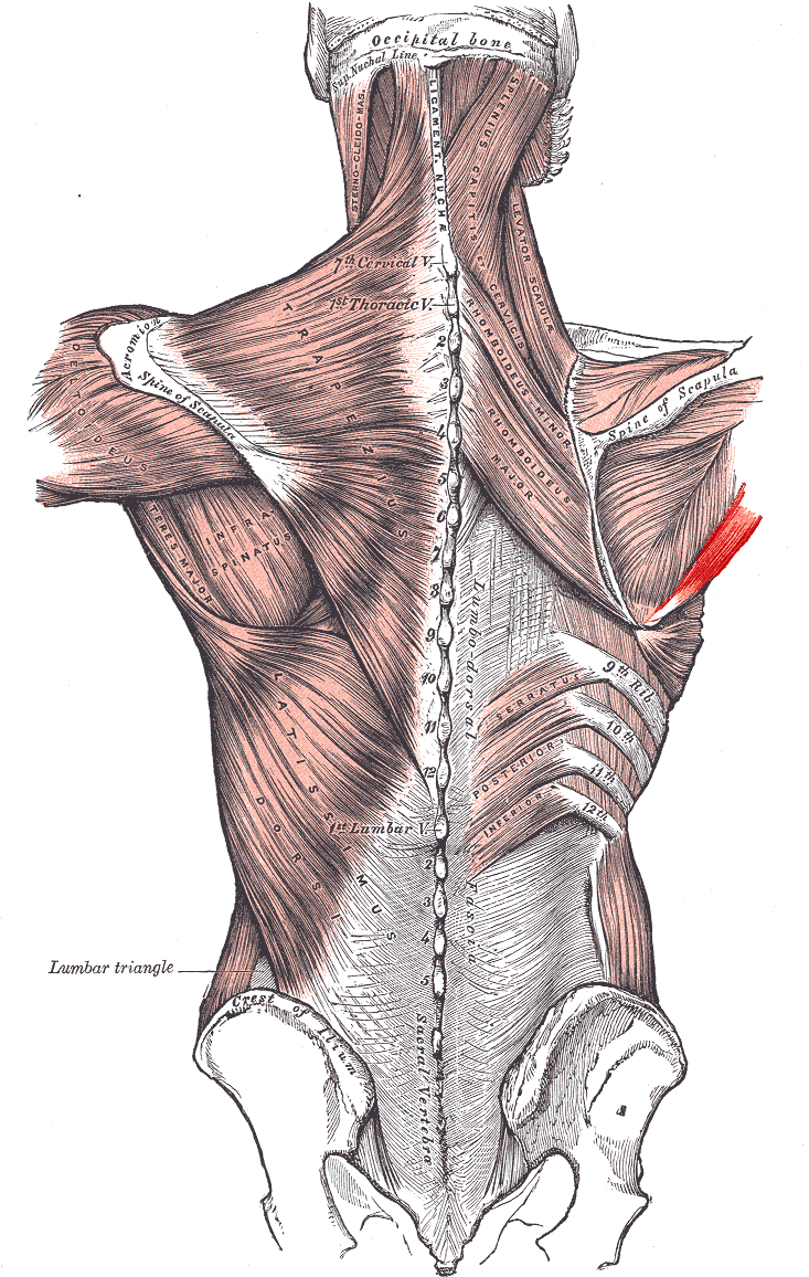
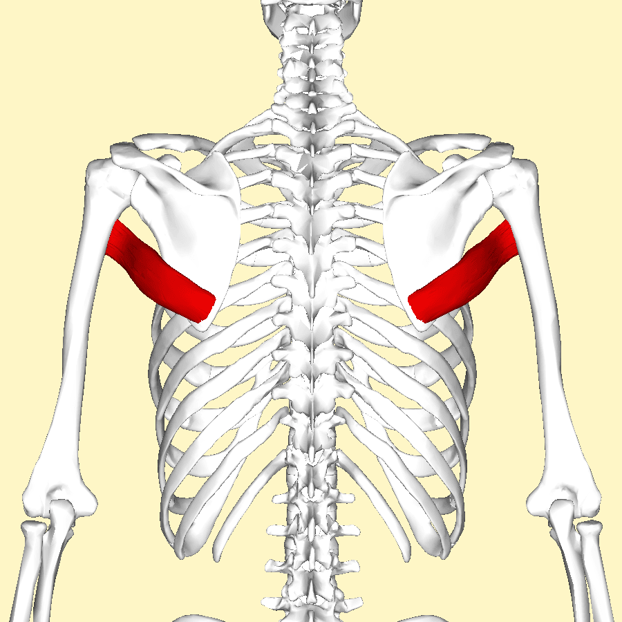
- Origin: oval area on the dorsal surface of the inferior angle and lower third of the lateral border of the scapula.
- Insertion: medial lip of the intertubercular (bicipital) groove — the crest of the lesser tubercle.
- Innervation: lower subscapular nerve (C5, C6, sometimes C7) from the posterior cord, which also supplies the lower subscapularis.
- It forms the inferior boundary of both the quadrangular and triangular spaces, and the superior boundary of the triangular interval.
- With latissimus dorsi it forms the posterior axillary fold; its lower border marks where the axillary artery becomes the brachial artery.
- “Lady between two majors: the latissimus dorsi tendon inserts into the floor of the groove, between pectoralis major on the lateral lip and teres major on the medial lip.
- “Teres major is NOT a rotator cuff muscle — it does not insert on a tuberosity facet and does not blend with the capsule.
- “The radial nerve enters the triangular interval at the lower border of teres major, which is the nerve at risk during transfer harvest.
- “L'Episcopo transfers latissimus dorsi and teres major from the medial lip to the posterolateral humerus, converting internal rotators into external rotators.
Overview
The teres major runs from the inferior angle of the scapula to the medial lip of the bicipital groove, sandwiched between the teres minor above and the latissimus dorsi wrapping around it below and in front. Functionally it is a modest muscle: electromyographic studies show it is essentially silent during unresisted movement and fires only against load, which is why it is described as "latissimus dorsi's little helper".
Anatomically and surgically it is anything but modest. It is the floor of the quadrangular space and the triangular space, and the roof of the triangular interval — three named spaces containing, respectively, the axillary nerve, the circumflex scapular artery and the radial nerve. Its lower border is the boundary at which the axillary artery becomes the brachial artery and at which the radial nerve leaves the axilla for the spiral groove. And with the latissimus dorsi it forms the posterior axillary fold and the standard donor pair for restoring external rotation to a shoulder that has lost it.
Examiners set this trap constantly, and the answer must be given with reasons rather than as an assertion.
- It does not insert on a tuberosity facet. The cuff muscles insert on the three facets of the greater tuberosity (supraspinatus, infraspinatus, teres minor) or the lesser tuberosity (subscapularis). Teres major inserts on the medial lip of the bicipital groove, distal and anterior to all of them.
- It does not blend with the joint capsule. Every cuff tendon is inseparable from the capsule and contributes to the cuff-capsule hood. Teres major has a distinct tendon separated from the joint by the latissimus dorsi tendon and an intervening bursa.
- It does not compress the head. Its line of pull produces adduction, extension and internal rotation of the shaft; it does not contribute to concavity compression and therefore plays no part in the transverse force couple.
- Its innervation is different. The cuff is supplied by the suprascapular, subscapular and axillary nerves; teres major shares the lower subscapular nerve with the inferior subscapularis, a detail that means a lower subscapular nerve injury weakens internal rotation twice over.
Lady between two MajorsThe Bicipital Groove Insertions
Hook:Also fixes the harvest anatomy: at the groove the latissimus tendon lies immediately anterior and superficial to teres major, separated by a bursa.
Attachments, Innervation and Relations
Origin
- An oval area on the dorsal (posterior) surface of the inferior angle of the scapula and the adjacent lower third of the lateral border.
- Fibres also arise from the fibrous septa separating it from the teres minor above and from the infraspinatus fascia.
- The muscle runs superolaterally and anteriorly, spiralling forward to reach the front of the humerus, which is why a posteriorly originating muscle produces internal rotation.
Insertion
- Medial lip of the intertubercular (bicipital) groove of the humerus — also described as the crest of the lesser tubercle.
- The tendon is short and broad, roughly 5 cm long and 1.5 to 2 cm wide, and lies posterior and slightly distal to the latissimus dorsi tendon at the groove.
- A constant bursa separates the teres major and latissimus dorsi tendons at the insertion. Finding and opening this bursa is the reliable way to separate the two tendons during a transfer harvest.
- The relationship at the groove, from lateral to medial: pectoralis major on the lateral lip, latissimus dorsi in the floor, teres major on the medial lip.
The posterior axillary fold
- The latissimus dorsi and teres major together form the posterior axillary fold, the palpable ridge that closes the axilla behind. This is the structure released with Z-plasty or flap reconstruction in post-burn axillary contracture, and the structure grasped when a surgeon palpates the muscles for a transfer.
At the upper border.
- Runs in the quadrangular space, whose floor is the upper border of teres major.
- At risk when the interval between teres minor and teres major is opened, and during proximal mobilisation of a transfer.
- Loss costs deltoid, teres minor and lateral arm sensation.
At the lower border.
- Enters the triangular interval at the lower border of teres major and passes into the spiral groove.
- The nerve at greatest risk during latissimus dorsi and teres major transfer harvest, lying only a few centimetres from the tendon insertions.
- Loss produces wrist drop and a catastrophic functional result in a patient operated on for shoulder weakness.
Action and Biomechanics
Actions by plane
- Adduction of the humerus.
- Extension of the humerus from a flexed position.
- Internal (medial) rotation of the humerus.
- Assists scapular stabilisation indirectly: because it originates on the mobile inferior angle rather than on the thorax, strong contraction with a fixed humerus rotates the scapula, and in climbing or pulling activity it contributes to drawing the trunk toward the arm.
The electromyographic point
- Teres major is electrically silent during unresisted movement. It recruits only against resistance, which distinguishes it from latissimus dorsi and explains why isolated teres major weakness or rupture is often clinically silent.
- This has two consequences. First, a teres major tear rarely produces measurable weakness in daily life. Second, after transfer, the patient must actively be taught to recruit it, because the muscle is not habitually engaged in low-load movement.
Moment arm and length-tension
- The moment arm for internal rotation is greatest with the arm abducted, since abduction brings the line of pull perpendicular to the humeral shaft.
- Excursion is short — the muscle is broad but not long, and the tendon is stiff. This is the mechanical reason teres major alone rarely provides sufficient excursion for a transfer, and why it is combined with the longer, more compliant latissimus dorsi.
Synergists and antagonists
- Synergists: latissimus dorsi (the dominant partner in every action), pectoralis major (adduction and internal rotation), subscapularis (internal rotation), posterior deltoid (extension).
- Antagonists: deltoid and supraspinatus (abduction), infraspinatus and teres minor (external rotation), the anterior deltoid and pectoralis major clavicular head (flexion).
The rationale is a vector change, and it must be explained as such.
- In their native position both muscles insert anterior to the axis of humeral rotation — latissimus into the floor of the bicipital groove, teres major on the medial lip. Contraction therefore produces internal rotation.
- Detaching them and reattaching them posterolateral to the axis, on the greater tuberosity or the posterolateral humeral shaft, reverses the moment arm. The same contraction now produces external rotation.
- Neither muscle has been made stronger; the lever has been moved. This is the essential concept behind the whole L'Episcopo family, and it also explains the two prerequisites: an adequate passive arc of external rotation (the transfer cannot move a stiff joint) and a patient who can be re-educated to fire an internal rotator as an external rotator.
- Teres major supplies power over a short excursion; latissimus supplies excursion. Together they cover the arc that either alone would not.
Surface Anatomy and Examination
Palpation and positioning
- Grasp the posterior axillary fold between thumb and fingers with the patient's hand on the hip and the arm pressed inward against resistance. The fold is composed of latissimus dorsi superficially and teres major deep to it; teres major is felt as the firmer, more cord-like structure closer to the humerus.
- Follow the lateral border of the scapula downward to the inferior angle with the arm across the chest; the origin becomes prominent as the patient adducts against resistance.
- The lower border of teres major can be traced as the oblique line that marks the top of the triangular interval, roughly at the level where the posterior axillary fold meets the arm.
Named tests and clinical assessment
- How to perform
- Patient presses the hand into the hip; palpate the posterior axillary fold
- Positive finding
- Palpable contraction of the fold; weakness or a defect
- What it means
- Latissimus dorsi and teres major integrity
- False positives
- Pectoralis major substitutes; palpate posteriorly to isolate
- How to perform
- Compare fold contour with the arm abducted to 90 degrees
- Positive finding
- Loss of fold bulk or a step in the contour
- What it means
- Rupture, atrophy, or previous transfer harvest
- False positives
- Constitutional asymmetry in a dominant-arm athlete
- How to perform
- Ask the patient to bring the hand to the mouth
- Positive finding
- The elbow must be abducted to 90 degrees
- What it means
- External rotation deficit; the indication for a transfer, not a teres major test itself
- False positives
- Elbow contracture; stiff shoulder
- How to perform
- With the arm at the side and at 90 degrees abduction, measure passive external rotation
- Positive finding
- Restricted passive arc
- What it means
- A transfer will not work; release the contracture first
- False positives
- Guarding and pain in an acute presentation
- How to perform
- Passive external rotation with the scapula stabilised, arm at the side
- Positive finding
- Passive external rotation less than 0 degrees
- What it means
- Internal rotation contracture of brachial plexus birth palsy; risk of glenohumeral dysplasia
- False positives
- Failure to stabilise the scapula produces a falsely reassuring arc
Adduction and internal rotation are produced by pectoralis major, latissimus dorsi, teres major and subscapularis together, so no strength test isolates teres major. The only reliable clinical assessment is palpation of the posterior axillary fold during resisted adduction, comparing the two sides. This is why teres major pathology is almost always an imaging diagnosis or an intra-operative finding rather than a clinical one.
Pitfalls
- The scapula must be stabilised when measuring passive external rotation, particularly in an infant with a birth palsy. An unstabilised scapula rotates on the chest wall and creates the illusion of a mobile glenohumeral joint.
- Do not mistake latissimus for teres major on palpation. Latissimus is the broad sheet reaching the iliac crest; teres major is the short thick cord confined to the scapular angle.
Complications
Radial nerve injury
- Mechanism: blind dissection at the lower border of teres major during transfer harvest, or traction on the nerve when routing the tendon.
- Consequence: wrist drop and loss of finger and thumb extension — a devastating result in a patient operated on to improve shoulder rotation.
- Avoidance: abduct and externally rotate the arm, release under direct vision from the humerus outward, and never sweep blindly inferior to the tendons.
Axillary nerve injury
- Mechanism: dissection in the quadrangular space at the upper border of teres major, or forceful passage of the tendon through a tunnel deep to the posterior deltoid.
- Consequence: deltoid and teres minor loss, which in a cuff-deficient shoulder removes the option of a reverse arthroplasty.
- Avoidance: create the tunnel bluntly with a finger under direct palpation, and confirm it is free before pulling the tendon.
Failure of the transfer
- Insufficient passive motion: the commonest cause. A transfer cannot move a stiff joint; the contracture must be released first.
- Inadequate tensioning: tensioning in internal rotation leaves the transfer slack. Tension with the arm in external rotation and neutral abduction, and immobilise in that position.
- Failure of re-education: teres major is habitually silent in unresisted motion, so the patient must be taught to fire it. Biofeedback and a structured programme are essential.
- Loss of internal rotation: rebalancing an internal rotation contracture can overshoot, leaving the patient unable to reach the midline or perform perineal care. This is a real and underappreciated trade-off, particularly in birth palsy.
Donor site morbidity
- Loss of latissimus dorsi and teres major function reduces adduction and extension power. Most patients do not notice, but it matters for wheelchair users, crutch users, climbers and swimmers, who depend on these muscles for transfers and propulsion.
- Seroma in the harvest bed, particularly with a wide latissimus dissection.
- Loss of the posterior axillary fold contour is a visible cosmetic change patients should be warned about.
Recurrence of contracture in the growing child
- After release and transfer for birth palsy, contracture can recur as the child grows if the underlying imbalance persists.
- Glenoid dysplasia may progress despite an apparently successful soft-tissue operation, which is why serial imaging and long-term follow-up to skeletal maturity are required rather than discharge at one year.
Clinical Relevance
Pathologies of the muscle itself
- Typical Patient
- Baseball pitchers and overhead throwers
- Presentation
- Sudden posterior axillary pain during the acceleration phase, ecchymosis, palpable defect
- Imaging
- MRI shows myotendinous junction disruption and oedema
- Management
- Non-operative in almost all cases; return to throwing over several months
- Typical Patient
- Elite throwers, water skiers
- Presentation
- Loss of the posterior axillary fold contour, weakness of resisted adduction
- Imaging
- MRI shows retraction of one or both tendons from the groove
- Management
- Non-operative for most; repair considered in elite throwers
- Typical Patient
- Infant with C5-C6 palsy
- Presentation
- Fixed internal rotation, loss of passive external rotation, posterior subluxation
- Imaging
- MRI or ultrasound shows glenoid retroversion and posterior humeral head subluxation
- Management
- Release of subscapularis, latissimus and teres major with transfer for external rotation
- Typical Patient
- Any age after axillary burn
- Presentation
- Restricted abduction with a taut band across the posterior axilla
- Imaging
- Clinical diagnosis
- Management
- Z-plasty or local flap release of the fold
- Typical Patient
- Overhead athletes with muscular hypertrophy
- Presentation
- Posterolateral pain, isolated teres minor atrophy
- Imaging
- MRI shows teres minor fatty change with a normal deltoid
- Management
- Activity modification; decompression only for refractory symptomatic cases
This is where teres major matters most in paediatric practice, and the mechanism must be described as an imbalance, not simply as weakness.
- The injury is typically an upper trunk (C5-C6, sometimes C7) lesion, weakening the external rotators — infraspinatus, teres minor and the deltoid.
- The internal rotators — subscapularis, latissimus dorsi, teres major and pectoralis major — retain or recover more of their function and become relatively overpowering.
- The consequence is a progressive internal rotation and adduction contracture. Because the humeral head is held internally rotated and posteriorly directed, it subluxates posteriorly, and the growing glenoid remodels into retroversion and a biconcave or pseudoglenoid shape — the deformity graded on MRI by the Waters classification.
- The window matters. Early, this is a soft-tissue imbalance and is reversible by releasing the contracted internal rotators and rebalancing with a transfer. Late, the glenoid is deformed and soft-tissue surgery alone will not reduce or remodel the joint; a humeral derotation osteotomy or glenoid procedure may be required.
- Surgical response: anterior release of the subscapularis (a subscapularis slide rather than a tenotomy preserves function) with release of the latissimus dorsi and teres major from the groove, and transfer of those two muscles posterolaterally to restore active external rotation.
The Waters classification concept
Imaging
- MRI demonstrates the teres major muscle and tendon well on axial and coronal oblique sequences; the tendon is identified at the medial lip of the groove posterior to the latissimus dorsi tendon, separated by the bursa.
- In brachial plexus birth palsy, MRI or ultrasound assesses glenoid version, humeral head subluxation and the state of the internal rotators. Ultrasound is preferred in infants because it avoids anaesthesia.
- Rupture appears as myotendinous junction disruption with fluid signal and, in chronic cases, retraction and fatty change of the belly.
- Look for teres major hypertrophy in throwers as a contributor to quadrangular space narrowing.
Surgical Relevance
The transfer family
- What is moved
- Latissimus dorsi and teres major tendons
- Where it goes
- Rerouted around the humerus to the posterolateral humeral shaft
- Indication
- External rotation deficit in obstetric brachial plexus palsy
- Key caveat
- Requires an adequate passive external rotation arc; a contracture must be released first
- What is moved
- Latissimus dorsi and teres major tendons
- Where it goes
- Sutured to the rotator cuff insertion at the greater tuberosity
- Indication
- Brachial plexus birth palsy with internal rotation contracture
- Key caveat
- Attaches the transfer nearer the axis, improving the external rotation vector
- What is moved
- Latissimus dorsi and teres major
- Where it goes
- Posterolateral humerus at the time of reverse total shoulder arthroplasty
- Indication
- Combined loss of active elevation and active external rotation in cuff-deficient adults
- Key caveat
- The reverse restores the fulcrum; the transfer restores the rotation
- What is moved
- Latissimus dorsi alone
- Where it goes
- Greater tuberosity, posterosuperior
- Indication
- Irreparable posterosuperior cuff tear with an intact subscapularis - a DEFICIENT subscapularis is a contraindication, not merely an adverse factor: Gerber saw no improvement at all in that group and advised against the operation
- Key caveat
- Teres major is deliberately left behind; separating the two tendons at the bursa is the key step. Expect pain relief and elevation, not rotation - external rotation gained only 7 degrees
- What is moved
- Teres major alone
- Where it goes
- Posterosuperior greater tuberosity
- Indication
- Irreparable posterosuperior cuff tear
- Key caveat
- Excursion is short; less commonly used than latissimus transfer
- What is moved
- Subscapularis slide with latissimus and teres major release
- Where it goes
- Nothing transferred; release only
- Indication
- Early contracture without a fixed deformity
- Key caveat
- Often combined with transfer at the same sitting
Whether you are performing an isolated latissimus transfer or taking both muscles, the operation turns on correctly separating the two tendons at the insertion.
- At the groove, the latissimus dorsi tendon lies anterior and slightly proximal, inserting into the floor of the bicipital groove; the teres major tendon lies posterior and slightly distal, on the medial lip.
- A constant bursa lies between them. Opening this bursa is the reliable landmark; blunt dissection elsewhere risks tearing into either tendon or straying toward the radial nerve.
- The classic error is to harvest the two together when only latissimus was intended, which shortens excursion and alters the transfer vector, or to divide teres major believing it to be latissimus, which leaves the intended donor behind.
- Trace proximally to confirm. Latissimus is a broad sheet extending to the thoracolumbar fascia and iliac crest; teres major ends at the inferior angle of the scapula. Follow the muscle belly before dividing anything.
The radial nerve enters the triangular interval at the lower border of teres major, lying only a few centimetres from the latissimus and teres major insertions on the humerus. Cadaveric studies consistently place it within roughly 2 to 3 cm of the superior edge of the latissimus dorsi insertion.
- Release the tendons under direct vision, working from the humerus outward, with the arm abducted and externally rotated to move the nerve away from the field.
- Do not use blind blunt finger dissection inferior to the tendons.
- The axillary nerve guards the other end: it lies in the quadrangular space at the upper border of teres major and is at risk during proximal mobilisation and when the transferred tendon is routed posteriorly beneath the deltoid.
- Route the transfer deep to the posterior deltoid and superficial to the long head of triceps, and pass a finger to confirm the tunnel is free before pulling the tendon through.
Approaches in which teres major is the landmark
- Posterior approach to the humerus: the interval used is between the long head and lateral head of triceps, and the radial nerve is found in the spiral groove after it emerges from the triangular interval at the lower border of teres major. Identifying that border orients the surgeon to the nerve.
- Approach to the axillary artery: the transition from axillary to brachial artery is at the lower border of teres major, which defines whether exposure is an axillary or a brachial exploration.
- Posterior axillary fold flaps: the fold, composed of latissimus and teres major, is the basis of Z-plasty and local flap release of axillary burn contracture.
- Latissimus dorsi flap harvest: teres major is the muscle the surgeon must not take, and its recognition prevents inadvertent inclusion.
- 1Confirm the deficit is active, not passiveMeasure passive external rotation with the scapula stabilised. If the passive arc is restricted, no transfer will work; release the contracture first.
- 2Establish the causeBrachial plexus birth palsy with an internal rotation contracture, irreparable posterosuperior cuff tear, or cuff tear arthropathy with a degenerate teres minor.
- 3Assess the fulcrumIf the glenohumeral joint still provides a stable fulcrum, a transfer alone suffices. If the fulcrum is lost with pseudoparalysis and arthropathy, the transfer must be combined with a reverse arthroplasty.
- 4Select the donor constructChild with birth palsy: release plus Hoffer-type transfer of latissimus and teres major to the cuff. Adult with an irreparable posterosuperior tear and intact subscapularis: latissimus transfer. Cuff-deficient adult with hornblower's sign: reverse arthroplasty with modified L'Episcopo.
Guidelines, Registries & Global Practice
Anatomical variation across populations
- Fusion of teres major with the latissimus dorsi tendon at the insertion is described in cadaveric series with variable frequency, and where present it makes the transfer harvest considerably more difficult and increases the risk of taking both when only one is intended.
- Accessory slips from the inferior angle of the scapula to the latissimus dorsi (the axillary arch or Langer's arch when it crosses the axilla) are a recognised variant with reported prevalence differences between populations, and can compress the axillary neurovascular bundle or complicate axillary lymph node dissection.
- The level at which the lower subscapular nerve enters the muscle varies, which matters for the arc of rotation achievable at transfer.
Side-by-side guidance
- Position Relevant to Teres Major
- Recognises tendon transfer as an option for irreparable posterosuperior cuff tears in younger patients with an intact subscapularis, and reverse arthroplasty for the cuff-deficient older shoulder.
- Position Relevant to Teres Major
- Recommend early specialist referral for brachial plexus birth palsy, serial assessment of passive external rotation, and imaging when passive external rotation is lost.
- Position Relevant to Teres Major
- Emphasises identification of the lower border of teres major as the landmark orienting the surgeon to the radial nerve in posterior humeral exposure.
- Position Relevant to Teres Major
- Support combining an external rotation transfer with reverse arthroplasty when hornblower's sign is positive and the posterior cuff is degenerate.
Registry and outcome signals
- Tendon transfers about the shoulder are performed in low volume and are not captured by national joint registries; the evidence base is institutional cohorts and long-term case series rather than randomised trials.
- Where reverse arthroplasty is combined with a transfer, registries capture the arthroplasty component only, so the functional benefit attributable to the transfer is not visible at registry level.
- Brachial plexus birth palsy outcomes are tracked by specialist multidisciplinary networks rather than registries. The consistent message across those cohorts is that early recognition of the internal rotation contracture and rebalancing before fixed glenoid dysplasia develops is the single most important determinant of long-term shoulder function.
High-resource versus limited-resource practice
- Well-resourced settings: MRI assessment of glenoid morphology in infants, botulinum toxin to the internal rotators as a temporising measure, arthroscopically assisted release, and combined reverse arthroplasty with transfer in adults.
- Limited-resource settings: the essential assessment, passive external rotation with the scapula stabilised, requires no equipment and identifies the child who needs surgery. Open release and Hoffer-type transfer is an equipment-light operation with a strong track record.
- The L'Episcopo family of transfers remains globally relevant precisely because it requires no implants: the operation changes a lever, and the cost is surgical skill rather than technology.
MCQ Practice Points
Q: Where does teres major insert? A: The medial lip of the intertubercular (bicipital) groove, also called the crest of the lesser tubercle. Latissimus dorsi inserts into the floor of the groove and pectoralis major on the lateral lip.
Q: Which nerve supplies teres major and what else does it supply? A: The lower subscapular nerve (C5, C6), from the posterior cord, which also supplies the inferior part of the subscapularis.
Q: Why is teres major not a rotator cuff muscle? A: It inserts on the medial lip of the bicipital groove rather than a tuberosity facet, it does not blend with the joint capsule, and it contributes nothing to concavity compression or to the transverse force couple.
Q: What happens to the axillary artery at the lower border of teres major? A: It becomes the brachial artery. The axillary vein correspondingly becomes the basilic vein, and the radial nerve enters the triangular interval at the same level.
Q: In how many named posterior spaces does teres major form a boundary, and which? A: Three. It is the floor of the quadrangular space (axillary nerve), the floor of the triangular space (circumflex scapular artery), and the roof of the triangular interval (radial nerve).
Q: Which two muscles form the posterior axillary fold? A: Latissimus dorsi and teres major. This fold is released with Z-plasty in post-burn axillary contracture.
Q: Why is isolated teres major weakness often clinically silent? A: Because teres major is electrically silent during unresisted movement and recruits only against load. It is also completely overlapped in function by latissimus dorsi, pectoralis major and subscapularis.
Q: How does transferring latissimus dorsi and teres major restore external rotation? A: By moving the insertion from anterior to posterior relative to the axis of humeral rotation, reversing the moment arm. The muscles are not made stronger; the lever is moved.
Q: Which nerve is most at risk during latissimus dorsi and teres major transfer harvest, and how far away is it? A: The radial nerve, entering the triangular interval at the lower border of teres major, typically within 2 to 3 cm of the tendon insertions.
Q: What single pre-operative finding contraindicates a tendon transfer for external rotation deficit? A: A restricted passive external rotation arc. A transfer moves a lever; it cannot move a stiff joint. Release the contracture first.
Exam Viva Scenarios
Practise clinical reasoning and management decisions out loud
“An 11-month-old with a right C5-C6 brachial plexus birth palsy has recovered some biceps and deltoid function but holds the arm internally rotated, and passive external rotation with the scapula stabilised is minus 10 degrees. What is happening at the joint and what do you do?”
“You are performing a latissimus dorsi transfer for an irreparable posterosuperior cuff tear. At the humeral insertion you find two flat tendons adjacent to one another. How do you decide which is which, and what is immediately at risk?”
“Point to the lower border of teres major and tell me everything that changes there.”
Attachments
- Origin: dorsal surface of the inferior angle and lower lateral border of the scapula
- Insertion: medial lip of the bicipital groove
- Tendon about 5 cm long, posterior and distal to latissimus
- A constant bursa separates it from latissimus dorsi
- With latissimus it forms the posterior axillary fold
Nerve and Vessels
- Lower subscapular nerve, C5-C6 (sometimes C7), posterior cord
- Also supplies the inferior subscapularis
- Nerve and pedicle enter the deep anterior surface at mid-belly
- Supplied by thoracodorsal, circumflex scapular and posterior circumflex humeral arteries
- Not a free flap donor; the pedicle is short and shared
Spaces
- Quadrangular space: teres major is the FLOOR — axillary nerve
- Triangular space: teres major is the FLOOR — circumflex scapular artery
- Triangular interval: teres major is the ROOF — radial nerve
- Lower border: axillary artery becomes brachial artery
- Lower border: radial nerve enters the spiral groove
Function
- Adduction, extension, internal rotation
- Electrically silent in unresisted motion
- Short excursion, stiff tendon
- No role in concavity compression
- Not a rotator cuff muscle
Transfers
- L'Episcopo: latissimus plus teres major to the posterolateral humerus
- Hoffer: same muscles onto the rotator cuff insertion
- Modified L'Episcopo with reverse arthroplasty for combined deficit
- Prerequisites: passive external rotation arc, stable fulcrum, re-educable patient
- Radial nerve 2 to 3 cm away at harvest
Evidence Base
Latissimus Dorsi and Teres Major Transfer to Rotator Cuff for Erb's Palsy
- 56 children with Erb's palsy operated on over 20 years; only 35 were followed beyond 2 years (mean 5 years) and those 35 are the reported cohort
- Pre-operatively, PASSIVE external rotation averaged 5 degrees and external rotation strength was below grade 2 - so active external rotation was effectively absent
- Post-operative ACTIVE external rotation averaged 31 degrees, with only two recurrent internal rotation contractures
- Active abduction improved from a mean of 74 to 120 degrees; external rotation strength improved in 29 of 35 and abduction strength by one or more grades in 13 of 35
- Pre-operative abduction strength was grade 2 or 3 in 33 of the 35, so abduction was weak but present - unlike external rotation, which was absent
- Four complications: three children continued to LOSE external rotation and needed further surgery, and one had a superficial infection
- The authors conclude that the procedure 'compares well with the classic transfers to the proximal humerus' - a COMPARATIVE claim made without a comparison group, since no alternative transfer was performed or measured in this series
Glenohumeral Deformity Secondary to Brachial Plexus Birth Palsy
- 94 patients entered prospectively; 42 had CT or MRI as part of PRE-OPERATIVE PLANNING for a reconstructive operation, with function rated on the Mallet classification
- Mean glenoscapular angle (glenoid retroversion) minus 25.7 degrees on the affected side against minus 5.5 degrees on the unaffected side
- 26 of the 42 shoulders (62 per cent) showed POSTERIOR SUBLUXATION, with a mean of only 25 per cent of the humeral head intersected by the scapular line (range 0 to 50 per cent)
- Deformity was PROGRESSIVE with increasing age (p less than 0.001)
- The authors' conclusion: the natural history of untreated birth palsy with residual weakness is progressive glenohumeral deformity driven by persistent MUSCLE IMBALANCE