The Medial Restraint and the Key to the Knee Approaches
- The vastus medialis obliquus (VMO) is the distal segment whose fibres run at roughly 50-55 degrees to the femoral shaft and insert 1.5-2 cm distal to the patellar base on the medial patellar border.
- The VMO is the only dynamic medial restraint to lateral patellar translation and is most important in the first 20-30 degrees of flexion, before the patella engages the trochlear groove.
- Innervated by the femoral nerve, posterior division; root values L2, L3, L4, with the nerve to vastus medialis running in the adductor canal on the muscle's deep surface alongside the saphenous nerve and the descending genicular artery.
- The subvastus (Southern) approach elevates the whole muscle off the medial intermuscular septum without cutting the extensor mechanism; the midvastus approach splits the VMO in line with its fibres.
- The infrapatellar branch of the saphenous nerve crosses the anteromedial knee transversely and is the commonest sensory nerve injured in any medial knee approach, producing numbness or a painful neuroma.
- “The VMO cannot be selectively strengthened in isolation — no exercise reliably recruits VMO preferentially over vastus lateralis; the correct rehabilitation answer is overall quadriceps and hip strengthening with motor control retraining.
- “VMO wasting in patellofemoral pain is largely a consequence of pain and effusion-mediated arthrogenic muscle inhibition, not a primary structural cause — an important reversal of the traditional teaching.
- “The subvastus approach preserves the extensor mechanism and the superior lateral genicular blood supply but limits exposure in the stiff, obese, or revision knee — its principal trade-off.
- “The descending genicular artery is encountered at the deep proximal corner of the subvastus plane, emerging from the adductor canal — the vessel that surprises the surgeon doing a subvastus approach for the first time.
Overview
The vastus medialis is the medial head of the quadriceps: a broad muscle arising along the medial femur from the intertrochanteric line to the medial supracondylar line and the medial intermuscular septum, converging on the medial side of the quadriceps tendon and the medial border of the patella. It is monoarticular — its length depends only on knee angle — but it is far from a simple extensor. Its distal oblique segment (the VMO) is the only muscle that actively resists lateral patellar translation, making it the single most discussed muscle in patellofemoral medicine.
Surgically it is the muscle you must decide what to do with every time you open a knee. The three medial arthrotomies — medial parapatellar, midvastus and subvastus — are distinguished precisely by how they treat the vastus medialis: cut around it, split it, or lift it. Its deep surface is the roof of the distal adductor canal, so the nerve to vastus medialis, the saphenous nerve and the descending genicular artery all lie against it.
The patella is a sesamoid held in a force balance. Understanding the vector is the concept examiners will push on.
- The quadriceps pulls the patella along the line of the femoral shaft, while the patellar tendon pulls it along the line of the tibia. The angle between these — the Q-angle — produces a net laterally directed force on the patella.
- The vastus lateralis obliquus fibre angle is shallow (roughly 30-35 degrees to the shaft) and adds to the lateral pull. The VMO fibre angle is steep (roughly 50-55 degrees) and is the only dynamic force opposing it.
- Passive restraints — the medial patellofemoral ligament (MPFL), which provides the majority of passive medial restraint, and the trochlear groove itself — do the rest.
- Timing is everything. In 0-20 to 30 degrees of flexion the patella is not yet engaged in the trochlea, so bony containment contributes almost nothing and the burden falls on the MPFL and the VMO. Beyond about 30 degrees the trochlea captures the patella and the dynamic contribution becomes progressively less important.
- Clinical corollary: patellar dislocation almost always occurs in early flexion, MPFL reconstruction restores the passive restraint in that arc, and VMO rehabilitation targets the dynamic restraint in the same arc. A quadriceps that is strong at 90 degrees but weak and poorly controlled at 10 degrees does not protect the patella.
CUT / SPLIT / LIFTThree Medial Arthrotomies — What Happens to Vastus Medialis
Hook:Cut gives most exposure, Lift gives most preservation, Split sits between them.
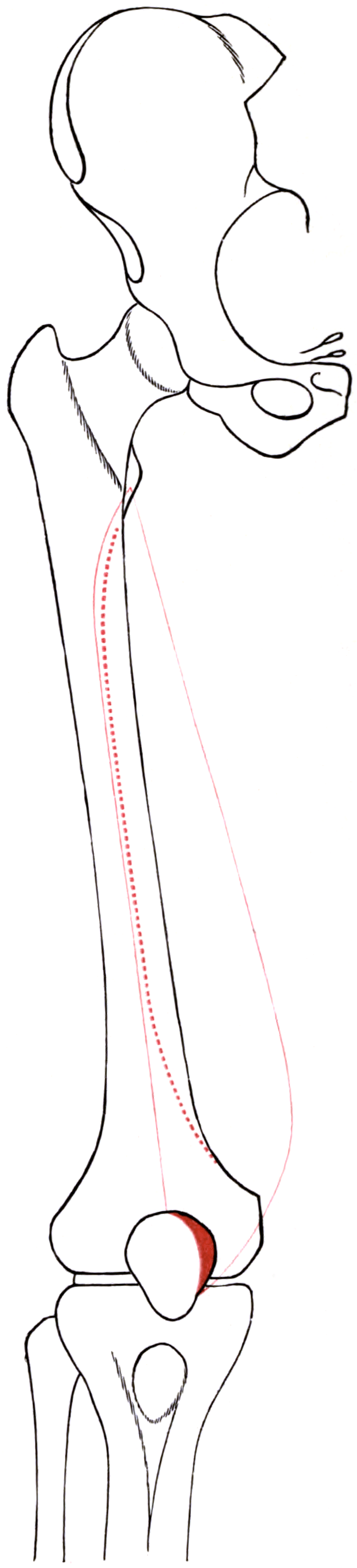
Attachments, Innervation and Relations
Origin — a continuous medial line
- Lower part of the intertrochanteric line.
- Spiral (pectineal) line of the femur.
- Medial lip of the linea aspera.
- Medial supracondylar line, extending almost to the adductor tubercle.
- Tendon of adductor magnus and the medial intermuscular septum.
- The muscle therefore has an extensive, distal-reaching origin — it extends further distally on the femur than any other quadriceps head, which is why it is the muscle that must be dealt with in any medial knee arthrotomy.
Insertion — and the VMO distinction
- The main bulk forms the medial portion of the quadriceps tendon, inserting on the medial aspect of the patellar base.
- Vastus medialis obliquus (VMO): the distal oblique segment, with fibres running at approximately 50-55 degrees to the femoral shaft (compared with roughly 15-20 degrees for the proximal, longitudinal vastus medialis longus fibres). The VMO inserts on the medial border of the patella, extending approximately 1.5-2 cm distal to the patellar base — commonly quoted as covering the proximal third to proximal half of the medial patellar border.
- Fibres continue into the medial retinaculum and blend with the superficial layer overlying the medial patellofemoral ligament (MPFL). The VMO's aponeurosis lies superficial to the MPFL, which is why the MPFL is found by elevating or retracting the VMO in MPFL reconstruction.
- A distal expansion reaches the medial tibial condyle and medial capsule.
Named landmarks and measurements
- Relationship
- Distal limit of the origin along the medial supracondylar line
- Practical use
- Landmark for MPFL femoral origin (just distal and posterior to it) and for the distal extent of a subvastus elevation
- Relationship
- 1.5-2 cm distal to the patellar base, covering the proximal third to half of the medial border
- Practical use
- Reference for the apex of a midvastus split and for medial reefing
- Relationship
- Origin and posterior boundary; the plane elevated in the subvastus approach
- Practical use
- Subvastus interval; deep to it lie the adductor canal contents
- Relationship
- Lies deep to the VMO aponeurosis, between the adductor tubercle region and the superomedial patella
- Practical use
- Found by retracting the VMO; provides most passive medial restraint
- Relationship
- Deep to the muscle, roof formed by vastoadductor membrane
- Practical use
- Site of the adductor canal block; contains the nerve to vastus medialis and the saphenous nerve
The commonest nerve injury in knee surgery.
- The infrapatellar branch of the saphenous nerve crosses the anteromedial knee transversely, so any longitudinal medial incision crosses it and any transverse anteromedial incision transects several branches.
- Consequence: an area of numbness lateral and distal to the incision, or a painful neuroma with a positive Tinel sign — a recognised cause of persistent anteromedial pain after knee arthroscopy, arthroplasty, tibial nailing and hamstring graft harvest.
- Answer: consent for it, use a midline or medial longitudinal incision, raise full-thickness flaps, and avoid transverse anteromedial incisions where possible.
The traditional teaching is wrong.
- No exercise has been shown to reliably recruit the VMO preferentially over the vastus lateralis; electromyographic studies show the two activate together.
- VMO wasting in patellofemoral pain is largely a consequence of pain and effusion-mediated arthrogenic muscle inhibition, not a primary cause.
- Answer: prescribe whole-quadriceps and hip strengthening with motor control and movement retraining, and treat the effusion and the pain, rather than promising selective VMO training.
Action and Biomechanics
Actions by plane
- Action
- Knee extension
- Arc of greatest importance
- Throughout flexion range
- Note
- Fibre angle roughly 15-20 degrees to the shaft
- Action
- Medial patellar stabilisation and terminal extension
- Arc of greatest importance
- 0 to 20-30 degrees of flexion
- Note
- Fibre angle roughly 50-55 degrees; the only dynamic medial restraint
- Action
- Eccentric control of knee flexion in loading response
- Arc of greatest importance
- Early stance
- Note
- Monoarticular, so unaffected by hip position
- Action
- Contributes to the medial retinaculum and medial capsule
- Arc of greatest importance
- All arcs
- Note
- Superficial to the MPFL
Force contribution and architecture
- Cross-sectional area: roughly 25-30 percent of the quadriceps — second to vastus lateralis.
- Architecture: the muscle is regionalised. The proximal longus portion is nearly longitudinal, the distal obliquus portion strongly oblique. The transition is gradual, not a discrete boundary, which is why anatomists differ on whether the VMO is a separate muscle.
- The VMO has a small extension moment but a substantial medial patellar moment, precisely the opposite priority to the rest of the quadriceps.
The patellofemoral force balance
- Passive medial restraint: the MPFL provides the majority (commonly quoted at around 50-60 percent) of the passive restraint against lateral patellar translation; the medial patellomeniscal and patellotibial ligaments contribute the remainder.
- Dynamic medial restraint: the VMO alone.
- Bony restraint: the trochlear groove, effective only once the patella engages at roughly 20-30 degrees of flexion.
- Therefore: the vulnerable arc is 0 to 30 degrees, and this is the arc in which lateral patellar dislocation occurs, in which apprehension is elicited, and in which the J-sign appears.
Length-tension and terminal extension
- Being monoarticular, VM length depends only on knee angle. Like the other vasti, its mechanical disadvantage in terminal extension means the last 15-20 degrees demands the greatest force — so extension lag and terminal extension weakness are the earliest signs of VM deficiency.
Synergists and antagonists
- Synergists: vastus lateralis, vastus intermedius, rectus femoris for extension; the MPFL and trochlea as passive partners for medial patellar restraint.
- Antagonists: hamstrings and gastrocnemius for knee flexion; vastus lateralis obliquus, the lateral retinaculum and the iliotibial tract oppose the VMO's medial patellar pull.
What happens when it fails
- VMO dysfunction or wasting: relative lateral patellar tracking, a J-sign, apprehension, anterior knee pain, and reduced confidence in single-leg tasks in the 0-30 degree arc.
- Whole-muscle weakness: extension lag, quadriceps avoidance gait, difficulty with stair descent, and inability to control the knee in loading response.
- Detachment or non-healing of a divided VMO tendon after medial parapatellar arthrotomy: extensor lag, medial patellar tracking loss, and rarely a frank extensor mechanism dehiscence.
- Over-tightening (medial reefing performed too aggressively): medial overload, loss of lateral patellar glide, medial facet pain and iatrogenic medial patellar instability.
The examiner will ask why the VMO wastes in patellofemoral pain and in knee effusion. The modern answer is arthrogenic muscle inhibition (AMI).
- Joint effusion, pain and altered mechanoreceptor afferent input produce reflex inhibition of the quadriceps motor pool at spinal and supraspinal levels.
- Experimentally, injecting saline into a normal knee to create an effusion immediately reduces quadriceps activation — the muscle is not damaged, it is inhibited.
- The VMO appears to be affected earliest and most visibly, partly because it is superficial and its bulk is easy to see, and partly because it has a small cross-section and a distal position.
- Practical consequences:
- Treat the effusion and the pain — aspiration where appropriate, load management, analgesia — because inhibition will otherwise defeat strengthening.
- Do not interpret VMO wasting as the primary cause of anterior knee pain. It is usually downstream.
- Motor control and neuromuscular retraining, including hip abductor and external rotator strengthening to control femoral internal rotation and adduction, addresses the mechanics that the VMO cannot fix alone.
Surface Anatomy and Examination
Palpation and positioning
- VMO: supine with the knee in slight flexion over a bolster, ask for an isometric quadriceps contraction with the knee near extension. The VMO appears as a distinct oblique bulge on the anteromedial thigh immediately proximal and medial to the patella — the most distal quadriceps bulk on either side.
- VM longus: the longitudinal mass along the medial thigh proximal to the VMO.
- Adductor tubercle: palpate the medial femoral condyle and run proximally to the small prominence at the distal end of the medial supracondylar line — the reference point for the MPFL femoral origin and the distal limit of the VM origin.
- Medial patellar border: the VMO insertion is palpable along the proximal third to half of the medial border, roughly 1.5-2 cm distal to the patellar base.
- Infrapatellar branch territory: map the area of altered sensation anterolateral and distal to a medial incision, over the anteromedial proximal tibia and inferior patellar region.
Named tests and their interpretation
- How to perform
- Knee near extension, ask for isometric quadriceps contraction; observe and palpate both VMOs
- Positive finding
- Reduced bulk, delayed or absent VMO contour on the affected side
- What it means
- Quadriceps inhibition or wasting, usually secondary to pain or effusion
- False positives
- Limb dominance, body habitus, examiner expectation bias
- How to perform
- Measure passive then active terminal extension with a goniometer
- Positive finding
- Active extension short of passive
- What it means
- Quadriceps power or continuity deficit
- False positives
- Fixed flexion deformity; effusion causing arthrogenic inhibition
- How to perform
- Knee at 20-30 degrees flexion, quadriceps relaxed; push the patella laterally
- Positive finding
- Apprehension, guarding or quadriceps contraction
- What it means
- Lateral patellar instability in the arc where the VMO and MPFL are the restraints
- False positives
- Pain from any cause; a very anxious patient
- How to perform
- Knee extended and relaxed; translate the patella laterally, expressed in quadrants
- Positive finding
- Greater than two quadrants, or the patella can be perched on the lateral trochlear edge
- What it means
- Deficient medial restraint (MPFL, VMO)
- False positives
- Generalised hypermobility; guarding masking laxity
- How to perform
- Observe the patella through active extension from flexion
- Positive finding
- Patella deviates laterally near terminal extension
- What it means
- Maltracking in the arc where the trochlea does not contain the patella
- False positives
- Voluntary substitution; observation from the wrong angle
- How to perform
- Observe a single-leg squat to about 45-60 degrees
- Positive finding
- Dynamic knee valgus, femoral internal rotation, contralateral pelvic drop
- What it means
- Proximal (hip) control deficit contributing to patellofemoral loading, not a VMO problem alone
- False positives
- Ankle stiffness, fear-avoidance, footwear
- How to perform
- Patellar tap for larger effusion; wipe/bulge test for small effusion
- Positive finding
- Fluid displacement or a ballottable patella
- What it means
- Effusion, the driver of arthrogenic muscle inhibition — must be treated for rehabilitation to work
- False positives
- Prepatellar bursitis mistaken for intra-articular fluid; obesity
- How to perform
- Tap along the expected oblique course over the anteromedial knee
- Positive finding
- Radiating electric pain reproducing the patient's symptom
- What it means
- Infrapatellar branch neuroma after a medial incision
- False positives
- Local tenderness from scar or pes anserine bursitis
Pitfalls in examination
- Do not equate VMO bulk with VMO function. The bulge is what you see, but timing and activation are what matter, and both are dominated by pain and effusion.
- Always measure passive extension before interpreting a lag; a fixed flexion deformity is a capsular problem.
- Assess the hip. Dynamic valgus and femoral internal rotation during a single-leg squat shift the patella laterally regardless of VMO status, and hip strengthening improves patellofemoral pain in trials.
- Map the sensory deficit before revision knee surgery and document it — patients frequently attribute pre-existing infrapatellar branch numbness to the most recent operation.
Complications
- Mechanism
- The nerve crosses the anteromedial knee transversely and is divided by any medial incision
- Avoidance / management
- Midline or medial longitudinal incision, full-thickness flaps, explicit consent; block for diagnosis, neurectomy with stump burial for the refractory case
- Mechanism
- Encountered at the deep proximal corner during subvastus elevation
- Avoidance / management
- Identify and ligate deliberately; preserve if a medial femoral condyle flap may be needed
- Mechanism
- Distal branches of the nerve to vastus medialis divided by an over-long split
- Avoidance / management
- Limit the split to 3-5 cm; prefer subvastus where muscle preservation is the priority
- Mechanism
- Division of the quadriceps tendon and VMO insertion
- Avoidance / management
- Meticulous layered repair; early quadriceps activation; expect resolution by three months
- Mechanism
- Failure of the arthrotomy repair, often in a diabetic, obese or steroid-exposed patient
- Avoidance / management
- Robust repair, avoid excessive early flexion loading; formal repair or reconstruction if it fails
- Mechanism
- Persisting with a subvastus or midvastus approach in a knee that needed a parapatellar arthrotomy
- Avoidance / management
- Assess the knee before deciding; convert early without hesitation
- Mechanism
- Medial arthrotomy plus lateral release dividing the superior lateral genicular artery, plus fat pad excision
- Avoidance / management
- Avoid unnecessary lateral release, preserve the superior lateral genicular vessels, retain the fat pad where possible
- Mechanism
- Over-aggressive medial reefing or VMO advancement, often combined with a lateral release
- Avoidance / management
- Do not over-tighten; assess lateral glide intraoperatively; prefer MPFL reconstruction to reefing
- Mechanism
- Adductor canal block anaesthetising the nerve to vastus medialis; a block placed too proximally blocks more motor branches
- Avoidance / management
- Confirm block level, assess quadriceps power before first mobilisation, use a knee immobiliser or supervision
- Mechanism
- Blind dissection deep to the medial intermuscular septum into the adductor canal
- Avoidance / management
- Recognise the canal as the deep limit; never dissect posterior to the septum blindly
- Mechanism
- Direct blow with an intramuscular haematoma
- Avoidance / management
- Ice and compression in flexion, gentle range of motion, avoid deep massage, delay any excision until mature
- Mechanism
- Untreated effusion and arthrogenic muscle inhibition, or unrecognised structural risk factors
- Avoidance / management
- Treat the effusion, reassess for trochlear dysplasia, patella alta, TT-TG and torsion
Denervation pattern
- The nerve to vastus medialis runs distally on the deep surface of the muscle from the adductor canal. Complete denervation produces visible loss of the VMO bulge, terminal extension weakness and a maltracking patella; on MRI, selective fatty atrophy of vastus medialis with preserved vastus lateralis and rectus femoris.
- Because the saphenous nerve travels with it in the adductor canal, an adductor canal lesion produces vastus medialis weakness plus anteromedial leg and medial foot border numbness — a combination that localises the lesion precisely.
Clinical Relevance
Patellofemoral pain and VMO wasting
The traditional model (now substantially revised): VMO weakness or delayed VMO activation causes lateral patellar tracking, which causes increased lateral facet pressure and pain. Selective VMO strengthening was therefore prescribed.
The current understanding
- VMO wasting in patellofemoral pain is predominantly secondary, driven by pain and effusion-mediated arthrogenic muscle inhibition, deconditioning and altered movement strategies.
- Selective VMO training is not achievable. Electromyographic studies of the commonly recommended manoeuvres — terminal extension, adduction with extension, hip adduction added to squats, taping — have not demonstrated reproducible preferential VMO recruitment over vastus lateralis.
- What does work: whole-quadriceps strengthening, hip abductor and external rotator strengthening, motor control and movement retraining, load management, and — where indicated — taping or bracing for short-term symptom relief and orthoses for selected patients.
- The proximal (hip) contribution is important: femoral internal rotation and adduction during weight-bearing effectively moves the trochlea under the patella, producing lateral maltracking that no amount of VMO training corrects.
Structural risk factors that must be assessed before blaming the VMO
- How measured
- Lateral radiograph crossing sign; axial MRI/CT trochlear morphology (Dejour classification)
- Threshold commonly quoted
- Crossing sign, supratrochlear spur, double contour
- How measured
- Insall-Salvati, Caton-Deschamps, Blackburne-Peel indices
- Threshold commonly quoted
- Caton-Deschamps greater than 1.2; Insall-Salvati greater than 1.2
- How measured
- Axial CT or MRI
- Threshold commonly quoted
- Greater than 20 mm is abnormal; 15-20 mm borderline
- How measured
- CT torsional profile
- Threshold commonly quoted
- Anteversion greater than about 30 degrees may warrant derotation
- How measured
- Patellar tilt test, medial glide
- Threshold commonly quoted
- Lateral border cannot be brought to neutral
- How measured
- Beighton score
- Threshold commonly quoted
- Beighton 6 or more suggests generalised laxity
Patellar dislocation and the VMO
- First-time lateral patellar dislocation: the MPFL is the structure that fails, most commonly at its femoral attachment, and the VMO aponeurosis overlying it is disrupted. Osteochondral fragments from the medial patellar facet or the lateral femoral condyle must be sought.
- Management of a first dislocation is generally non-operative unless there is an osteochondral fragment requiring fixation, an irreducible dislocation, or a large loose body — with rehabilitation targeting quadriceps activation, hip control and graded return.
- Recurrent instability: MPFL reconstruction restores the passive restraint; the graft is placed deep to the VMO aponeurosis and the femoral origin is identified just distal and posterior to the adductor tubercle (radiographically the Schöttle point). Bony correction (tibial tubercle transfer, trochleoplasty, derotation) is added for the structural abnormalities above.
- Isolated medial reefing (VMO advancement) has largely been superseded by MPFL reconstruction because of higher recurrence rates and the risk of medial overload.
Surgical Relevance
Approach-by-approach summary
- How vastus medialis is handled
- VMO tendon divided at its patellar attachment along the medial border of quadriceps tendon and patella
- Structures at risk with location
- Medial peripatellar vessels; infrapatellar branch of saphenous nerve in the skin incision; extensor mechanism continuity
- How vastus medialis is handled
- VMO split in line with fibres for 3-5 cm from the superomedial patella
- Structures at risk with location
- Distal branches of the nerve to vastus medialis if the split is extended; intramuscular bleeding
- How vastus medialis is handled
- Whole muscle elevated off the medial intermuscular septum and retracted laterally
- Structures at risk with location
- Descending genicular artery at the deep proximal corner; adductor canal contents deep to the septum
- How vastus medialis is handled
- VMO aponeurosis elevated or retracted to expose the MPFL layer
- Structures at risk with location
- Femoral origin just distal and posterior to the adductor tubercle; saphenous nerve if dissection strays posteriorly
- How vastus medialis is handled
- VMO advanced distally and laterally onto the patella
- Structures at risk with location
- Over-tightening produces medial overload and iatrogenic medial instability
- How vastus medialis is handled
- Not directly, but the incision crosses the infrapatellar branch territory
- Structures at risk with location
- Infrapatellar branch of saphenous nerve; saphenous nerve itself with a proximally directed stripper
- How vastus medialis is handled
- The muscle forms the anterolateral wall of the canal
- Structures at risk with location
- Nerve to vastus medialis blocked, producing partial quadriceps weakness; femoral artery deep to the target
- How vastus medialis is handled
- Muscle elevated to expose the descending genicular artery pedicle
- Structures at risk with location
- Descending genicular artery must be preserved; medial collateral ligament and joint line distally
- How vastus medialis is handled
- Muscle retracted anteriorly with the intermuscular septum incised
- Structures at risk with location
- Femoral artery and vein in the adductor canal; saphenous nerve
Distances and landmarks worth quoting
- VMO insertion: medial patellar border, extending approximately 1.5-2 cm distal to the patellar base, covering the proximal third to half of the border. This defines the safe apex for a midvastus split.
- Midvastus split length: approximately 3-5 cm proximomedially from the superomedial patellar corner.
- MPFL femoral origin: a small depression just distal and posterior to the adductor tubercle, between the adductor tubercle and the medial epicondyle; radiographically the Schöttle point on a true lateral.
- Descending genicular artery: arises from the femoral artery within the adductor canal just proximal to the adductor hiatus, and is encountered at the deep proximal corner of the subvastus plane.
- Infrapatellar branch of the saphenous nerve: crosses the anteromedial knee obliquely, most branches lying at and below the level of the joint line and medial to the patellar tendon, having pierced or passed deep to the distal sartorius.
- Adductor canal: from the apex of the femoral triangle to the adductor hiatus; the vastus medialis is its anterolateral wall.
Do not defend one approach dogmatically. Defend a decision rule.
- Exposure is non-negotiable. A malaligned, maltracking or malrotated implant caused by inadequate exposure costs far more than a few weeks of extensor lag.
- Choose the least invasive approach that gives adequate exposure for this knee. Assess flexion range, body habitus, patellar height, previous incisions and the anticipated complexity before deciding.
- Subvastus or midvastus for the straightforward primary knee where early quadriceps recovery matters.
- Medial parapatellar for the stiff knee, significant obesity, revision, patella baja, or where an extensile option (quadriceps snip, tubercle osteotomy) may be needed.
- Be prepared to convert — recognising inadequate exposure early and converting is good surgery, not failure.
- Quote the evidence honestly: quadriceps-sparing approaches give earlier straight-leg raise and modestly better very early function, with no consistent difference beyond three to six months.
Guidelines, Registries & Global Practice
Variation and prevalence
- Whether the VMO is a distinct muscle or simply the distal oblique portion of a single vastus medialis remains debated. Anatomical studies differ on whether a separate fascial plane and separate innervation reliably define it. The functional description — steeply oblique distal fibres providing the only dynamic medial patellar restraint — is consistent across sources and is what examiners want.
- The course of the infrapatellar branch of the saphenous nerve varies in the number and level of branches, which is why no anteromedial incision is entirely safe and why mapping the deficit preoperatively matters.
- The descending genicular artery varies in calibre and in the level at which it arises from the femoral artery, and occasionally its muscular and articular branches arise separately — relevant when raising a medial femoral condyle flap.
Differences in described technique
- Emphasis relating to vastus medialis
- Medial parapatellar arthrotomy remains the default in most published series; midvastus and subvastus are described as options in selected primary knees with explicit exclusion of obesity, stiffness and prior surgery.
- Emphasis relating to vastus medialis
- Emphasises exposure adequacy and implant position over approach novelty; strong emphasis on quadriceps rehabilitation and on early recognition of extensor mechanism problems.
- Emphasis relating to vastus medialis
- MPFL reconstruction rather than medial reefing for recurrent instability; structured assessment of trochlear dysplasia, patellar height, TT-TG and torsion before choosing the operation; lateral release only for demonstrable tilt.
- Emphasis relating to vastus medialis
- Recommend combined hip and knee exercise therapy as first-line; explicitly do not support selective VMO training; taping and orthoses as short-term adjuncts.
- Emphasis relating to vastus medialis
- Adductor canal block preferred over femoral nerve block for knee arthroplasty because of reduced quadriceps weakness, with explicit acknowledgement that some weakness persists via the nerve to vastus medialis and that block level determines the degree.
Registry and outcome signals
- National joint registries (Australian, UK, American and Nordic) do not record the arthrotomy used, so registry data cannot adjudicate approach. What they do capture is revision for malalignment, instability and infection — the outcomes that inadequate exposure jeopardises, which is the strongest practical argument for choosing exposure over minimalism.
- Randomised and meta-analytic comparisons of subvastus and midvastus against medial parapatellar arthrotomy consistently show earlier straight-leg raise and modestly better function in the first few weeks, converging by three to six months, with no difference in radiographic alignment when exposure was adequate.
- Patellofemoral instability cohorts show clearly lower recurrence after MPFL reconstruction than after isolated soft-tissue medial reefing, which has shifted international practice.
High- versus limited-resource practice
- Well-resourced settings: ultrasound-guided adductor canal blocks with defined injection level, MRI and CT for TT-TG and torsional assessment, trochleoplasty available in specialist centres, and formal gait and movement analysis in patellofemoral clinics.
- Limited-resource settings: the medial parapatellar approach requires no special instrumentation and remains the safest default. Patellofemoral instability can be assessed with plain radiographs including a true lateral (for the crossing sign and patellar height) and an axial view, and treated with MPFL reconstruction using an autograft, which needs no implants beyond simple fixation. Patellofemoral pain is managed with supervised exercise, which is the intervention with the best evidence and the lowest cost anywhere.
- Universal principles: never operate for instability without assessing trochlear dysplasia, patellar height, TT-TG and torsion; never promise selective VMO training; treat the effusion before blaming the muscle; and consent every anteromedial knee incision for infrapatellar branch numbness.
MCQ Practice Points
Q: What is the fibre angle of the vastus medialis obliquus and how does it compare with the vastus lateralis? A: Roughly 50-55 degrees to the femoral shaft, compared with roughly 30-35 degrees for the vastus lateralis (and 15-20 degrees for the proximal vastus medialis longus fibres). The steep angle gives the medial patellar vector.
Q: Where does the VMO insert? A: The medial border of the patella, extending approximately 1.5-2 cm distal to the patellar base, covering the proximal third to half of the medial border.
Q: In which arc of knee flexion is the VMO most important, and why? A: 0 to 20-30 degrees, because the patella has not yet engaged the trochlear groove, so bony containment contributes little and the MPFL passively and the VMO dynamically are the only restraints. This is the arc in which dislocation occurs.
Q: How do the medial parapatellar, midvastus and subvastus approaches differ? A: Parapatellar cuts the VMO tendon at the patella; midvastus splits the VMO in line with its fibres for 3-5 cm; subvastus lifts the whole muscle off the medial intermuscular septum without dividing the extensor mechanism.
Q: Which artery is encountered in the subvastus plane and where? A: The descending genicular (supreme genicular) artery, at the deep proximal corner of the plane, emerging from the adductor canal just proximal to the adductor hiatus. It is also the pedicle of the medial femoral condyle vascularised bone flap.
Q: Name the contents of the adductor canal and its walls. A: Contents: femoral artery, femoral vein, saphenous nerve, nerve to vastus medialis (and the descending genicular artery arising within it). Walls: vastus medialis anterolaterally, adductor longus and magnus posteromedially, and the vastoadductor membrane deep to sartorius as the roof.
Q: Why does an adductor canal block still cause some quadriceps weakness? A: It anaesthetises the nerve to vastus medialis, which runs in the canal. It is more quadriceps-sparing than a femoral nerve block but not motor-neutral — a falls-risk consideration on the ward.
Q: Can the VMO be selectively strengthened? A: No. No exercise has been shown to reliably recruit the VMO preferentially over vastus lateralis. Prescribe whole-quadriceps and hip strengthening with motor control retraining.
Q: Why does the VMO waste in patellofemoral pain and knee effusion? A: Arthrogenic muscle inhibition — effusion, pain and altered afferent input reflexly inhibit the quadriceps motor pool. Treat the effusion and the pain or strengthening will fail.
Q: Where is the femoral origin of the MPFL and what is its relation to the VMO? A: In a small depression just distal and posterior to the adductor tubercle (radiographically the Schöttle point). The MPFL lies deep to the VMO aponeurosis, so it is exposed by elevating or retracting the VMO.
Q: Why is the infrapatellar branch of the saphenous nerve injured in almost every medial knee approach? A: Because it crosses the anteromedial knee transversely, while surgical incisions here run longitudinally — so any medial incision crosses it. Use full-thickness flaps and consent for it.
Exam Viva Scenarios
Practise clinical reasoning and management decisions out loud
“You are about to perform a primary total knee arthroplasty. Your registrar asks whether you will use a medial parapatellar, midvastus or subvastus approach, and why. How do you answer?”
“A 17-year-old has had four lateral patellar dislocations. The VMO is visibly wasted, there is a J-sign, and lateral glide is three quadrants. What is the role of the VMO here, and how do you plan treatment?”
“A 34-year-old man is four months after a medial meniscal repair through an accessory anteromedial incision. He describes a numb patch below and lateral to the scar and an electric pain when the area is touched or when trousers rub it. What has happened and how do you manage it?”
Anatomy
- Origin: intertrochanteric line, spiral line, medial linea aspera, medial supracondylar line to adductor tubercle, adductor magnus tendon, medial intermuscular septum
- Insert: medial quadriceps tendon and medial patellar border
- VMO: 50-55 degree fibre angle, inserts 1.5-2 cm below patellar base
- Nerve: femoral, posterior division, L2-L4, via the adductor canal
- Supply: descending genicular artery, medial superior genicular artery
Patellar Stability
- VMO is the ONLY dynamic medial restraint
- Critical arc 0-30 degrees, before trochlear engagement
- MPFL gives most passive restraint, deep to VMO aponeurosis
- MPFL femoral origin just distal and posterior to adductor tubercle
- VMO cannot be selectively strengthened
Approaches
- Medial parapatellar: cut VMO tendon; widest, extensile
- Midvastus: split 3-5 cm from superomedial patella
- Subvastus: lift whole muscle off medial intermuscular septum
- Subvastus contraindications: obese, stiff under 90 degrees, patella baja, prior surgery
- Descending genicular artery at the deep proximal corner
Adductor Canal
- Apex of femoral triangle to adductor hiatus
- Anterolateral wall: vastus medialis
- Roof: vastoadductor membrane deep to sartorius
- Contents: femoral artery and vein, saphenous nerve, nerve to vastus medialis
- Block is quadriceps-sparing but NOT motor-neutral
Pitfalls
- Infrapatellar branch of saphenous nerve crosses transversely — consent for it
- VMO wasting is secondary: arthrogenic muscle inhibition
- Treat the effusion before strengthening
- Lateral release treats tilt not translation
- Exposure is non-negotiable — convert early
Evidence Base
Quadriceps Function: An Anatomical and Mechanical Study Using Amputated Limbs
- Anatomical and mechanical study of quadriceps function carried out on amputated limbs
- Indexed subject headings cover muscle anatomy and physiology, knee physiology, movement, the femur and the patella
- No abstract is indexed for this 1968 paper, so only the title and subject headings are cited here rather than specific numerical results
- It is conventionally cited as the source for the differing fibre orientations of the individual quadriceps heads and for the disproportionate quadriceps force demanded by terminal knee extension
Subvastus (Southern) Approach for Primary Total Knee Arthroplasty
- A NARRATIVE ANATOMICAL DESCRIPTION - the abstract reports no series, no cohort, no numbers and no outcome data of any kind
- Notes that the subvastus or Southern approach was described as early as 1929 and had dropped out of standard orthopaedic textbooks
- The two stated advantages are anatomical, not measured: the integrity of the EXTENSOR MECHANISM is preserved, and the VASCULAR SUPPLY TO THE PATELLA is maintained
- The paper's own argument is that a complete understanding of the surgical anatomy is required before the benefits can be realised
A Midvastus Muscle-Splitting Approach for Total Knee Arthroplasty
- A PROSPECTIVE RANDOMISED TRIAL, not a technique description: 118 consecutive total knee arthroplasties by a single surgeon randomised to medial parapatellar or midvastus muscle-splitting approach
- The split separates vastus medialis in the direction of its fibres beginning at the SUPERIOR POLE OF THE PATELLA
- Measured outcomes were frequency of lateral retinacular release, patellar tilt and translation, and quadriceps strength
- THE RESULT WAS EQUIVALENCE, NOT SUPERIORITY: patellar stability and quadriceps strength were EQUIVALENT for the two approaches
- The midvastus approach provided excellent exposure to all knees, and the authors conclude it is an efficacious ALTERNATIVE to the medial parapatellar approach
Nerve Supply of the Human Vastus Medialis Muscle
- THIRTY human vastus medialis muscles dissected, with the nerves traced back to the lumbar plexus in three cadavers
- A CONSISTENT BIPARTITE nerve supply from the posterior division of the femoral nerve: a short slender LATERAL branch to the upper lateral portion, and a MEDIAL branch to the middle and lower portion
- There is a DISTALWARD INCREASE in the number of nerve fibres - the LOWERMOST muscle fibres receive the RICHEST nerve supply, which is the opposite of what a simple proximal-to-distal branching pattern would give
- The two branches carry DIFFERENT SPINAL SEGMENTS: the lateral branch L3-L4, the medial branch L1, L2 and L3 - and the lateral branch sometimes arises from the nerve to vastus intermedius, sharing its root supply
- The authors conclude the upper portion is closely aligned with vastus intermedius while the lower third has a richer innervation and a distinct gross morphology, so the muscle is FUNCTIONALLY TRIPARTITE
The Surgical Anatomy of the Infrapatellar Branch of the Saphenous Nerve in Relation to Incisions for Anteromedial Knee Surgery
- TWENTY embalmed knees dissected, with a computer-assisted surgical anatomy mapping tool used to calculate risk zones and the location-dependent direction of the nerve
- THE HEADLINE FINDING IS A NEGATIVE ONE: the location of the infrapatellar branch is HIGHLY VARIABLE and NO DEFINITE SAFE ZONE could be identified
- The nerve runs NEITHER a purely horizontal NOR a purely vertical course - its direction depends on where you are. MEDIALLY it is nearly VERTICAL; medial to the patellar tendon it runs at about MINUS 45 degrees distal-lateral; over the patella and patellar tendon it is close to HORIZONTAL-lateral
- Three LOW RISK - not safe - zones were identified: medially at the level of the tibial tuberosity, where a minus 45 degree oblique incision is least damaging; and two zones medial to the patellar apex, cranial and caudal, where close-to-horizontal incisions are least damaging
- The operative rule the authors draw: make the incision PARALLEL to the direction of the nerve at that location, whenever technically possible
Adductor Canal Block Versus Femoral Nerve Block and Quadriceps Strength: A Randomized, Double-Blind, Placebo-Controlled, Crossover Study in Healthy Volunteers
- Double-blind, placebo-controlled, randomised CROSSOVER study in HEALTHY YOUNG MEN - 12 randomised, 11 analysed - with placebo in the opposite limb on each of two study days
- THE NUMBERS THAT MATTER: mean reduction in quadriceps strength from baseline was 8 PER CENT with adductor canal block against 49 PER CENT with femoral nerve block
- Adductor canal block still reduced quadriceps strength significantly compared with placebo (p = 0.02) - it is quadriceps-SPARING, not quadriceps-neutral
- Adductor strength was significantly reduced only by the femoral nerve block, not by the adductor canal block
- Performance in all three standardised ambulation tests was worse after femoral nerve block than after adductor canal block (p less than 0.05)
Defining the Location of the Adductor Canal Using Ultrasound
- TWENTY-TWO volunteers examined by ultrasound. The proximal end of the adductor canal was defined where the medial border of SARTORIUS crosses the medial border of ADDUCTOR LONGUS; the distal end is the adductor hiatus
- Mean distance from ASIS to the MIDPOINT OF THE THIGH: 22.9 cm (range 20.3 to 24.9)
- Mean distance from ASIS to the PROXIMAL END OF THE ADDUCTOR CANAL: 27.4 cm (range 24.0 to 31.4)
- So the conventional midthigh needle insertion point sits a mean 4.6 cm PROXIMAL to the canal (range 2.3 to 7.0 cm)
- IN ALL 22 VOLUNTEERS the midpoint of the thigh was proximal to the beginning of the canal - the authors conclude that an injection at that level is in fact a FEMORAL TRIANGLE BLOCK, and that 'adductor canal block' is a misnomer for it
Exercise for Treating Patellofemoral Pain Syndrome
- 31 heterogeneous randomised and quasi-randomised trials, 1,690 participants - and THE EVIDENCE FOR EVERY ONE OF THE SEVEN MAIN OUTCOMES WAS GRADED VERY LOW QUALITY, so the authors state they are 'very unsure about the estimates'
- Exercise versus control, pain during activity at 3 months or less: mean difference MINUS 1.46 (95 per cent CI minus 2.39 to minus 0.54) on a 0-10 scale - the CI INCLUDES the minimal clinically important difference of 1.3, so a clinically important effect is possible but not established
- Function short-term: SMD 1.10, re-expressed as an Anterior Knee Pain Score 12.21 points higher (CI 6.44 to 18.09) - again the CI INCLUDES the MCID of 10
- HIP PLUS KNEE versus knee exercise alone, pain during activity short-term: mean difference MINUS 2.20 (CI minus 3.80 to minus 0.60), a clinically important effect - but FUNCTION and RECOVERY showed neither approach superior
- THE RECOVERY FINDING IS WEAKER THAN THE AUTHORS' OWN CONCLUSION SUGGESTS: against a control recovery of 250 per 1000, exercise gave 88 more recovered per 1000 - but the interval runs from 2 FEWER to 210 more, so it crosses no effect, and it pools just two studies and 166 participants
- Eight trials were at high risk of selection bias, six of them quasi-randomised, and most were at high risk of performance and detection bias from absent blinding
- The authors' bottom line: there is INSUFFICIENT EVIDENCE TO DETERMINE THE BEST FORM of exercise therapy