The Small Muscle That Solves Big Problems
- Origin: posterior aspect of the LATERAL EPICONDYLE of the humerus, deep to the common extensor origin; insertion: the lateral aspect of the OLECRANON and the proximal quarter of the posterior ULNA.
- Innervation: the RADIAL nerve (C7, C8) via the terminal continuation of the nerve to the medial head of triceps, which passes THROUGH the medial head to reach it.
- Vascular pedicle: the RECURRENT POSTERIOR INTEROSSEOUS ARTERY, entering the deep surface distally, with a mean pedicle length of 31.3 mm and a mean diameter of 1.9 mm — long and wide enough for microsurgical anastomosis.
- The Kocher interval lies between the anconeus (radial nerve) and extensor carpi ulnaris (posterior interosseous nerve) — it is the standard lateral approach to the radial head and lateral collateral ligament complex.
- Basmajian and Griffin's electromyographic study is the classic reference for anconeus function; the conclusion attributed to it — that activity is not confined to extension — underpins the muscle's characterisation as a joint stabiliser rather than a significant extensor.
- “The lateral ulnar collateral ligament lies at the FLOOR of the Kocher interval — dissecting too posteriorly and dividing it iatrogenically creates posterolateral rotatory instability.
- “To avoid the lateral ulnar collateral ligament, enter the Kocher interval ANTERIOR to the equator of the radial head with the forearm PRONATED, and keep the capsulotomy anterior to the midline of the head.
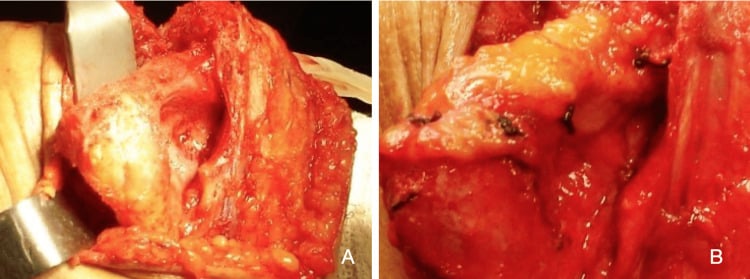
- “Elhassan and colleagues covered 20 chronic posterior elbow soft-tissue defects (up to 7 x 4 cm) with local pedicled anconeus flaps: all healed at a mean of 3 weeks with no flap complications and full range of motion.
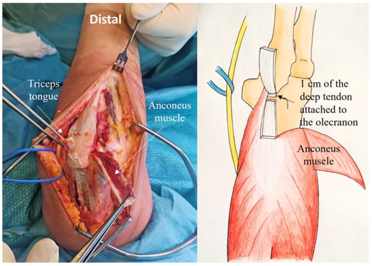
- “The anconeus is one of the three Mayo reconstruction options for triceps insufficiency after total elbow arthroplasty, alongside direct suture and Achilles tendon allograft.
Overview
Anconeus is a small triangular muscle spanning the posterolateral elbow from the lateral epicondyle to the proximal ulna. Functionally it is almost negligible as an extensor. Surgically it is one of the most useful muscles in the upper limb, for four separate reasons:
- Its anterior border defines the Kocher interval, the most commonly used lateral approach to the elbow.
- It provides a reliable local pedicled rotation flap for posterior elbow soft-tissue cover, on a pedicle long enough to permit free transfer.
- It is a dynamic lateral stabiliser of the ulnohumeral joint and features in every discussion of posterolateral rotatory instability.
- It is rotated proximally to reconstruct a deficient triceps after total elbow arthroplasty.
Attachments, Innervation and Relations
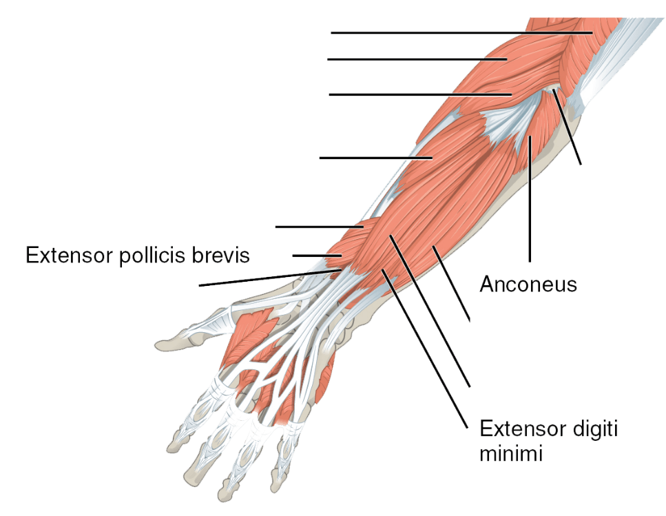
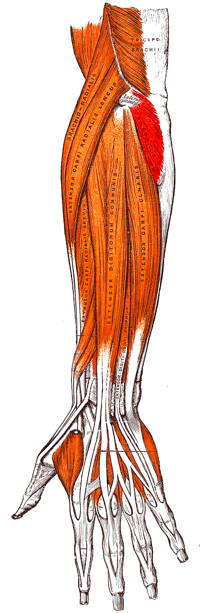
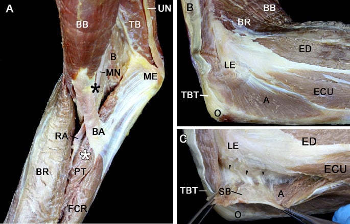
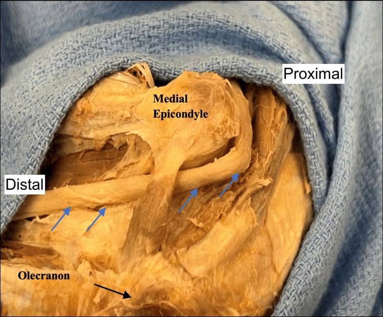
Origin. The posterior (and posteroinferior) aspect of the lateral epicondyle of the humerus, by a short thick tendon deep to the common extensor origin and deep to the lateral collateral ligament complex. The origin is intimately related to the lateral ulnar collateral ligament and the posterolateral capsule, which is the relationship that matters enormously in lateral elbow approaches.
Insertion. The lateral surface of the olecranon and the proximal quarter of the posterior (dorsal) surface of the ulna, along and just lateral to the subcutaneous border; the subcutaneous border of the ulna forms the medial edge of the muscle and is the palpable landmark for its entire length. The muscle fans out from a narrow humeral origin to a broad ulnar insertion, hence its triangular shape. The fascia over the anconeus is continuous with the triceps aponeurosis proximally, which is the anatomical basis for using it to reinforce a deficient triceps.
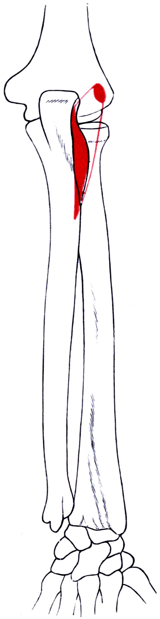
Dimensions. Ng and colleagues measured the anconeus in eight cadaveric upper extremities: mean fibre length 88.0 plus or minus 9.9 mm, mean muscle area 1341.9 plus or minus 230.4 mm squared and mean fibre angle 70.5 plus or minus 11.9 degrees. Practically, the muscle is a triangle roughly 5 to 7 cm along the ulna (the proximal quarter of the bone) and 3 to 4 cm from epicondyle to ulna, capable of covering a defect of similar dimensions when rotated.
Action and Biomechanics
Extension. Weak extension of the elbow, a small contribution alongside the triceps and essentially never the limiting factor in extension power.
What it is really for. Basmajian and Griffin's electromyographic study is the classic reference for anconeus function, and the conclusion consistently attributed to it is that anconeus activity is not confined to elbow extension. Activity that persists irrespective of the direction of movement is the signature of a stabiliser, not a prime mover.
The vector. Its fibres run obliquely from the lateral epicondyle inferomedially to the proximal ulna, so its line of pull has a medially directed vector at the proximal ulna. Contraction therefore resists varus angulation and, critically, resists posterolateral rotatory displacement of the ulna on the humerus, and it is continuously active on electromyography throughout elbow motion. Its remaining functions follow:
- Abduction of the ulna during pronation. As the forearm pronates the ulna undergoes a small obligate lateral (abduction) movement; the anconeus assists and controls this, maintaining ulnohumeral congruity.
- Joint compression. It pulls the olecranon into the trochlear notch, contributing to the compressive stability of the ulnohumeral articulation.
- Prevention of posterior capsular impingement. By tensioning the posterolateral capsule during extension it helps prevent the capsule being pinched in the olecranon fossa.
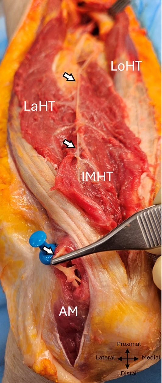
The anconeus-brachialis sling. Leonello and colleagues, describing the two-headed brachialis, observed that the radial-innervated inferolateral fibres of the deep brachialis head run in a direction similar to the anconeus. Brachialis anterolaterally and anconeus posterolaterally therefore form a muscular sling around the ulnohumeral joint, both radial-innervated, both oblique, both spanning the joint: a dynamic stabilising complex that complements the static lateral ligamentous restraints.
Where it fits in posterolateral rotatory instability. The lateral ulnar collateral ligament is the static primary restraint; the anconeus is a dynamic secondary restraint acting in the same vector as the deficient ligament. It does not substitute for the ligament, but it explains why some patients with a demonstrably deficient ligament remain functionally stable and why clinical severity varies, and it is the reason to preserve and reattach the muscle during lateral elbow surgery rather than treating it as disposable.
- Nerve
- Radial C6-C8
- Extension contribution
- Essentially all elbow extension torque
- Stabilising role
- Compression across the ulnohumeral joint
- Expendable?
- NO
- Nerve
- Radial C7-C8
- Extension contribution
- Small and clinically insignificant
- Stabilising role
- Dynamic lateral stabiliser; resists varus and PLRI
- Expendable?
- YES — as a flap, with negligible functional cost
- Nerve
- -
- Extension contribution
- None
- Stabilising role
- STATIC primary restraint to PLRI
- Expendable?
- NO — its loss defines PLRI
- Nerve
- PIN and radial
- Extension contribution
- None
- Stabilising role
- Secondary static and dynamic lateral restraint
- Expendable?
- Partially — released and repaired in lateral approaches
What its loss costs. Isolated anconeus loss, by denervation or by harvest as a flap, causes no measurable loss of extension power and no clinically apparent instability in a normal elbow. Loss combined with a deficient lateral ulnar collateral ligament removes the dynamic backup to an already deficient static restraint, and this is the setting that matters: a patient with a partially attenuated lateral ligament complex may be held stable by the anconeus, and lose that compensation if the muscle is detached or denervated.
Surface Anatomy and Examination
The soft spot. The anconeus fills the soft triangular hollow bounded by the lateral epicondyle, the olecranon tip and the radial head. This triangle is the point for elbow aspiration and injection and for the direct lateral (soft spot) arthroscopic portal: the anconeus is traversed to enter the joint here, which is why it is the safest portal on the lateral side.
Palpation. With the elbow flexed to 90 degrees and the forearm pronated, palpate two fingerbreadths distal to the lateral epicondyle along the lateral border of the ulna. Resisted elbow extension makes the muscle firm under the finger.
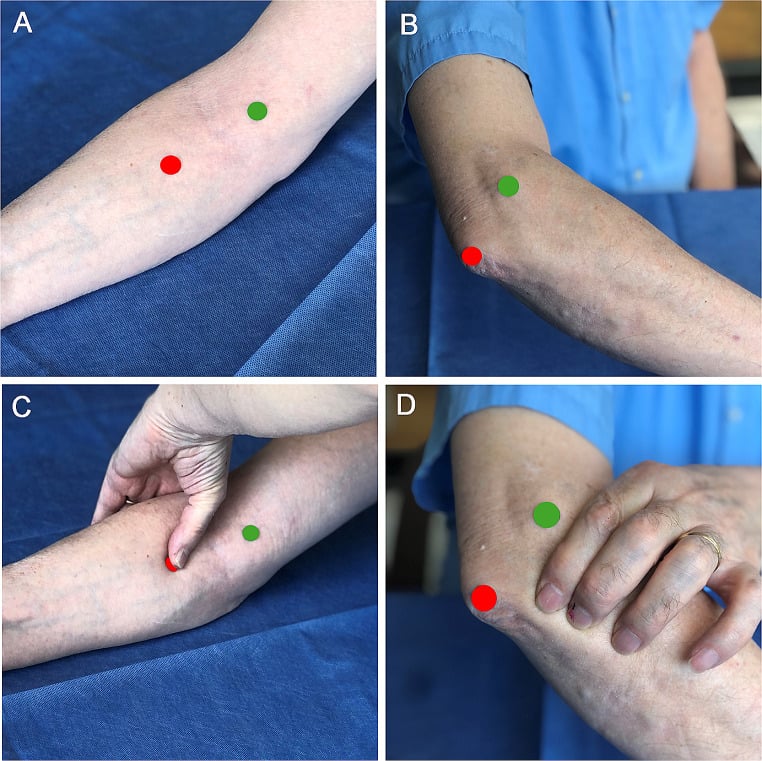
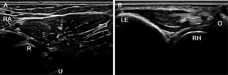
The tests. Those below are the tests for posterolateral rotatory instability, the condition in which the anconeus is a supporting player, together with aspiration through the soft spot.
- How to perform
- Supine, arm overhead, forearm supinated; apply valgus and axial compression while flexing from full extension
- Positive finding
- Apprehension awake; a visible clunk of reduction at about 40 degrees of flexion under anaesthesia
- What it means
- Posterolateral rotatory instability from lateral ulnar collateral ligament insufficiency
- False positives
- Frequently negative in the awake patient because of guarding — apprehension alone is a positive awake test
- How to perform
- Elbow at 40 degrees flexion; translate the proximal forearm posterolaterally like a drawer
- Positive finding
- Posterolateral rotation of the ulna and radius away from the humerus
- What it means
- Posterolateral rotatory instability
- False positives
- Generalised ligamentous laxity
- How to perform
- Ask the patient to push up out of a chair with the forearms supinated and elbows abducted
- Positive finding
- Apprehension or refusal as the elbow passes about 40 degrees
- What it means
- Posterolateral rotatory instability — an awake test the patient can perform
- False positives
- Pain from any cause of lateral elbow pathology
- How to perform
- Patient pushes up from a table edge with the forearm supinated; repeat with the examiner's thumb over the radial head
- Positive finding
- Apprehension relieved by thumb pressure on the radial head
- What it means
- Posterolateral rotatory instability, confirmed by relocation
- False positives
- None significant; this is a specific test
- How to perform
- Palpate the triangle of lateral epicondyle, olecranon and radial head; aspirate through it
- Positive finding
- Effusion obtained
- What it means
- Joint effusion or haemarthrosis; a fat globule suggests an intra-articular fracture
- False positives
- A dry tap does not exclude an effusion in a loculated or thickened joint
Assessing extension power. Anconeus weakness is not clinically detectable; assessment of elbow extension power is assessment of the triceps. Extension must be tested against gravity, with the shoulder flexed to 90 degrees and the arm forward, because testing with the arm dependent allows gravity to extend the elbow and conceals a genuine extensor deficit.
Complications
Donor-site morbidity. Negligible in an elbow with an intact lateral ulnar collateral ligament. Where the lateral complex is marginal the muscle may be the compensation keeping the elbow stable, so the ligament should be assessed before the muscle is committed.
Wound problems at the posterior elbow. The thin, tension-loaded skin over the olecranon breaks down readily. Raise full-thickness flaps on the fascia, avoid multiple parallel incisions, and offset the skin incision laterally around the olecranon tip so that the scar does not sit directly on the bony prominence.
Heterotopic ossification and stiffness. The posterolateral elbow is a high-risk site. Early active motion, avoidance of forced passive stretching and prophylaxis in high-risk cases are the mitigations.
Clinical Relevance
The essential lesion. Posterolateral rotatory instability is the commonest pattern of chronic elbow instability. The lesion is insufficiency of the lateral ulnar collateral ligament; without it the ulna, and the radius with it since the proximal radioulnar joint is intact, rotates externally and subluxates posterolaterally away from the humerus, with the radial head subluxating posteriorly.
The Horii circle describes the sequential soft-tissue failure of an elbow dislocation, progressing from lateral to medial in three stages:
- Stage 1: lateral ulnar collateral ligament disruption, producing posterolateral rotatory subluxation.
- Stage 2: the remaining lateral structures and the anterior and posterior capsule fail, producing an incomplete (perched) dislocation.
- Stage 3: the medial collateral ligament fails, producing a complete dislocation. Stage 3A spares the anterior band of the medial collateral ligament; 3B disrupts it.
Aetiology. Trauma is the commonest cause: an elbow dislocation, or a fall on the outstretched hand with valgus, supination and axial load, in which the lateral ulnar collateral ligament fails first (Horii stage 1). Iatrogenic injury is the second commonest, and the one under a surgeon's control:
- An overly aggressive lateral epicondylitis release extending posterior to the midline of the radial head, taking the lateral ulnar collateral ligament with the extensor carpi radialis brevis origin
- A Kocher approach with the capsulotomy carried posterior to the equator of the radial head
- Repeated corticosteroid injection into the lateral epicondyle, attenuating the ligament complex
- Radial head excision without appreciating that the radial head is a secondary stabiliser to valgus and posterolateral forces
Chronic cubitus varus after a malunited paediatric supracondylar fracture shifts the mechanical axis and progressively attenuates the lateral complex, the so-called tardy posterolateral rotatory instability.
Presentation. Mechanical symptoms (clicking, clunking, snapping or locking) on extension with the forearm supinated, and difficulty pushing up out of a chair or down a flight of stairs, both of which load the elbow in the provocative position. Recurrent lateral elbow pain misattributed to tennis elbow is the classic diagnostic trap.
Imaging. Radiographs may be normal, or show subtle posterior radial head subluxation on the lateral view, or a stress lateral view under fluoroscopy demonstrating the subluxation. MRI is best for the lateral ulnar collateral ligament, particularly with the elbow in extension and supination; look for discontinuity or attenuation of the ligament at its humeral origin. Examination under anaesthesia with fluoroscopy remains the definitive assessment.
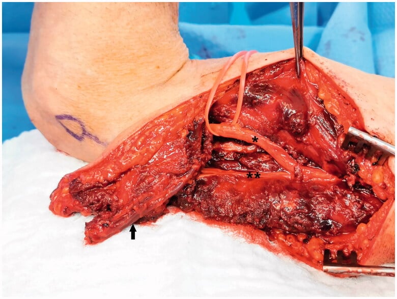
Management. An acute ligament avulsion is repaired to the isometric point on the lateral epicondyle with an anchor or transosseous sutures. Chronic insufficiency needs reconstruction with a tendon graft (palmaris longus, gracilis or triceps fascia) from the isometric point to the supinator crest of the ulna, usually in a docking or figure-of-eight configuration. At closure, preserve and reattach the anconeus and the common extensor origin: the dynamic restraints matter.
Surgical Relevance
The interval. Between anconeus (radial nerve) and extensor carpi ulnaris (posterior interosseous nerve): a true internervous plane, and the standard lateral approach to the elbow. The skin incision runs from just proximal to the lateral epicondyle, curving distally toward the subcutaneous border of the ulna, roughly 6 to 8 cm.
Finding it. Look for the fine fat stripe running obliquely between the anconeus and extensor carpi ulnaris. It is the reliable landmark, and it is easy to miss if the dissection is too proximal, because the two muscles are fused near the epicondyle.
Deep dissection and exposure. The joint capsule is entered anterior to the equator of the radial head with the forearm pronated. The approach exposes the radial head and neck, the capitellum, the lateral collateral ligament complex and, with additional anterior capsular release, the anterolateral coronoid.
The single most important hazard of the Kocher approach is iatrogenic posterolateral rotatory instability created by dividing the lateral ulnar collateral ligament, which lies at the floor of the interval. Three rules prevent it:
- Enter the interval and perform the capsulotomy anterior to the equator (midline) of the radial head. The ligament attaches posteriorly; staying anterior to the midline keeps you in front of it.
- Pronate the forearm. Pronation moves the posterior interosseous nerve away distally and also relaxes and displaces the lateral structures favourably.
- If the ligament must be released for exposure, for example in a terrible triad or a complex fracture-dislocation, release it from the humeral epicondyle with a bony or soft-tissue tag and repair it back to the isometric point with a bone anchor or transosseous sutures at closure.
- Where
- Floor of the Kocher interval
- Distance from a landmark
- Attaches POSTERIOR to the equator of the radial head
- Protection
- Keep the capsulotomy ANTERIOR to the midline of the radial head
- Where
- Within the supinator, crossing the radial shaft
- Distance from a landmark
- Mean 52.0 plus or minus 7.8 mm distal to the radiocapitellar joint in PRONATION (minimum 38 mm); falls to a mean 33.4 plus or minus 5.7 mm (minimum 22 mm) in supination
- Protection
- PRONATE the forearm; do not dissect beyond about 3.8 cm distal to the joint even in pronation
- Where
- Anterior, in the brachialis-brachioradialis interval
- Distance from a landmark
- Proximal extension of the approach
- Protection
- Do not extend proximally beyond the epicondyle without identifying it
- Where
- Subcutaneous, posterolateral
- Distance from a landmark
- Crosses the field superficially
- Protection
- Spread rather than cut in the subcutaneous plane
The pronation rule, and why it is the opposite of Henry. Diliberti and colleagues performed this posterolateral approach between anconeus and extensor carpi ulnaris in 32 cadaveric specimens and measured the posterior interosseous nerve safe zone from the capitellum in each forearm position; the distances are in the table above. The angle between the nerve and the radial shaft was 47.4 plus or minus 6.8 degrees in supination and fell to 27.8 plus or minus 6.7 degrees in pronation, and their conclusion was that approaching the lateral proximal radius is safest in pronation.
The exam trap is that the volar Henry approach requires supination to move the nerve away, whereas the posterolateral Kocher or Thompson approach requires pronation. The directions are opposite because you are approaching from opposite sides, and candidates confuse this constantly.
Guidelines, Registries & Global Practice
Anatomical Variation
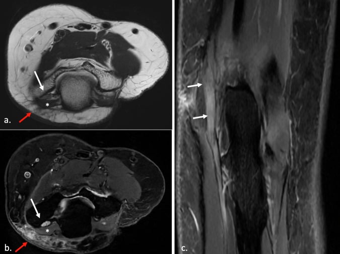
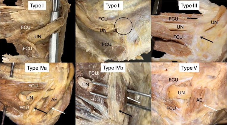
- Anconeus epitrochlearis is present in roughly 10 to 30 per cent of limbs across published series, with substantial variation between populations and between imaging-based and cadaveric estimates. It is common enough that its absence should not be assumed at cubital tunnel decompression.
- The anconeus itself is anatomically constant in position and attachment, which is one reason the flap is so reliable — the published series reported no flap failures across 20 consecutive cases.
- Accessory slips between the anconeus and the extensor carpi ulnaris can obscure the Kocher interval; the fat stripe rather than a visible gap is the reliable landmark.
- The anconeus epitrochlearis should not be confused with the dorsoepitrochlearis (a latissimus dorsi slip to the medial epicondyle or triceps), which is a different variant with different consequences.
Side-by-Side Guidance
- Position relevant to the anconeus and lateral elbow
- Describes the Kocher interval between anconeus and extensor carpi ulnaris, with explicit instruction to pronate the forearm to protect the posterior interosseous nerve and to keep the capsulotomy anterior to the radial head equator to protect the lateral ulnar collateral ligament.
- Position relevant to the anconeus and lateral elbow
- Originated both the anconeus rotation reconstruction for triceps insufficiency and the largest published anconeus flap series; regards the muscle as the first-line local option for posterior elbow cover.
- Position relevant to the anconeus and lateral elbow
- Universal agreement that the lateral ulnar collateral ligament is the primary static restraint to posterolateral rotatory instability, that repair or reconstruction must be to the isometric point, and that iatrogenic injury during lateral release is a leading cause.
- Position relevant to the anconeus and lateral elbow
- Where an anconeus epitrochlearis is found, excision alone in combination with in situ decompression is generally sufficient; the finding is not by itself an indication for transposition.
Global Practice Differences
- Soft-tissue cover of the posterior elbow is where the greatest practical difference lies. In centres with microsurgical capability the choice includes free tissue transfer; in centres without it the anconeus rotation flap is the definitive single-stage solution and requires no specialised equipment, no vessel sacrifice and no second operative site. It is arguably one of the highest-value techniques in limited-resource upper limb surgery.
- Similarly for triceps reconstruction: Achilles tendon allograft is unavailable in most of the world, which makes anconeus rotation and triceps turn-down the practical options and raises the value of knowing this anatomy well.
- Elbow arthroscopy availability varies widely; the soft spot remains the universally available access point for diagnostic aspiration regardless of resource setting, and it is the single most useful piece of anconeus surface anatomy.
Registry and Outcome Signals
- Neither anconeus flaps nor lateral collateral ligament reconstructions are captured in national registries; the evidence base is institutional series. The Elhassan series of 20 consecutive flaps remains the largest published clinical experience.
- Total elbow arthroplasty is captured in national joint registries, but triceps insufficiency is systematically under-represented because it is treated by soft-tissue reconstruction rather than component revision and therefore does not register as a revision event.
Rehabilitation Consensus
- After lateral collateral ligament repair or reconstruction, the elbow is protected in pronation and flexion initially, then mobilised in a hinged brace preventing terminal extension and supination for around six weeks. Supination with the elbow extended is the position that stresses the reconstruction.
- After an anconeus flap, early active motion is encouraged; the published series achieved full range of motion in all patients, so there is no rationale for prolonged immobilisation.
Related pages: Posterolateral Rotatory Instability of the Elbow is the condition the lateral ulnar collateral ligament beneath this muscle exists to prevent, and the reason the Kocher capsulotomy must stay anterior to the equator of the radial head; Elbow Dislocations is how that ligament is usually lost in the first place. Posterior Interosseous Nerve Anatomy and Posterior Interosseous Syndrome are the structure the pronation safe zone protects and what happens when it is not. Radial Head Fractures and Radial Head Arthroplasty are the operations the anconeus-ECU interval is most often opened for, and Olecranon Fractures is the injury whose wound breaks down over the muscle used to cover it. Triceps Brachii Anatomy shares this muscle's nerve supply from the branch to the medial head, Triceps Tendon Rupture and Total Elbow Arthroplasty are the setting for anconeus rotation, and Cubital Tunnel Syndrome is where the separately named anconeus epitrochlearis - a different muscle with a different nerve - actually matters. Lateral Elbow Pain and Lateral Epicondylitis are the differential for the posterolateral tenderness this muscle can produce.
MCQ Practice Points
Q: Where does anconeus arise and insert? A: Posterior aspect of the lateral epicondyle (deep to the common extensor origin) to the lateral olecranon and the proximal quarter of the posterior ulna.
Q: How is anconeus innervated? A: The radial nerve (C7, C8), via the terminal continuation of the nerve to the MEDIAL HEAD OF TRICEPS, which passes through the medial head to reach it.
Q: What are the two muscles of the Kocher interval and their nerves? A: Anconeus (radial nerve) and extensor carpi ulnaris (posterior interosseous nerve) — a true internervous plane.
Q: What lies at the floor of the Kocher interval and why does it matter? A: The lateral ulnar collateral ligament. Dividing it creates iatrogenic posterolateral rotatory instability. Keep the capsulotomy anterior to the equator of the radial head.
Q: Which forearm position protects the posterior interosseous nerve in a Kocher approach? A: PRONATION — safe zone mean 52 mm (minimum 38 mm) from the capitellum, versus 33 mm (minimum 22 mm) in supination. The volar Henry approach requires the opposite.
Q: What is the pedicle of the anconeus flap? A: The recurrent posterior interosseous artery, entering the deep surface distally. Pedicle length 31.3 mm, diameter 1.9 mm — enough for microsurgical anastomosis.
Q: What outcomes are reported for the local pedicled anconeus flap? A: 20 of 20 elbows healed at a mean of 3 weeks, no flap complications, all regained full range of motion, with defects up to 7 by 4 cm.
Q: What are the three Mayo options for triceps insufficiency after total elbow arthroplasty? A: Direct suture repair (7 of 16), ANCONEUS ROTATION (4 of 16) and Achilles tendon allograft (4 of 16). Extension against gravity restored in 15 of 16.
Q: What is the anconeus epitrochlearis and what does it cause? A: An accessory muscle from the medial epicondyle to the olecranon across the roof of the CUBITAL TUNNEL, supplied by the ULNAR nerve. It is a cause of cubital tunnel syndrome and is excised if found.
Q: What is the soft spot and what traverses it? A: The triangle of lateral epicondyle, olecranon tip and radial head. The direct lateral portal traverses the anconeus — the safest portal at the elbow, and the site for aspiration and injection.
Q: What is the anconeus principally for? A: Dynamic stabilisation — resisting varus and posterolateral rotatory displacement, and abducting the ulna during pronation. Its extension contribution is negligible.
Exam Viva Scenarios
Practise clinical reasoning and management decisions out loud
“You are fixing a Mason type III radial head fracture through a Kocher approach. Describe the interval and tell me the two structures you are most worried about and how you protect each.”
“A 62-year-old man has an olecranon fracture fixed with a tension band eight months ago. The wound has broken down over the olecranon; the plate and bone are exposed through a 5 by 3 cm defect. He has had three debridements and two attempted closures. Deep tissue cultures are negative. What do you do?”
“A 45-year-old woman had an open lateral epicondylitis release 18 months ago. She now describes clicking and a sense of the elbow giving way when she pushes up out of a chair. What is your diagnosis, how do you confirm it, and what is your management?”
Anatomy
- Origin: posterior lateral epicondyle, deep to the common extensor origin
- Insertion: lateral olecranon and proximal QUARTER of the posterior ulna
- Triangular, subcutaneous, fascia continuous with the triceps aponeurosis
- Fills the SOFT SPOT triangle: lateral epicondyle, olecranon, radial head
Innervation and Supply
- Radial nerve C7, C8 via the nerve to the MEDIAL HEAD of triceps
- The branch passes THROUGH the medial head to reach it
- Pedicle: recurrent posterior interosseous artery, deep surface distally
- Pedicle 31.3 mm long, 1.9 mm diameter - suitable for free transfer
Kocher Approach
- Anconeus (radial) / ECU (PIN) - true internervous plane
- Find the FAT STRIPE; muscles are fused near the epicondyle
- LUCL at the FLOOR - stay ANTERIOR to the radial head equator
- PRONATE: PIN safe zone 52 mm (min 38); supination only 33 mm (min 22)
Flap
- Covers posterior elbow defects up to 7 x 4 cm
- 20 of 20 healed at a mean of 3 weeks, no flap complications
- All patients regained FULL range of motion
- One of the three Mayo options for triceps insufficiency
Function and Pathology
- Dynamic stabiliser - resists varus and PLRI; extension role negligible
- EMG activity not confined to extension (Basmajian and Griffin)
- Forms a sling with the deep brachialis head around the ulnohumeral joint
- Anconeus epitrochlearis: ULNAR-innervated accessory muscle causing cubital tunnel syndrome
Evidence Base
Outcome of Local Anconeus Flap Transfer to Cover Soft Tissue Defects over the Posterior Aspect of the Elbow
- Twenty elbows in 20 patients with chronic posterior elbow soft-tissue defects managed with a local pedicled anconeus flap
- Mean defect size 4 by 3 cm, range 2 by 3 cm up to 7 by 4 cm; mean symptom duration 11 months with a mean of 3 previous debridements and attempted closures
- Wound healing was achieved in ALL elbows at a mean of 3 weeks (range 2 to 5)
- No flap complications occurred either intra-operatively or postoperatively
- At a mean 17 months, subjective elbow value improved from 70 to 95 per cent and Mayo Elbow Performance Score from 74 to 90 (both p equals 0.001); all patients had full range of motion and all were satisfied
Anatomical Considerations Regarding the Posterior Interosseous Nerve During Posterolateral Approaches to the Proximal Radius
- The posterolateral approach BETWEEN ANCONEUS AND EXTENSOR CARPI ULNARIS was performed in 32 cadaveric specimens and the posterior interosseous nerve exposed
- Pronation allowed safe exposure of at least the proximal 38 mm of the lateral radius, with a mean proximal safe zone of 52.0 plus or minus 7.8 mm
- Supination reduced the safe zone to as little as 22 mm, mean 33.4 plus or minus 5.7 mm
- The angle formed by the nerve and the radial shaft averaged 47.4 plus or minus 6.8 degrees in supination, falling to 27.8 plus or minus 6.7 degrees in pronation
- Approaching the lateral aspect of the proximal radius is safest in pronation
Function of Anconeus Muscle: An Electromyographic Study
- The classic electromyographic study of anconeus function, and the reference from which the muscle's modern characterisation derives
- No abstract is indexed for this 1972 paper; the specific recordings are not reproduced here
- The conclusion consistently attributed to it, and reflected in all subsequent elbow literature, is that anconeus activity is not confined to elbow extension
- That activity pattern is the basis for describing anconeus as a dynamic stabiliser of the ulnohumeral joint rather than a meaningful contributor to extension torque
Triceps Insufficiency Following Total Elbow Arthroplasty
- Review of 887 total elbow arthroplasties (1982 to 2001); 16 elbows in 14 patients underwent a subsequent triceps procedure
- Three reconstruction techniques were used, selected on tissue quality, tendon retraction and the state of the olecranon
- ANCONEUS ROTATION was used in 4 of 16 elbows, alongside direct suture in 7 and Achilles tendon allograft in 4
- Capacity to extend against gravity was restored in 15 of 16 elbows
- Mayo Elbow Performance Score at a mean 67 months: 11 excellent, 3 good, 2 clinical failures
Functional Anconeus Free Flap for Thenar Reconstruction: A Cadaveric Study
- Eight cadaveric upper extremities dissected to characterise the anconeus and its neurovasculature against the abductor pollicis brevis
- Anconeus fibre length 88.0 plus or minus 9.9 mm and area 1341.9 plus or minus 230.4 mm squared, both larger than abductor pollicis brevis (57.7 mm and 987.7 mm squared)
- No significant difference in fibre angle (70.5 versus 78.4 degrees), artery diameter (1.9 versus 2.0 mm) or nerve diameter (1.7 versus 2.1 mm)
- The vascular pedicle — the recurrent posterior interosseous artery — measured 31.3 plus or minus 6.9 mm in length and 1.9 plus or minus 0.2 mm in diameter, with venae comitantes of 1.0 mm
- Pedicle length and calibre are sufficient for microsurgical anastomosis