A lucency centred on the epiphysis or subarticular bone — the site itself is the strongest discriminator in bone tumour radiology
- The epiphysis (and its adult equivalent, the subarticular bone) is a restrictive location: the credible list is short and age does most of the work.
- Skeletally immature: chondroblastoma, infection (Brodie's abscess), Langerhans cell histiocytosis, rarely aneurysmal bone cyst.
- Skeletally mature: giant cell tumour, intraosseous ganglion, degenerative geode, infection, metastasis/myeloma, clear cell chondrosarcoma.
- Chondroblastoma characteristically has florid perilesional marrow and soft tissue oedema on MRI, out of proportion to a small lesion — do not call it aggressive.
- Giant cell tumour abuts the subchondral plate, is eccentric, has no matrix and no sclerotic rim, and is lytic and geographic.
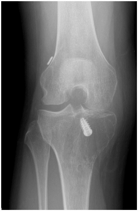
- A subarticular lucency in an adult with joint space narrowing and osteophytes is a geode until proven otherwise — do not biopsy osteoarthritis.
- “Say the age, say the physeal status, then say the location relative to the physis — that sequence wins marks before you name a lesion.
- “Multiple subarticular lucencies with erosions and preserved joint space suggests inflammatory arthropathy, gout or amyloid, not a tumour.
- “Any epiphyseal lesion with a periosteal reaction or cortical breach must have infection on the list.
- “Clear cell chondrosarcoma is the low-grade epiphyseal chondrosarcoma of the proximal femur in the 30–50 age group — treat it as chondroblastoma's dangerous twin.
Chondroblastoma and osteoid osteoma both generate enormous perilesional marrow oedema and a sympathetic effusion. Oedema is a reaction, not a grade. Judge aggressiveness on the margin, the cortex and the periosteum on the radiograph, not on MRI signal.
"Epiphyseal" only means something with reference to the growth plate. In an adult you are describing a subarticular lesion. Announcing chondroblastoma in a 55-year-old is a classic failure — that lesion is clear cell chondrosarcoma or giant cell tumour.
Subchondral cysts in osteoarthritis, rheumatoid disease and avascular necrosis are common and benign. The clue is on the joint, not the lesion: cartilage loss, sclerosis, osteophytes or a collapsed articular surface. Look at the joint before you look at the hole.
A subacute epiphyseal osteomyelitis in a child, and tuberculosis in an adult, both present as a painless-to-mildly-painful subarticular lucency. Any lesion crossing the physis or with an adjacent effusion and raised inflammatory markers must be aspirated before it is treated as a tumour.
Recognising the Pattern
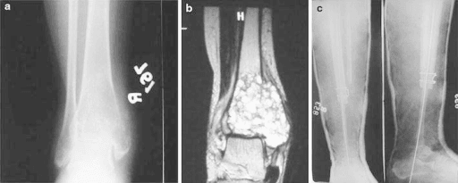
Definition. A lesion whose epicentre lies within the secondary ossification centre (skeletally immature) or within the subarticular bone between the articular surface and the fused physeal scar (skeletally mature). Typical hosts are the proximal humerus, proximal and distal femur, proximal tibia, distal radius, talus, calcaneus and patella.
Confirming the pattern is genuine.
- Identify the physis or the physeal scar on both views. If the epicentre is on the metaphyseal side, this is a metaphyseal lesion that has merely extended across, and the differential changes entirely.
- Get two orthogonal views. An apparently epiphyseal lucency on an anteroposterior film is frequently a metaphyseal or cortical lesion projected over the epiphysis on the lateral.
- Check the lesion abuts the subchondral plate. Giant cell tumour characteristically extends to within a few millimetres of the articular surface; chondroblastoma usually does not need to.
- Assess the margin: geographic with a thin sclerotic rim (type IA), geographic with a sharp non-sclerotic edge (IB), or ill-defined (IC or moth-eaten).
Words to use out loud. "This is an anteroposterior and lateral radiograph of the left knee in a skeletally immature patient — the physes are open. There is a geographic lucent lesion centred on the epiphysis of the proximal tibia, eccentric and abutting but not breaching the subchondral plate. It has a thin sclerotic margin, measures approximately two centimetres, and there are punctate internal calcifications. There is no cortical destruction, no periosteal reaction and no soft tissue mass. My leading diagnosis is chondroblastoma."
Mimics and false positives.
- Normal variants: the dorsal defect of the patella, and the pseudocyst of the humeral head at the greater tuberosity insertion, are both asymptomatic well-corticated subarticular lucencies.
- Overlying gas or bowel on pelvic and shoulder films.
- Focal marrow heterogeneity on MRI in the proximal humerus and femur — no radiographic correlate, no oedema, no discrete margin.
- Physiological red marrow islands and bone islands (enostoses), which are sclerotic rather than lucent but are often the cause of an "abnormal epiphysis" referral.
- Kissing subchondral cyst pairs either side of a degenerate joint, which is a joint diagnosis masquerading as a bone lesion.
Next Investigation
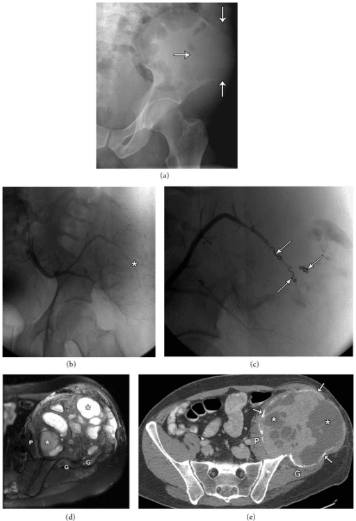
The Differential
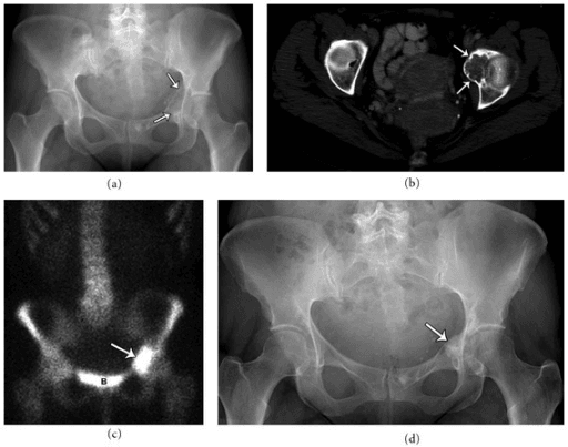
- Typical age / setting
- 30–50, proximal femoral head, male predominance
- Discriminating feature
- Looks exactly like a chondroblastoma but the physes are long fused; often larger than 4 cm with expansion of the femoral head
- What confirms it
- MRI shows cartilage-type lobular high T2 signal without the florid oedema; biopsy shows clear cells with a low-grade chondroid matrix
- Typical age / setting
- Children under 10 with transphyseal vessels; any age in immunosuppression
- Discriminating feature
- Serpiginous 'penumbra sign' — a T1 hyperintense rim of granulation tissue lining the cavity; may cross the physis
- What confirms it
- MRI penumbra sign plus target rim enhancement; aspiration and culture; raised CRP
- Typical age / setting
- Adults, endemic exposure, hip and knee
- Discriminating feature
- Juxta-articular osteopenia with marginal erosions and preserved joint space until late (Phemister triad), minimal sclerosis
- What confirms it
- Aspirate for acid-fast bacilli, culture and PCR; synovial biopsy
- Typical age / setting
- Over 40, known primary, subarticular lucency
- Discriminating feature
- Ill-defined margin with cortical destruction and no reactive rim; often multiple when the skeletal survey is reviewed
- What confirms it
- CT chest–abdomen–pelvis, serum electrophoresis, whole-body imaging; biopsy if solitary
- Typical age / setting
- 20–45, physes closed, distal femur, proximal tibia, distal radius, sacrum
- Discriminating feature
- Eccentric, purely lytic, no matrix, non-sclerotic margin, extends to the subchondral plate; narrow zone of transition
- What confirms it
- Radiograph plus MRI for soft tissue extension; biopsy shows osteoclast-like giant cells with H3F3A G34W mutation
- Typical age / setting
- 10–20, open physes, proximal humerus, femur, tibia, talus, calcaneus
- Discriminating feature
- Small (usually less than 4 cm), thin sclerotic rim, punctate chondroid calcification, with disproportionate perilesional marrow oedema and joint effusion
- What confirms it
- MRI oedema pattern with lobulated low-to-intermediate T2 signal; biopsy shows chicken-wire calcification and S100-positive chondroblasts
- Typical age / setting
- Over 50, weight-bearing joint
- Discriminating feature
- Sits directly beneath a denuded articular surface with joint space narrowing, sclerosis and osteophytes; often multiple and bilateral
- What confirms it
- No further imaging needed if joint changes are convincing; CT if the diagnosis is doubted
- Typical age / setting
- 30–60, medial malleolus, distal tibia, femoral head, carpal bones
- Discriminating feature
- Well-corticated, thick sclerotic rim, normal adjacent joint space — unlike a geode there is no arthritis
- What confirms it
- MRI shows uniform fluid signal with rim-only enhancement; no solid component
- Typical age / setting
- Steroids, alcohol, sickle cell, trauma; femoral head and knee
- Discriminating feature
- Serpiginous sclerotic demarcation line and a subchondral crescent lucency parallel to the articular surface, not a round lesion
- What confirms it
- MRI double-line sign on T2; radiographs to stage collapse
- Typical age / setting
- Under 15, may be anywhere; epiphyseal involvement uncommon
- Discriminating feature
- Aggressive-appearing but painful and rapidly evolving lucency; may show a lamellated periosteal reaction and heals spontaneously
- What confirms it
- Skeletal survey for other lesions; biopsy shows CD1a and langerin positive cells
- Typical age / setting
- Under 20; usually metaphyseal, epiphyseal in about a tenth
- Discriminating feature
- Expansile with a thin shell and multiple fluid–fluid levels filling the whole lesion
- What confirms it
- MRI fluid–fluid levels; biopsy to exclude a secondary ABC within giant cell tumour or telangiectatic osteosarcoma
- Typical age / setting
- 10–25, femoral neck, elbow, talar neck
- Discriminating feature
- Night pain relieved by NSAIDs with a synovitis and little sclerosis (intra-articular lesions do not produce the usual reactive sclerosis)
- What confirms it
- Thin-slice CT showing a lucent nidus less than 1.5 cm with a central calcified dot
- Typical age / setting
- Over 40, chronic gout, long-term dialysis
- Discriminating feature
- Para-articular erosion with an overhanging edge and preserved joint space; adjacent soft tissue density
- What confirms it
- Dual-energy CT for monosodium urate; serum urate; dialysis history for beta-2 microglobulin amyloid
- Typical age / setting
- Children and young adults, hand and long bones
- Discriminating feature
- Rings-and-arcs chondroid matrix without oedema or endosteal scalloping greater than two-thirds of cortical thickness
- What confirms it
- Radiograph often sufficient; MRI to exclude aggressive features if painful
Narrowing It Down
- 1Step 1 — Are the physes open or closed?
This single question splits the list in half. Open physes point to chondroblastoma, subacute osteomyelitis, Langerhans cell histiocytosis and aneurysmal bone cyst. Closed physes point to giant cell tumour, intraosseous ganglion, geode, metastasis, myeloma and clear cell chondrosarcoma. A patient in their thirties with a chondroid epiphyseal lesion is clear cell chondrosarcoma until proven otherwise.
Physeal status alone halves the differential - look at it before anything else
- 2Step 2 — Is it solitary or are there others?
Multiple subarticular lucencies with preserved joint space and marginal erosions suggests inflammatory arthropathy, gout or dialysis amyloid. Multiple lytic lesions in an adult over 40 suggests metastasis or myeloma and mandates serum electrophoresis and staging imaging. A single lesion keeps the primary bone tumour list alive.
Multiple redirects to biochemistry and staging; solitary keeps the tumour list open
- 3Step 3 — Where exactly within the epiphysis?
Abutting the subchondral plate and eccentric favours giant cell tumour. Central within the ossification centre with a sclerotic rim favours chondroblastoma. Directly beneath a denuded articular surface with an arthritic joint means geode. Crescentic and parallel to the articular surface means avascular necrosis, not a mass.
Position within the epiphysis separates GCT from chondroblastoma from geode
- 4Step 4 — What is the matrix?
Punctate or rings-and-arcs calcification means cartilage: chondroblastoma if immature, clear cell chondrosarcoma or enchondroma if mature. Ground-glass suggests fibrous dysplasia. A total absence of matrix in a lytic subarticular lesion in a young adult is the signature of giant cell tumour. Fluid–fluid levels throughout mean aneurysmal bone cyst — but exclude a secondary component.
Cartilage matrix in a MATURE skeleton is the finding that must not be filed as benign
- 5Step 5 — How aggressive is the margin?
Grade with the Lodwick system on the radiograph, not the MRI. A thin sclerotic rim (IA) supports chondroblastoma, ganglion or Brodie's abscess. A sharp non-sclerotic edge (IB) supports giant cell tumour. Moth-eaten or permeative destruction with cortical breach in an epiphysis means infection, metastasis or, rarely, an epiphyseal osteosarcoma or lymphoma — arrange staging and biopsy.
Grade I is 94 per cent benign and grade III 81 per cent malignant; IC decides nothing
- 6Step 6 — Are there systemic or joint features?
Fever, raised CRP and an effusion push infection to the top and demand aspiration before biopsy. Night pain abolished by NSAIDs points to osteoid osteoma. Steroid, alcohol or sickle cell history points to avascular necrosis. Osteoarthritic joint changes make a geode the answer and no further work-up is needed.
An arthritic joint ends the investigation; a fever changes the order of it
- 7Step 7 — Decide: observe, aspirate, or biopsy at a tumour unit
Confident geode, ganglion or normal variant with a corroborating clinical picture: reassure and discharge. Suspected infection: aspirate. Anything with cartilage matrix in an adult, anything Lodwick IB or worse, or any diagnostic doubt: refer to a specialist sarcoma service for staging and image-guided biopsy along the definitive surgical approach.
Refer BEFORE biopsy - the tract must be excisable with the specimen
MCQ Practice Points
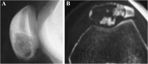
Q: A 16-year-old has a 3 cm lucency in the talus with a sclerotic rim and punctate calcification. MRI shows extensive marrow oedema. What is the diagnosis?
A: Chondroblastoma. The talus and calcaneus are classic hindfoot sites. The disproportionate oedema is characteristic and must not be interpreted as an aggressive feature. Expect roughly one in five to recur by two years after curettage and grafting — Turcotte's Mayo series reported 82 per cent local control — so this is a lesion to follow, not discharge.
Q: Which single radiographic feature best separates giant cell tumour from chondroblastoma?
A: Physeal status. Giant cell tumour occurs after physeal closure; chondroblastoma occurs before it. Secondary features: giant cell tumour has no matrix and no sclerotic rim, chondroblastoma has both. Where the case straddles closure or sits at an odd site, the molecular answer is definitive — H3F3B K36M in 95 per cent of chondroblastomas against H3F3A G34W in 92 per cent of giant cell tumours.
Q: A 55-year-old has a 1 cm lucency in the medial tibial plateau with joint space narrowing, subchondral sclerosis and marginal osteophytes. What is the next step?
A: Reassurance and management of the osteoarthritis. This is a degenerative geode. No cross-sectional imaging and no biopsy — the arthritic joint is the diagnosis, and imaging it further generates anxiety and unnecessary intervention.
Q: What is the penumbra sign and what does it indicate?
A: A rim of T1-hyperintense granulation tissue lining a lytic cavity on unenhanced T1-weighted MRI. It is a strong indicator of subacute osteomyelitis or Brodie's abscess and helps distinguish it from a tumour. It is a positive sign only: its absence does not exclude infection, and where fever, a raised CRP or an effusion is present the lesion is aspirated before it is biopsied.
Q: A 40-year-old man has a lytic lesion of the femoral head with rings-and-arcs calcification, measuring 5 cm. Which diagnosis must be excluded and how is it treated?
A: Clear cell chondrosarcoma. It is a low-grade malignancy that mimics chondroblastoma in an older patient. Treatment is wide en bloc resection — curettage carries an unacceptable local recurrence rate. The discriminator is skeletal maturity: cartilage matrix in an epiphysis with a CLOSED physis is malignant until proven otherwise, and a negative H3F3 immunostain does not make it benign.
Exam Viva Scenarios
Practise clinical reasoning and management decisions out loud
“You are shown an anteroposterior radiograph of the left shoulder in a 14-year-old boy with six months of aching shoulder pain and a small effusion. There is a 2.5 cm geographic lucency in the humeral head with a thin sclerotic rim and faint internal calcification. The physis is open.”
“You are shown radiographs of a knee in a 32-year-old woman with three months of pain and a recent effusion. There is an eccentric, purely lytic lesion in the medial femoral condyle extending to within 2 mm of the subchondral plate. The margin is sharp but not sclerotic. No matrix. The physes are fused.”
“You are shown an anteroposterior radiograph of the knee in a 6-year-old with a two-week limp and low-grade fever. There is an ill-defined lucency in the distal femoral epiphysis extending towards the physis, with a faint surrounding sclerotic halo. CRP is 48.”
Skeletally immature
- Chondroblastoma — under 4 cm, sclerotic rim, chondroid matrix, florid oedema
- Subacute osteomyelitis — penumbra sign, may cross the physis, raised CRP
- Langerhans cell histiocytosis — rapidly evolving, painful, may be multifocal
- Aneurysmal bone cyst — expansile with fluid–fluid levels throughout
Skeletally mature
- Giant cell tumour — eccentric, lytic, no matrix, no sclerotic rim, abuts subchondral plate
- Intraosseous ganglion — thick sclerotic rim, normal joint
- Degenerative geode — arthritic joint above it
- Metastasis / myeloma — over 40, ill-defined, cortical destruction
- Clear cell chondrosarcoma — chondroid matrix in the femoral head, 30–50 years
Red flags demanding referral
- Lodwick IB margin or worse
- Cortical breach, periosteal reaction or soft tissue mass
- Cartilage matrix in a skeletally mature patient
- Any lesion greater than 5 cm or rapidly enlarging
- Age over 40 with a destructive subarticular lucency
Investigation sequence
- Orthogonal radiographs of the whole bone first — physeal status, margin, matrix, joint
- MRI with contrast for extent, oedema, fluid levels and penumbra sign
- Thin-slice CT for matrix, sequestrum and nidus
- Aspirate if infection is plausible, before biopsy
- Biopsy only at the definitive treating unit, along the surgical approach
- No further imaging for geodes, dorsal patellar defect and humeral pseudocyst
Viva phrases that score
- 'The physes are open, so this is a lesion of the secondary ossification centre'
- 'The margin is Lodwick IA with a narrow zone of transition'
- 'The perilesional oedema is reactive and does not imply aggression'
- 'I would exclude infection with inflammatory markers and aspiration before biopsy'
- 'Biopsy would be performed at the sarcoma unit along the planned resection approach'
Evidence Base
Distinct H3F3A and H3F3B Driver Mutations Define Chondroblastoma and Giant Cell Tumour of Bone
- CHONDROBLASTOMA: p.Lys36Met (K36M) alterations, predominantly in H3F3B, in 73 of 77 cases (95 per cent).
- GIANT CELL TUMOUR: histone H3.3 alterations exclusively in H3F3A, giving p.Gly34Trp (G34W) - or in one case G34L - in 49 of 53 cases (92 per cent).
- The two lesions that share this page are separated by which of two nearly identical genes is mutated, and at which residue.
- THE MUTATIONS WERE RESTRICTED TO THE STROMAL CELL POPULATION and were not detected in osteoclasts or their precursors.
- Sets these alongside the H3F3A p.Lys27Met and p.Gly34Arg/Val alterations of childhood brain tumours - the same histone, different residues, entirely different diseases.
Chondroblastoma - the Mayo Series
- Affects the EPIPHYSEAL ENDS of long bones, in men, during the second and third decades.
- The tumour is located more frequently at OTHER sites in older patients - the epiphyseal rule is an age-dependent rule, not an absolute one.
- Treatment of choice is complete curettage with bone grafting.
- That provided local control in 82 per cent of patients at two years' follow-up.
- Recurrent tumours can usually be treated the same way again.
- Pulmonary metastasis is indexed on the record - the rare 'benign metastasising' behaviour is real.
Denosumab in Patients with Giant-Cell Tumour of Bone - an Open-Label, Phase 2 Study
- 37 patients with RECURRENT OR UNRESECTABLE giant cell tumour received subcutaneous denosumab 120 mg monthly with loading doses on days 8 and 15.
- 30 of 35 evaluable patients (86 per cent, 95 per cent CI 70-95) had a tumour response.
- But the response was defined as elimination of at least 90 per cent of GIANT CELLS or no radiological progression by week 25 - and the two arms of that definition performed very differently: 20 of 20 responded on histology against only 10 of 15 on radiology.
- The rationale is that the mononuclear stromal cells express RANKL, and denosumab is a monoclonal antibody against RANKL.
- The authors' own conclusion is that further investigation is WARRANTED - this is a signal, not a licence to replace surgery.
Which Treatment Is Best for Giant Cell Tumours of the Distal Radius? A Meta-Analysis
- Six studies pooled: 80 curettage cases against 59 en bloc excisions.
- Intralesional excision carried a HIGHER recurrence rate (RR 2.80, 95 per cent CI 1.17-6.71), and markedly so in Campanacci grade 3 (RR 4.90, CI 1.36-17.66).
- But intralesional excision carried FEWER major complications (RR 0.21, CI 0.09-0.54).
- POLYMETHYLMETHACRYLATE VERSUS BONE GRAFT MADE NO DIFFERENCE TO RECURRENCE (RR 0.98, CI 0.44-2.17).
- Concludes intralesional excision suits grades 1 and 2, and is less appropriate for grade 3.