Echinococcus granulosus in bone — a parasitic infection that must be treated with oncological surgical discipline
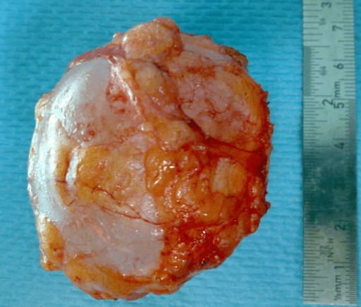
- Bone hydatid is not liver hydatid: no fibrous pericyst forms, so the parasite grows along paths of least resistance through cancellous bone and Haversian systems — the true extent always exceeds the radiological extent.
- The classical liver approach of percutaneous puncture-aspiration-injection-reaspiration (PAIR) does NOT apply to bone.
- Uncontrolled aspiration or open biopsy risks anaphylaxis and local seeding; if tissue is essential, do it with anaesthetic anaphylaxis readiness, scolicidal precautions and drug cover, through a line that can be excised.
- Serology is far less sensitive for osseous than hepatic disease — a negative test never excludes the diagnosis.
- Curettage is palliation. The only realistic cure is wide en-bloc excision, planned exactly as for a locally aggressive bone tumour, plus prolonged benzimidazole therapy.
- “Multiloculated honeycomb or 'bunch of grapes' osteolysis WITHOUT periosteal reaction and without sclerotic rim in an endemic-region patient — think hydatid.
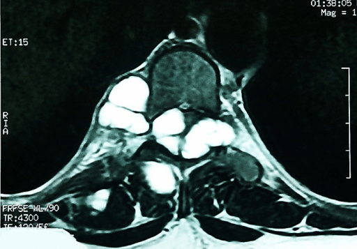
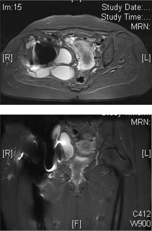
- “MRI: thin-walled cysts, low signal on T1, very high on T2, no or thin rim enhancement, daughter vesicles within a mother cyst; extension into psoas or epidural fat.
- “Spine: relative disc preservation with multilevel body and posterior element involvement helps separate it from tuberculous spondylitis.
- “Eosinophilia is inconstant and unreliable in bone disease.
- “Report the disease as chronic and relapsing — counsel the patient for decades of follow-up, not a single operation.
A needle into an unrecognised hydatid cyst can cause anaphylaxis and disseminates protoscolices along the tract. If biopsy is unavoidable, do it in theatre with anaesthetic support, adrenaline available, antihistamine and steroid cover, albendazole started beforehand, and a tract sited so it can be excised en bloc with the definitive specimen.
Intralesional treatment leaves microscopic vesicles in marrow beyond the visible cavity. Herrera's 26 patients, followed a mean of 12.8 years, make the consequence concrete: 73 per cent were disease-free at last review, but 9 of those 19 needed a second or further operation to get there, so only about 10 of 26 were cured by the index procedure. Of the 7 failures, 6 had chronic discharging sinuses. Ilium-and-hip disease was the hardest and always took multiple procedures. Their conclusion is the one to carry: results are satisfactory only where complete wide excision is possible, and in the pelvis and hip they are disappointing. Plan the case as a tumour resection, or accept that you are performing deliberate palliation and say so.
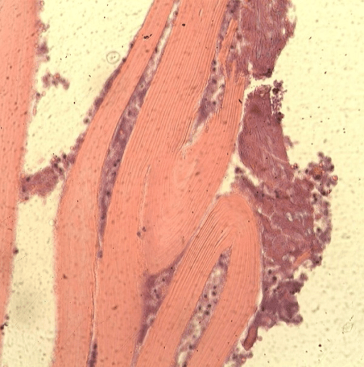
Sensitivity of indirect haemagglutination and ELISA is substantially lower for bone than liver disease because the parasite is walled by bone rather than exposed to systemic antigen presentation. Diagnosis is imaging plus context plus histology.
Cord compression demands urgent decompression, but leaving disease in the vertebral body and paraspinal tissue guarantees relapse. Aim for maximal safe excision, instrumented reconstruction, prolonged albendazole and indefinite imaging surveillance.
Parasite, Transmission and Global Epidemiology
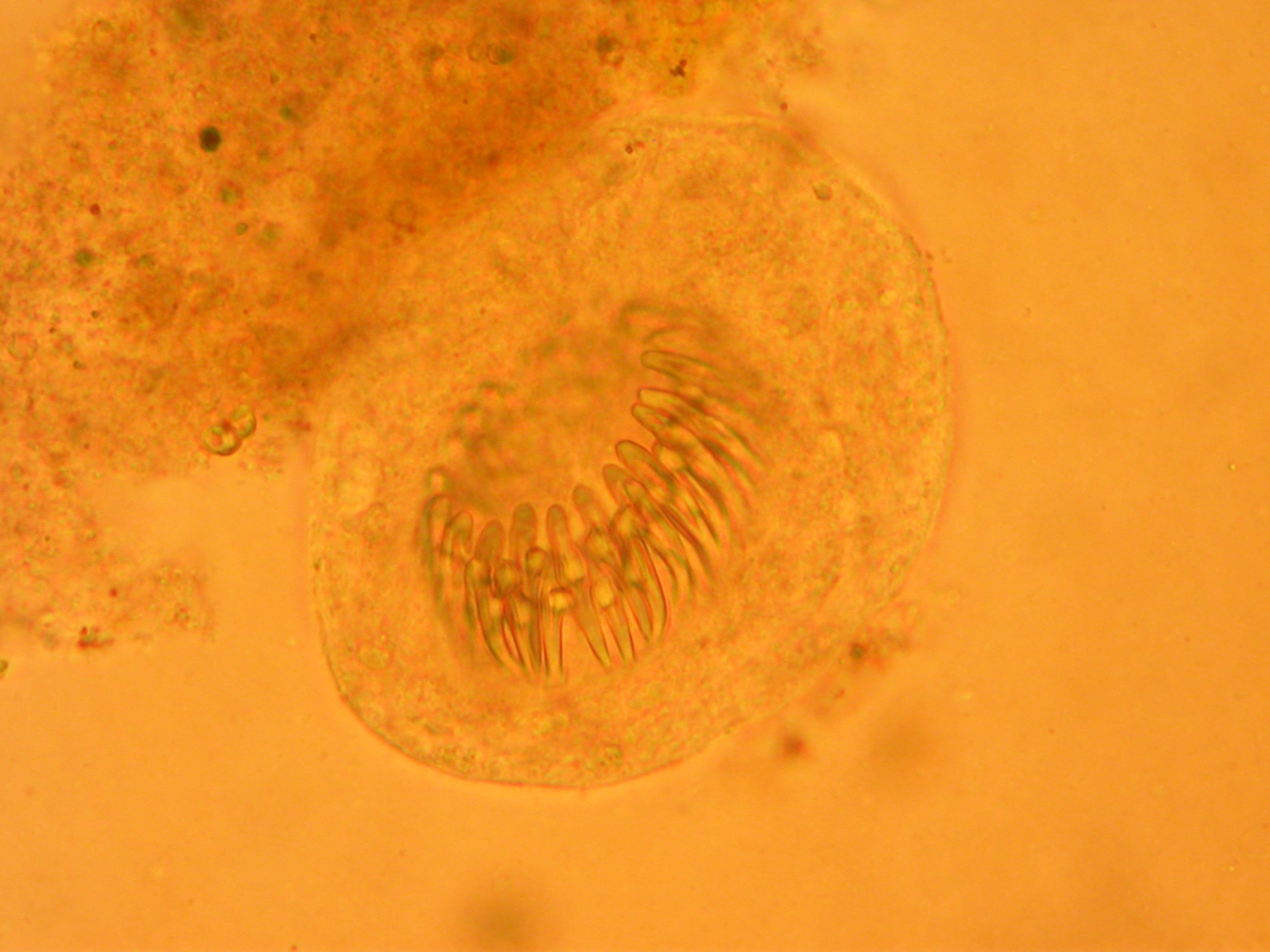
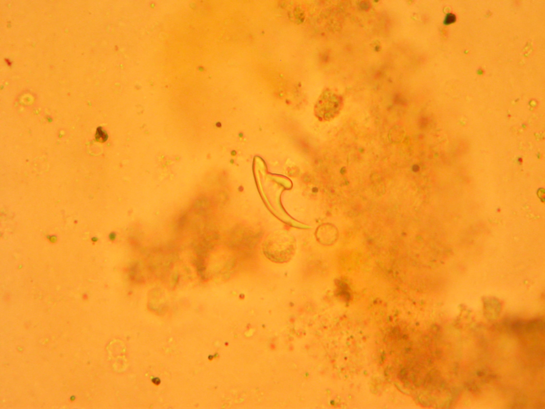
Organism. Echinococcus granulosus (cystic echinococcosis) causes almost all skeletal disease. E. multilocularis (alveolar echinococcosis) rarely involves bone but when it does behaves like an infiltrative malignancy without cyst walls at all.
Life cycle. The definitive host is the dog (and other canids), harbouring the adult tapeworm. Eggs are passed in faeces and ingested by an intermediate host — classically sheep, also goats, cattle, camels. Humans are accidental dead-end intermediate hosts, infected by hand-to-mouth contact with dog faeces or contaminated water and vegetables. Home slaughter of sheep with offal fed to dogs perpetuates the cycle.
Pathogenesis in humans. The oncosphere penetrates the duodenal mucosa, enters portal blood and is filtered first by the liver (around two-thirds of cases), then lung. Embryos escaping both filters seed systemically — bone accounts for only a small percentage of cases but a disproportionate share of morbidity.
Endemic regions. Mediterranean basin (Türkiye, Greece, Italy, Spain, North Africa), Middle East and Levant, Central Asia and western China, the Indian subcontinent, East Africa, and South America (Argentina, Peru, Uruguay, Chile). Diaspora and migration mean any surgeon anywhere may meet the disease — a patient who grew up herding sheep two decades ago remains at risk, because incubation is measured in years to decades.
Public health framing. Cystic echinococcosis is a WHO-listed neglected tropical disease. Control programmes rest on dog deworming with praziquantel, restricting home slaughter, offal disposal and livestock vaccination — surgical services alone never control the disease.
Why Bone Behaves Worse Than Liver
- Liver (soft organ)
- Fibrous pericyst forms and contains the parasite
- Bone
- No pericyst — rigid trabecular architecture prevents it
- Soft tissue after cortical breach
- Pericyst re-forms; conventional cysts in muscle and epidural fat
- Liver (soft organ)
- Spherical, unilocular, expansile
- Bone
- Exogenous budding vesicles creeping along marrow spaces, Haversian and Volkmann canals; multiloculated and irregular, following lines of least resistance
- Soft tissue after cortical breach
- Discrete daughter cysts in soft-tissue planes
- Liver (soft organ)
- Sharp shellable edge; clear plane for dissection
- Bone
- No visible edge, no dissection plane; lysis without sclerotic rim, no periosteal reaction until fracture or infection supervenes; microscopic extension beyond MRI signal
- Soft tissue after cortical breach
- Well-defined rounded cysts with a wall
- Liver (soft organ)
- PAIR or enucleation feasible
- Bone
- PAIR forbidden — no capsule to puncture safely; intralesional surgery gives near-universal recurrence; wide excision is the only cure
- Soft tissue after cortical breach
- Excision of cysts with intact wall, avoiding spillage
- Liver (soft organ)
- Not applicable
- Bone
- Pressure and osteoclastic resorption, cortical thinning, pathological fracture (femoral neck, humeral shaft); impaired healing and haematoma as a route of spread
- Soft tissue after cortical breach
- Extension into muscle and epidural fat; capsular reflections carry disease to adjacent bone
- Liver (soft organ)
- Not applicable
- Bone
- Cartilage resists the parasite so joint crossing is late; secondary bacterial infection mimics chronic osteomyelitis with sinuses and misleads first diagnosis
- Soft tissue after cortical breach
- Soft-tissue and capsular route dominates adjacent-bone spread
- Liver (soft organ)
- Curable with organ-sparing technique
- Bone
- Behaves as a locally aggressive benign tumour with satellite disease (analogues: recurrent giant cell tumour, low-grade chondrosarcoma); slow over years, no metastasis, albendazole suppressive not curative
- Soft tissue after cortical breach
- Recurrence unless every cyst is removed intact
BONEWhy Bone Hydatid Is Different
Hook:If you remember only one line: in BONE there is no bag, so you must take the bone.
Clinical Presentation
Tempo. Insidious. Median symptom duration before diagnosis is commonly measured in months to years. Any 'benign-looking' lytic lesion with a two-year history in an endemic-region patient deserves the thought.
Typical complaints by site
- Presentation
- Deep axial pain, then radicular pain, progressive paraparesis, sphincter disturbance; often presents already myelopathic
- Presentation
- Vague buttock or groin pain, palpable fluctuant mass, late acetabular collapse, lumbosacral plexus symptoms
- Presentation
- Hip pain, limp, then pathological subtrochanteric or neck fracture
- Presentation
- Aching, expansile swelling, pathological fracture
- Presentation
- Discharging sinus with clear vesicles ("grape skins" in the dressing) — pathognomonic when seen
Systemic features. Usually absent. Fever suggests secondary bacterial infection. Urticaria, wheeze or a documented anaphylactoid episode after minor trauma or a previous procedure is a strong clue to cyst rupture.
Examination discipline.
- Inspect the whole limb and trunk for old scars and sinuses — most patients are on their second or third episode.
- Palpate for a soft, non-tender, fluctuant paraspinal or gluteal mass.
- Full neurological examination with documented ASIA grade in spinal disease; record sphincter function and perianal sensation before any surgery.
- Examine the abdomen and request chest imaging: coexistent hepatic or pulmonary cysts are present in a substantial minority and change anaesthetic and systemic planning.
Imaging: How to Read It
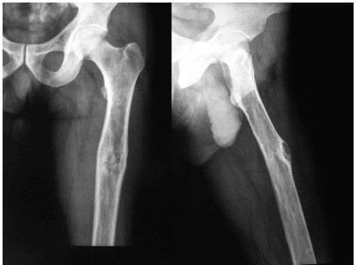
What to look for
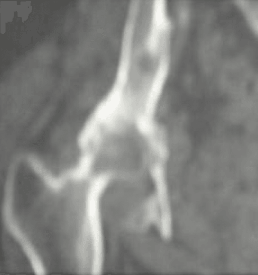
- Multiloculated, honeycomb or "bunch of grapes" osteolysis, coalescing round lucencies of varying size.
- Absent periosteal reaction and absent marginal sclerosis — this pairing is the single most useful plain-film discriminator against infection and against most tumours.
- Expansion with cortical thinning and scalloping from within; late cortical breach.
- No matrix mineralisation, no sequestrum.
- Fracture through thinned cortex.
- Spine: destruction of the vertebral body and pedicle with relative preservation of the disc space early — the reverse of pyogenic and (usually) tuberculous spondylitis.
Differential Diagnosis
- Features shared with hydatid
- Expansile lytic epiphyseal-metaphyseal lesion, no matrix, cortical thinning
- Discriminators
- Solid enhancing tissue not fluid signal on MRI; subarticular location; peak age 20 to 40; usually solitary and non-multiloculated
- Features shared with hydatid
- Multiloculated expansile lysis with septa, fluid content
- Discriminators
- Fluid-fluid levels with blood products of varying signal; thick enhancing septa; younger patients; rapid growth
- Features shared with hydatid
- Long-standing painless lytic-expansile bone lesion
- Discriminators
- Ground-glass matrix on CT, sclerotic rind, no fluid signal, no soft tissue mass
- Features shared with hydatid
- Purely lytic, no periosteal reaction, vertebral collapse
- Discriminators
- Older patient, paraprotein, solid enhancing marrow-replacing tissue, no cystic loculi
- Features shared with hydatid
- Endemic overlap, paraspinal collection, vertebral destruction
- Discriminators
- Disc destruction early, thick rim-enhancing abscess, calcified cold abscess, raised inflammatory markers, constitutional symptoms
- Features shared with hydatid
- Lysis, sinus, soft tissue collection
- Discriminators
- Sequestrum, involucrum, periosteal reaction, thick enhancing abscess wall, raised CRP
- Features shared with hydatid
- Lytic destruction, pathological fracture, older patient
- Discriminators
- Known primary, solid enhancement, avid on bone scan and PET, multiple lesions
Endemic exposure + multiloculated fluid-signal cysts with thin non-enhancing walls + absent periosteal reaction and absent sclerotic rim = hydatid until proven otherwise. Add coexisting hepatic cysts on staging CT and the diagnosis is essentially made without a needle.
If imaging suggests hydatid, the default is no percutaneous biopsy. Where tissue is genuinely required to exclude a sarcoma or myeloma:
- Start albendazole (weight-based, with fatty food, typically at least several days before) and continue post-procedure.
- Perform in theatre with an anaesthetist present, adrenaline drawn up, H1 and H2 antihistamines and corticosteroid immediately available, large-bore intravenous access.
- Site the tract so it lies entirely within the planned resection field, discussed with the surgeon who will do the definitive operation — never a radiology-convenience tract.
- Avoid transgressing uninvolved compartments, neurovascular sheaths or the epidural space.
- Isolate the field; do not decompress the cyst under pressure; avoid free spillage; irrigate with a scolicidal agent if the field is contaminated.
- Send fluid for microscopy (hooklets, protoscolices), send solid tissue for histology, and send samples for TB culture and routine culture to cover the differential.
- Document that this was a planned high-risk procedure and observe the patient afterwards.
PAIR (puncture, aspiration, injection, reaspiration), the accepted percutaneous treatment for hepatic cysts, is NOT a treatment for bone disease — there is no pericyst to inject into, no way to sterilise permeating marrow vesicles, and the intraosseous compartment cannot be safely irrigated.
SPILLPre-Biopsy Checklist
Hook:Everything about hydatid biopsy exists to prevent SPILL.
Medical Therapy
Benzimidazoles. Albendazole is first line; it is better absorbed and achieves higher tissue levels than mebendazole. Absorption is fat-dependent — give with a fatty meal. Praziquantel is sometimes added perioperatively for its protoscolicidal effect.
Role.
- Neoadjuvant: for several days to weeks before surgery to reduce cyst turgor and sterilise the contents, lowering the consequence of spillage.
- Adjuvant: continued for months after surgery — commonly cycles or continuous therapy for many months to years in skeletal disease, longer than the standard hepatic regimens because the residual microscopic burden is greater.
- Palliative: sole therapy for unresectable disease (extensive pelvic or multilevel spinal involvement, or the unfit patient), where it suppresses but does not eradicate.
Monitoring. Baseline and periodic full blood count and liver function. Liver function abnormalities are common but rarely treatment-limiting; white cell changes are less frequent but potentially more serious — that asymmetry is the reason both tests are done rather than just the liver panel. Alopecia is recognised. Avoid in pregnancy (teratogenic in animal models); counsel on contraception.
Honest framing for the exam. Albendazole alone will not cure bone hydatid. Anchor that to a number: in Horton's review of 12 years of use, albendazole produced an apparent cure in up to 30 per cent of cases and an objective short-term response in a further 40 to 50 per cent — and every one of those figures comes from liver and abdominal cysts, which have a pericyst and a defined wall. A bone lesion seeding vesicles through marrow beyond any visible cavity does worse than that, not better.
Two further cautions from the same review. Radiological response is slow, and some patients showing no early response were cured on follow-up years later — so do not read a stable radiograph as either success or failure. And the two questions clinicians most want answered, cyclical versus continuous dosing and the minimum effective dose, remain formally unstudied: 800 mg daily is convention, arrived at without a dose-ranging trial.
Surgical Management: Principles and Technique
- 1Rung 1 — Albendazole cover (never a stand-alone cure in bone)Started preoperatively and continued for months after surgery with liver function and blood count monitoring. Trigger to remain here alone: unresectable pelvic or multilevel spinal disease, or major comorbidity — then long-term albendazole with pain and neurological palliation. Superinfected cavity with sepsis: drainage and antibiotics first for source control, then definitive parasitic planning.
- 2Rung 2 — Urgent decompression for spinal cord compressionTrigger: neurological compromise from vertebral or epidural disease. Maximal safe excision of vertebral and epidural disease, instrumented stabilisation beyond involved levels into uninvolved bone, prolonged albendazole, indefinite surveillance. Goal hierarchy: neurological decompression greater than mechanical stability greater than oncological margin. Normal saline in the canal only — never hypertonic saline against dura or nerve roots (chemical neuritis, arachnoiditis).
- 3Rung 3 — Intralesional curettage, declared as palliationTrigger: disease that cannot be resected in a patient who needs symptom or structural relief, or selective debulking for cord or sciatic compression. Curettage of parasitic bone recurs — treating it as an aneurysmal bone cyst and curetting is the classic error. Pathological fracture: do NOT internally fix through parasitic bone as a stand-alone; plan resection, and if fixation is unavoidable as a temporising step, document it as palliative.
- 4Rung 4 — Wide en-bloc resection with reconstructionTrigger: resectable extremity or flat-bone disease in a fit patient — the only realistic cure. Osteotomy several centimetres beyond the MRI-visible marrow extent, biopsy tract and sinus excised in continuity, specimen delivered intact. Hypertonic saline (20% sodium chloride) packs with real contact time; avoid formalin and never inject scolicide into a cavity communicating with the epidural space or biliary tree. Polymethylmethacrylate cement fills contained defects. Reconstruct with endoprosthesis, allograft-prosthetic composite or intercalary graft; avoid biological graft into a bed where a cyst was breached.
- 5Rung 5 — Amputation, disarticulation or salvage arthrodesisTrigger: disease crossing critical neurovascular structures in the limb, or extensive multicompartmental involvement where margins are unobtainable. Legitimate and sometimes the kindest definitive answer. In the pelvis, resection arthroplasty or arthrodesis where reconstruction is unsafe. For local recurrence: re-image, restart or intensify albendazole, then re-resection or amputation — repeat curettage simply repeats the failure.
Decision thresholds
- Action
- Wide en-bloc resection with reconstruction plus prolonged albendazole — the only realistic cure
- Action
- Consider amputation or disarticulation — legitimate and sometimes the kindest definitive answer
- Action
- Urgent decompression, maximal safe excision of vertebral and epidural disease, instrumented stabilisation, prolonged albendazole, indefinite surveillance
- Action
- Do NOT internally fix through parasitic bone as a stand-alone; plan resection; if fixation is unavoidable as a temporising step, document it as palliative
- Action
- Long-term albendazole, pain and neurological palliation, selective debulking for cord or sciatic compression
- Action
- Drainage and antibiotics first for source control, then definitive parasitic planning
PIPADRAW — wide excision of an extremity lesion (proximal femur exemplar)
Supine on a radiolucent table with a sandbag under the buttock, or lateral for a posterior-based approach. Whole limb free-draped to the iliac crest so the resection can be extended proximally. Image intensifier available.
Current MRI and CT on screen with marked resection levels. Oscillating and Gigli saws, osteotomes, tumour retractors, high-volume suction with a closed collection system, hypertonic saline (20% sodium chloride) for scolicidal lavage, cement, and the reconstruction implant plus a backup. Anaesthetist briefed for possible anaphylaxis.
Albendazole running preoperatively. Anaphylaxis drugs drawn. Team brief: no uncontrolled cyst decompression, packs soaked in hypertonic saline surrounding the field, separate instruments for the contaminated stage, and a plan to change gloves and drapes before reconstruction.
Lateral or anterolateral approach incorporating any previous scar, sinus and biopsy tract as an ellipse to be excised with the specimen. The approach is dictated by the margin, not by familiarity.
Develop planes through normal muscle outside the reactive zone. Do not enter cyst collections. Ligate rather than tear vessels adjacent to soft-tissue cysts. Isolate and protect the sciatic nerve and femoral neurovascular bundle under direct vision.
Osteotomy at least a defined margin (commonly several centimetres) beyond the MRI-visible marrow extent, guided by intraoperative image intensifier and, where available, marrow margin frozen section. Deliver the specimen intact. If a cyst is inadvertently opened, stop, suction the contents into a closed system, pack with hypertonic-saline-soaked swabs for several minutes, and change gloves and instruments.
Sciatic nerve, femoral artery and vein, profunda branches, superior gluteal neurovascular bundle at the greater sciatic notch, obturator vessels in pelvic extension, ureter and iliac vessels in the pelvis, dura and nerve roots in the spine, radial nerve in the humerus.
Copious lavage with hypertonic saline (contact time matters — leave soaked packs in situ). Some surgeons use povidone-iodine or ethanol; avoid formalin and avoid injecting any scolicidal agent into a closed cavity communicating with the epidural space or a biliary system. Fill contained bony defects with polymethylmethacrylate cement, whose exotherm and space occupation are a useful local adjuvant and give immediate structural support.
Endoprosthetic replacement, allograft-prosthetic composite, or intercalary reconstruction depending on resource setting. In the pelvis, consider resection arthroplasty or arthrodesis where reconstruction is unsafe. Avoid biological grafts placed into a potentially contaminated bed if a cyst was breached.
Meticulous layered closure over drains; avoid dead space. Continue albendazole for months with liver function and blood count monitoring. Protected weight-bearing per reconstruction. Clinical review and MRI surveillance at intervals for years, indefinitely — relapse at five and ten years is well described.
Pitfalls: treating it as an ABC and curetting; unplanned biopsy tract outside the resection; inadequate marrow margin; cement into an unrecognised cortical breach. Salvage of local recurrence: re-imaging, restart or intensify albendazole, and consider re-resection or amputation — repeat curettage simply repeats the failure.
Spinal disease — modifications
- Goal hierarchy: neurological decompression greater than mechanical stability greater than oncological margin. A true en-bloc spondylectomy with clear margins is rarely achievable and carries formidable risk; most series describe piecemeal but maximal excision.
- Approach chosen by disease location: posterior decompression and instrumentation for posterior element and epidural disease; anterior or combined for vertebral body destruction with corpectomy and cage or strut reconstruction.
- Protect the dura from scolicidal agents — do not irrigate hypertonic saline against exposed dura or nerve roots (risk of chemical neuritis and arachnoiditis); use normal saline in the canal and reserve hypertonic lavage for the extradural bony bed.
- Instrument beyond the involved levels into uninvolved bone; expect to revise as disease progresses.
- Counsel explicitly: this is disease control, not cure, with recurrence in a third or more of patients and cumulative neurological deterioration over decades.
Complications
- Where it arises
- Biopsy, cyst decompression, any intraoperative rupture
- Mechanism
- Sudden release of cyst antigen into the circulation
- Avoid by
- Preoperative albendazole, no uncontrolled decompression, anaesthetist forewarned and ready
- If it happens
- Standard anaphylaxis algorithm — adrenaline, airway, fluids; abort or pause the case
- Where it arises
- Resection margin, biopsy or drain tract
- Mechanism
- Microscopic daughter vesicles beyond the visible margin, spillage, tract seeding
- Avoid by
- Wide margins, excision of the biopsy tract, scolicidal adjuvants, prolonged albendazole
- If it happens
- Re-imaging, re-resection or amputation; drug therapy alone rarely eradicates disease
- Where it arises
- Long bone or pelvis with extensive lytic destruction
- Mechanism
- Ongoing bone destruction and fixation placed into parasitic bone
- Avoid by
- Resect rather than fix; reconstruct with stable load-sharing implants
- If it happens
- Revision to a formal resection and reconstruction
- Where it arises
- Spine — epidural extension, kyphotic segment
- Mechanism
- Epidural cyst recurrence or progressive kyphotic collapse
- Avoid by
- Adequate instrumented stabilisation, surveillance MRI
- If it happens
- Repeat decompression and extension of fixation
- Where it arises
- Sinus tracts, previously operated or devascularised cavity
- Mechanism
- Contaminated dead space in a poorly vascularised cavity
- Avoid by
- Avoid open drainage without a plan, meticulous soft tissue handling
- If it happens
- Culture-directed antibiotics, debridement, staged reconstruction
- Where it arises
- Venous channels opened at resection
- Mechanism
- Intraoperative venous spillage of protoscolices
- Avoid by
- Closed suction, never pressurise a cyst
- If it happens
- Long-term drug therapy, staging imaging of liver and lung
- Where it arises
- Prolonged medical therapy
- Mechanism
- Hepatotoxicity, leucopenia, alopecia
- Avoid by
- Baseline and interval full blood count and liver function tests
- If it happens
- Interrupt dose, hepatology input, restart when counts and enzymes recover
- Mechanism
- Release of cyst antigen at biopsy, rupture or surgery
- Prevention
- Preoperative albendazole, no uncontrolled decompression, anaesthetic readiness
- Management
- Standard anaphylaxis algorithm — adrenaline, airway, fluids; abort or pause the case
- Mechanism
- Microscopic vesicles beyond margin, spillage, tract seeding
- Prevention
- Wide margins, tract excision, adjuvants, prolonged albendazole
- Management
- Re-imaging, re-resection or amputation; drug therapy alone rarely eradicates
- Mechanism
- Sinus, prior surgery, devascularised cavity
- Prevention
- Avoid open drainage without a plan, meticulous soft tissue handling
- Management
- Culture-directed antibiotics, debridement, staged reconstruction
- Mechanism
- Ongoing bone destruction, fixation in parasitic bone
- Prevention
- Resect rather than fix; reconstruct with stable implants
- Management
- Revision to a resection and reconstruction
- Mechanism
- Epidural cyst recurrence, kyphotic collapse
- Prevention
- Adequate instrumentation, surveillance MRI
- Management
- Repeat decompression, extension of fixation
- Mechanism
- Hepatotoxicity, leucopenia, alopecia
- Prevention
- Baseline and interval bloods
- Management
- Dose interruption, hepatology input, restart when recovered
- Mechanism
- Intraoperative venous spillage
- Prevention
- Closed suction, no cyst pressurisation
- Management
- Long-term drug therapy, staging imaging of liver and lung
Prognosis and Long-Term Care
- Skeletal hydatid is best conceptualised as a chronic relapsing surgical disease, not an infection that is cured with a course of drugs.
- After wide en-bloc resection with clear margins plus prolonged albendazole, long-term disease-free survival is achievable — this is the only route to cure.
- After intralesional surgery, recurrence is close to universal, often at 2 to 5 years.
- Spinal disease relapses in a third or more even after aggressive surgery, with cumulative neurological decline over decades in some patients.
- Surveillance: clinical review with MRI of the operated region, initially at 6 to 12 month intervals and thereafter annually or biennially, continued indefinitely. Include liver and lung imaging periodically.
- Counselling points: decades-long follow-up, possibility of further surgery, the reality that amputation may ultimately give better function than repeated salvage, contraception advice during albendazole, and household and community measures (dog deworming, avoiding feeding offal to dogs, hand hygiene, washing produce) to protect family members.
Guidelines, Registries & Global Practice
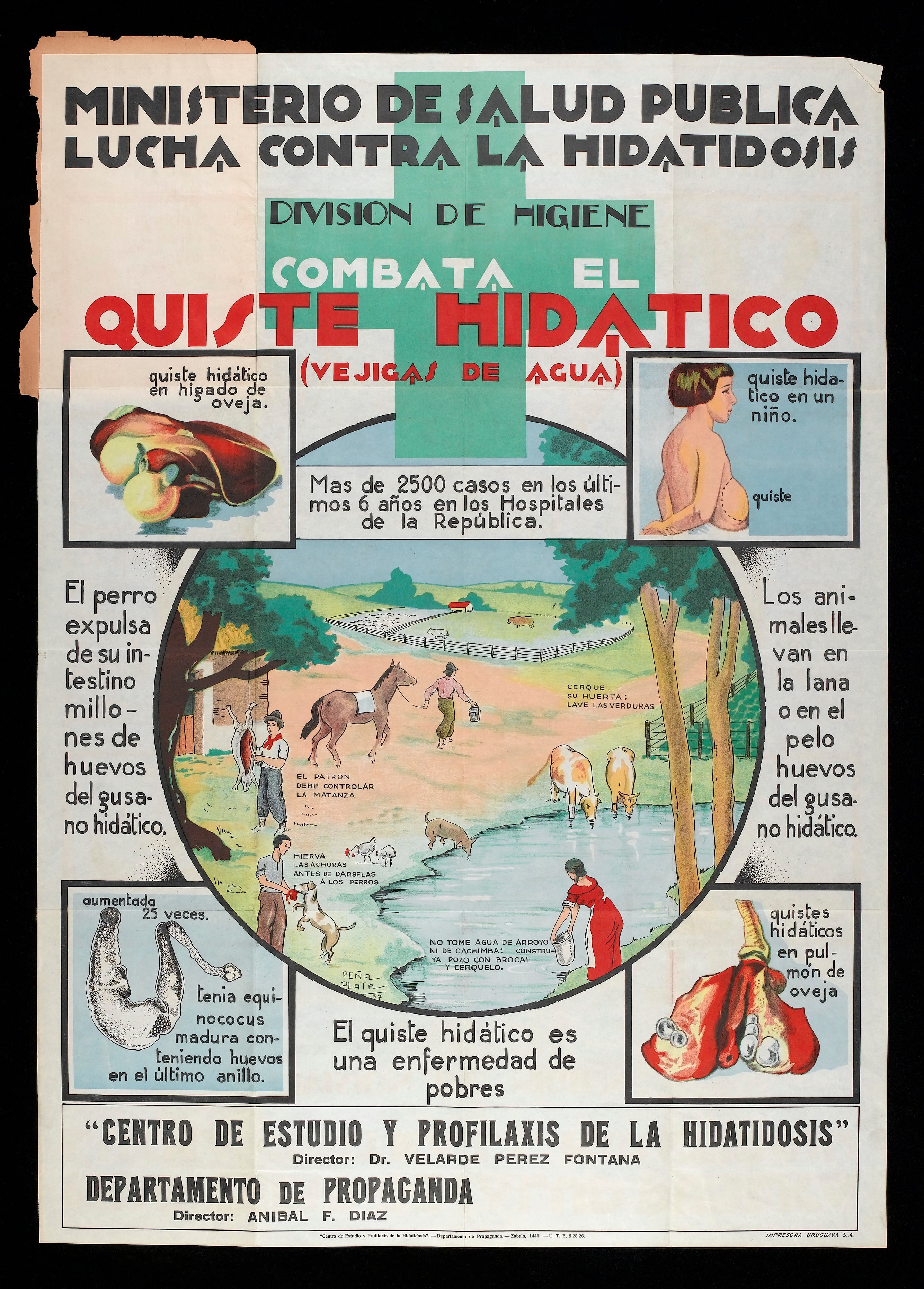
Global burden. Cystic echinococcosis is endemic on every inhabited continent where sheep and dogs coexist. WHO includes it among the neglected tropical diseases targeted for control; the highest human incidence figures come from pastoral communities in Central Asia, western China, the Andean region, North and East Africa and parts of the Mediterranean.
Society and body guidance — what genuinely applies
- What it addresses
- Cyst classification, PAIR, benzimidazole regimens, watch-and-wait for inactive cysts
- Relevance to bone
- Defines drug therapy; its percutaneous and observation arms are explicitly hepatic and do not transfer to bone
- What it addresses
- Dog deworming, livestock vaccination, slaughter hygiene, surveillance
- Relevance to bone
- The only intervention that reduces incidence; surgeons should notify cases where reporting systems exist
- What it addresses
- Referral of undiagnosed destructive bone lesions to a specialist unit before biopsy
- Relevance to bone
- Directly applicable — skeletal hydatid must be biopsied and resected by a unit that performs tumour surgery
- What it addresses
- Differential of destructive spinal lesions
- Relevance to bone
- Reinforces sending tissue for mycobacterial culture alongside parasitology
Registries. There is no implant registry evidence specific to hydatid reconstruction. Where endoprostheses are used, the relevant data are the general bone tumour endoprosthesis outcomes from national joint registries and tumour databases: revision is driven by infection and aseptic loosening, and infection risk in a previously contaminated or multiply operated field is higher than in primary sarcoma resection. Counsel accordingly.
Practice variation by resource setting
- High-resource, non-endemic centres see the disease rarely and misdiagnose it commonly; the failure mode is an unplanned biopsy or curettage of an assumed ABC or GCT. Mitigation: geographic history in every lytic-lesion clerking.
- High-resource endemic centres (Türkiye, Spain, Greece, Argentina, Iran) have mature pathways — serology, MRI, tumour-unit resection, endoprosthetic reconstruction, prolonged albendazole with pharmacy monitoring.
- Resource-limited endemic settings face late presentation with huge pelvic or multilevel spinal disease, limited MRI access, intermittent albendazole supply and no endoprosthetic reconstruction. Here amputation, resection arthroplasty, arthrodesis and cement-augmented reconstruction are legitimate, dignified, function-preserving answers, and continuous drug supply is often the binding constraint on outcome.
- Migration effect. Present in any country; incubation of years to decades means childhood exposure explains adult presentation far from the endemic region.
Controversies & Areas of Uncertainty
- Duration of albendazole. No trial defines the optimal postoperative course in bone. Practice ranges from three to six months to continuous multi-year therapy after incomplete excision. Longer courses are widely used in spinal disease on pathophysiological grounds, balanced against hepatotoxicity and adherence.
- Choice of scolicidal agent. Hypertonic saline, povidone-iodine, ethanol and silver nitrate have all been used. Comparative data are weak. Contact time is probably more important than agent. Formalin is abandoned; agents must not contact dura, nerve or a biliary communication.
- En-bloc spondylectomy versus maximal piecemeal excision. True Tomita-style en-bloc resection offers the best oncological logic but carries very high morbidity in a benign parasitic disease. Most units accept piecemeal excision plus instrumentation and prolonged drug therapy; the trade-off is unresolved.
- Amputation threshold. For extensive limb disease, early amputation may deliver cure and better function than repeated failed limb-salvage procedures. Timing this conversation is one of the hardest judgements in the disease.
- Role of any percutaneous therapy in bone. Occasional reports describe image-guided aspiration and instillation for soft-tissue components. This should be regarded as palliative and non-standard; there is no accepted percutaneous cure for intraosseous disease.
- Definition of cure. Because vesicles are microscopic and imaging response is slow, no reliable test of cure exists. Serological titres fall unpredictably. Most authorities treat this as a chronic relapsing condition requiring indefinite surveillance rather than a curable infection.
- Reconstruction choice in a contaminated field. Whether to use massive allograft or biological reconstruction after a case complicated by intraoperative spillage is contested; many prefer cement and metal for their resistance to colonisation and revisability.
CLEAROperative Plan for Skeletal Hydatid
Hook:You cannot see the edge of the disease, so make the operation CLEAR.
MCQ Practice Points
Q: Which plain radiographic feature best distinguishes skeletal hydatid from chronic pyogenic osteomyelitis?
A: Absence of periosteal reaction and absence of sequestrum. Hydatid produces multiloculated lysis by pressure resorption without a host reactive response; osteomyelitis characteristically shows periosteal new bone, involucrum and sequestrum.
Q: Why is bone hydatid disease more difficult to cure than hepatic disease?
A: No fibrous pericyst forms in rigid bone. Without a containing capsule the budding vesicles permeate marrow spaces and Haversian canals well beyond the radiologically visible lesion, so there is no plane to shell out and no reliable margin short of wide excision. The same fact explains why the WHO CE1-5 ultrasound staging has no skeletal equivalent — every stage in it describes the behaviour of a cyst inside a pericyst.
Q: A patient with a suspected hydatid bone lesion has negative echinococcal serology. What is the correct interpretation?
A: The diagnosis is not excluded. Serological sensitivity is markedly lower in osseous than hepatic disease because the intraosseous parasite presents limited antigen systemically. Management is driven by imaging pattern, endemic exposure and, where necessary, carefully planned tissue diagnosis.
Q: Is PAIR (puncture, aspiration, injection, reaspiration) an appropriate treatment for an intraosseous hydatid cyst?
A: No. PAIR sits within the WHO-IWGE algorithm, which was built around ultrasound-staged hepatic and soft-tissue cysts with a pericyst. In bone there is no capsule to inject, marrow vesicles cannot be sterilised percutaneously, and puncture risks anaphylaxis and tract seeding.
Q: Which intraoperative adjunct is most commonly used as a scolicidal agent in skeletal hydatid surgery, and what is the key caution?
A: Hypertonic (20 per cent) saline, with adequate contact time. The key caution is to avoid contact with dura, nerve roots or any biliary communication because of chemical neuritis and sclerosing cholangitis risk; use normal saline in the spinal canal.
Q: Which site accounts for the largest share of skeletal hydatid disease and what is its typical presentation?
A: The spine, roughly half of skeletal cases, most often thoracic, presenting with insidious back pain progressing to radiculopathy and cord compression, with vertebral body and posterior element destruction and relative early sparing of the intervertebral disc.
Q: A patient has hydatid involving the ilium and hip. What should the consent conversation contain?
A: That one operation is unlikely to be enough. In Herrera's 26-patient series followed a mean of 12.8 years, ilium-and-hip disease was the hardest site and always required numerous procedures; overall, 9 of the 19 patients who ended disease-free had needed repeat curettage or resection, and 6 of the 7 failures were left with chronic discharging sinuses. Where wide excision is anatomically impossible, say plainly that the aim is control rather than cure.
Exam Viva Scenarios
Practise clinical reasoning and management decisions out loud
“A 38-year-old man who grew up herding sheep in Central Asia presents with 18 months of hip pain and a recent low-energy subtrochanteric fracture. Radiographs show a large multiloculated lytic lesion of the proximal femur with cortical thinning, no periosteal reaction and no marginal sclerosis. He has been listed by a colleague for intramedullary nailing tomorrow morning.”
“A 45-year-old woman from a Mediterranean farming community has six weeks of progressive lower limb weakness, now ASIA D with a T7 sensory level and urinary hesitancy. MRI shows multiloculated fluid-signal lesions in the T6 and T7 vertebral bodies and posterior elements with epidural extension and paravertebral collections, thin non-enhancing walls, and preserved disc spaces. Inflammatory markers are normal.”
“During resection of a pelvic lesion you inadvertently enter a large cyst and clear fluid containing membranes floods the wound. Within two minutes the anaesthetist reports a fall in blood pressure, a rise in airway pressure and a developing rash.”
“A 52-year-old man had curettage and cement of a 'benign cyst' of the ilium two years ago in another country. Histology reportedly showed 'parasitic membranes'. He now has buttock pain and CT shows extensive multiloculated lysis of the ilium and sacroiliac region with a large gluteal soft tissue collection.”
Core concept
- Echinococcus granulosus; dog definitive host, sheep intermediate, human accidental
- Endemic: Mediterranean, Middle East, Central Asia, South Asia, East Africa, South America
- Bone involved in only a small percentage of cases but disproportionate morbidity
- No pericyst forms in bone — vesicles permeate marrow beyond imaging margins
- Behaves like a locally aggressive bone tumour; treat it like one
Distribution
- Spine about half of skeletal cases (thoracic commonest) — cord compression
- Pelvis and sacrum — large and silent until late
- Proximal femur and humerus — pathological fracture
- Ribs, scapula, skull less commonly
Imaging signature
- Radiograph: multiloculated honeycomb lysis, NO periosteal reaction, NO sclerotic rim
- CT: near-water density loculi; cortical breach; surgical planning of osteotomy levels
- MRI: T1 low, T2 very high, thin low-signal wall, minimal or thin rim enhancement
- Daughter vesicles within a mother cyst; detached membrane sign; modest marrow oedema
- Spine: relative disc preservation, multilevel body plus posterior element involvement
Differential
- Giant cell tumour and aneurysmal bone cyst — solid or blood-fluid levels, not clear fluid loculi
- Fibrous dysplasia — ground glass with sclerotic rind
- Plasmacytoma and metastasis — solid enhancing marrow replacement
- Tuberculous spondylitis — disc destruction, thick rim-enhancing abscess, raised markers
- Chronic osteomyelitis — sequestrum, involucrum, periosteal reaction
Safety rules
- Do not casually aspirate or biopsy — anaphylaxis and seeding risk
- If biopsy essential: albendazole cover, theatre with anaesthetist, adrenaline ready, tract within resection field
- PAIR is for liver, NOT for bone
- Do not internally fix a pathological fracture through undiagnosed parasitic bone
- Never irrigate scolicidal agents against dura or nerve roots
Treatment
- Cure = wide en-bloc resection with margins beyond MRI extent PLUS prolonged albendazole
- Curettage recurs almost universally — call it palliation if you do it
- Adjuncts: hypertonic saline lavage with contact time, cement filling of contained defects
- Spine: urgent decompression, maximal safe excision, instrumented fusion into uninvolved levels
- Amputation is a legitimate curative or functional answer in extensive limb disease
- Albendazole with fatty food; monitor liver function and blood count; avoid in pregnancy
Prognosis and follow-up
- Chronic relapsing disease — no reliable test of cure
- Spinal recurrence a third or more despite aggressive surgery
- Indefinite clinical and MRI surveillance; periodic liver and lung imaging
- Public health: dog deworming, safe slaughter and offal disposal, hand and food hygiene
Evidence Base
Extraspinal Bone Hydatidosis
- 26 patients treated over 26 years (1972 to 1998) at one Spanish unit, mean age 51.5 years, with a MEAN FOLLOW-UP OF 12.8 YEARS - unusually long, and necessary for a disease that recurs over decades
- Sites: ilium 4, ilium and sacral ala 2, ilium and hip 8, femoral head and acetabular roof 5, femoral shaft 1, distal femur 1, femoral head 1, scapula 2, ribs 2. All had curettage or wide resection; all but five also had chemotherapy
- 19 of 26 (73 per cent) were free of disease at last follow-up - BUT 9 of those 19 required repeated curettage or wide resection to get there (3 for wound infection, 6 for recurrence)
- Of the 7 not free of disease, 6 had persistent chronic productive sinuses and 1 a chronic wound infection
- Ilium-and-hip disease was the hardest to treat and always needed numerous procedures
- Conclusion in the authors' own words: results are satisfactory only where complete wide excision is possible, and in the pelvis and hip - where radical surgery is almost impossible - they are DISAPPOINTING