A narrow zone of transition means the host bone had time to wall the lesion off — the pattern is about growth rate, not about histology
- A sclerotic rim indicates a lesion growing slowly enough for reactive host bone to form — it is a rate marker, not a benignity guarantee.
- Lodwick grade IA (geographic, sclerotic margin) has the lowest rate of malignancy of any lytic pattern, but chondrosarcoma, low-grade central osteosarcoma and metastatic renal cell carcinoma can all be geographic.
- Age is the single most powerful discriminator: under 30 favours NOF, simple bone cyst, chondroblastoma, ABC, eosinophilic granuloma; over 40 favours metastasis, myeloma, chondrosarcoma, brown tumour and intraosseous ganglion.
- Site within the bone (epiphysis vs metaphysis vs diaphysis) and position in cross-section (central, eccentric, cortical, juxtacortical) narrows the list faster than any signal characteristic.
- Any lytic lesion with a rim PLUS pain at rest, night pain, a periosteal reaction or a soft tissue mass is not a leave-me-alone lesion.
- In a patient over 40 with multiple rimmed lucencies always exclude hyperparathyroidism (brown tumours) and myeloma before calling them metastases.
- “Say the Lodwick grade out loud: 'geographic lysis with a sclerotic margin, Lodwick IA'. It signals you understand aggressiveness grading.
- “Chondroblastoma is the epiphyseal lesion of the skeletally immature; giant cell tumour is the epiphyseal lesion once the physis has closed — and GCT classically has NO sclerotic rim.
- “A rimmed lucency in the anteromedial tibial diaphysis of a teenager with night pain relieved by NSAIDs is an osteoid osteoma until proven otherwise — look for the central nidus on CT.
- “Fibrous dysplasia gives ground-glass matrix with a thick rind of sclerosis; the rind is the giveaway, not the lucency.
- “Subchondral lucency with a rim next to a joint in a middle-aged adult: intraosseous ganglion, subchondral cyst (geode) or gout — check the adjacent joint before biopsying anything.
Metastatic renal cell and thyroid carcinoma, low-grade central osteosarcoma and grade 1 chondrosarcoma can all show a geographic margin with partial sclerosis. Look for cortical thinning greater than two-thirds, endosteal scalloping, or a soft tissue component before you relax.
Subacute osteomyelitis in the tibial or femoral metaphysis of a child or young adult is classically a lucency with a dense rim, often with a serpiginous channel to the physis. Missing it and biopsying without cover seeds the tract. Any rimmed lesion with warmth, raised CRP or a tortuous margin is infection until excluded.
Multiple rimmed lucencies with subperiosteal resorption at the radial border of the middle phalanges, a salt-and-pepper skull and distal clavicular erosion equals brown tumours. Check corrected calcium and PTH before oncology referral — biopsy of a brown tumour reads as giant cell rich lesion and misleads everyone.
A solitary rimmed lucency changes meaning completely if there are three more on the same radiograph. Polyostotic disease pushes you to fibrous dysplasia, Langerhans cell histiocytosis, enchondromatosis, brown tumours, myeloma or metastases — solitary favours NOF, SBC, chondroblastoma and osteoid osteoma.
Recognising the Pattern
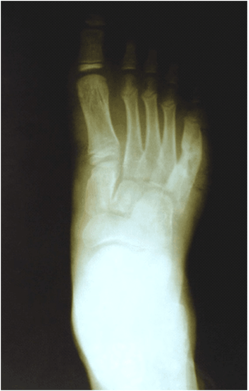
What the pattern actually is. A focal area of bone destruction (lucency) surrounded by a continuous or near-continuous band of increased density representing reactive host bone. The interface between lesion and normal bone can be traced with a pencil — this is a narrow zone of transition, Lodwick grade IA if the sclerotic rim is complete, IB if the margin is sharp but not sclerotic.
Confirming it is genuine.
- The rim must be seen on two orthogonal views. A single-view "rim" is usually the tangential edge of a normal cortex or a trabecular ridge summating over a lucency.
- The rim should be continuous around at least most of the circumference. Focal loss of the rim on one aspect, especially with adjacent cortical destruction, is the earliest sign of a lesion outgrowing its containment.
- Confirm the lucency is intramedullary, not a surface defect. A cortical fibrous defect and a subperiosteal lesion both mimic medullary lysis on an AP view alone.
- Check the cortex overlying the lesion: thinned but intact and expanded equals slow; interrupted equals aggressive regardless of rim.
How to describe it out loud. "There is a well-defined geographic lucency in the [distal femoral metaphysis], measuring approximately [3 by 2 cm], eccentrically placed, with a narrow zone of transition and a thin continuous sclerotic margin — Lodwick grade IA. There is no matrix mineralisation, no periosteal reaction, no cortical breach and no soft tissue mass. The appearances are of a non-aggressive lesion."
What mimics the pattern (false positives).
- Normal anatomy: the nutrient foramen and canal in the femoral or tibial diaphysis; the Ward triangle of relative lucency in the femoral neck; the pseudocyst of the greater tuberosity; the calcaneal pseudotumour between the trabecular groups; the herniation pit of the anterosuperior femoral neck.
- Overlapping structures: bowel gas over the pelvis, a skin fold, an air pocket in a plaster.
- Post-surgical: healing screw and pin tracks develop a dense rim; drill tracks after ACL reconstruction; bone graft harvest sites.
- Healing benign lesion: a treated or spontaneously involuting NOF, SBC or brown tumour fills in from the periphery and becomes densely rimmed — the rim is a healing sign, not a new lesion.
- Degenerative geode: a subchondral cyst with sclerotic margin in an arthritic joint. Look for joint space loss and osteophytes before working it up.
Next Investigation
The single most examinable rule: if the lesion is painful at rest or at night, is larger than 5 cm, has an incomplete rim, or shows deep endosteal scalloping, do not biopsy locally — stage and refer. An unplanned biopsy through the wrong compartment converts a limb-salvage case into an amputation.
F E G N O M A S H I CFEGNOMASHIC — the classic lytic lesion differential
The Differential
- Typical age / setting
- Children and young adults; tibial or femoral metaphysis
- Discriminating feature
- Tortuous or serpiginous margin with a channel directed towards the physis; pain unrelated to activity, raised CRP; penumbra sign on T1 MRI (thin high-signal granulation lining)
- What confirms it
- MRI showing central non-enhancing fluid with rim enhancement and marrow oedema; culture at open biopsy
- Typical age / setting
- Over 40; axial skeleton, proximal femur, humerus
- Discriminating feature
- Renal and thyroid deposits can be geographic and even expansile, but the rim is incomplete and cortical destruction is disproportionate to size; endosteal scalloping
- What confirms it
- CT chest/abdomen/pelvis, myeloma screen, whole-body bone scan or PET; biopsy after staging, never before
- Typical age / setting
- Over 40; proximal femur, humerus, pelvis, scapula
- Discriminating feature
- Ring-and-arc chondroid matrix with deep endosteal scalloping greater than two-thirds of cortical thickness, lesion length usually greater than 5 cm, and pain at rest — distinguishes from enchondroma
- What confirms it
- MRI for scalloping depth and soft tissue extension; refer to a sarcoma unit before biopsy
- Typical age / setting
- Adults; often multiple, jaw, pelvis, ribs, long bones
- Discriminating feature
- Coexisting subperiosteal resorption at the radial margin of the middle phalanges, distal clavicular and sacroiliac erosion, salt-and-pepper skull
- What confirms it
- Corrected calcium, phosphate, PTH, vitamin D, renal function — biochemistry, not biopsy
- Typical age / setting
- 5 to 20 years; distal femur, proximal and distal tibia, fibula
- Discriminating feature
- Eccentric, cortically based, multilocular with a lobulated scalloped sclerotic rim; asymptomatic incidental finding that sclerosis in from the periphery with age
- What confirms it
- Plain radiograph alone — no further imaging if classic and asymptomatic
- Typical age / setting
- 5 to 15 years; proximal humerus then proximal femur
- Discriminating feature
- Central, metaphyseal, abutting the physis, longitudinally orientated, no expansion beyond the width of the physis; fallen fragment sign if fractured
- What confirms it
- Radiograph; MRI shows uniform fluid signal without solid enhancing components
- Typical age / setting
- 10 to 20 years, open physis; epiphysis or apophysis of femur, humerus, tibia
- Discriminating feature
- Epiphyseal lucency with fine chondroid calcification and a thin sclerotic rim, plus marked surrounding marrow oedema on MRI out of proportion to lesion size
- What confirms it
- MRI oedema pattern; histology after curettage
- Typical age / setting
- 10 to 25 years; femoral neck, tibial diaphysis, posterior elements of spine
- Discriminating feature
- Nidus less than 1.5 cm with dense surrounding reactive sclerosis; night pain reliably relieved by NSAIDs; painful scoliosis if spinal
- What confirms it
- Thin-slice CT demonstrating the nidus and its central mineralisation
- Typical age / setting
- Any age, usually presents in the second and third decades; proximal femur, ribs, craniofacial, tibia
- Discriminating feature
- Ground-glass matrix with a thick sclerotic rind, bone expansion and bowing (shepherd's crook of the femoral neck); no periosteal reaction
- What confirms it
- Radiograph is usually diagnostic; CT for ground-glass density; bone scan for polyostotic extent
- Typical age / setting
- 20 to 50; small tubular bones of the hand, then proximal humerus and femur
- Discriminating feature
- Central, chondroid ring-and-arc calcification, endosteal scalloping less than two-thirds cortical thickness, painless unless fractured
- What confirms it
- Radiograph plus MRI if long bone or symptomatic; interval radiograph at 6 to 12 months if uncertain
- Typical age / setting
- Under 20; metaphysis of long bones, posterior spinal elements
- Discriminating feature
- Eccentric, markedly expansile with a thin shell, and fluid–fluid levels on MRI; grows rapidly for a benign lesion
- What confirms it
- MRI fluid–fluid levels; biopsy mandatory to exclude telangiectatic osteosarcoma and secondary ABC
- Typical age / setting
- 30 to 60; medial malleolus, carpal lunate, acetabulum, femoral head
- Discriminating feature
- Subchondral, juxta-articular, unilocular, thick sclerotic rim, and adjacent joint changes (geode) or a normal joint (true ganglion)
- What confirms it
- Radiograph plus MRI showing fluid signal with a communication to the joint capsule
- Typical age / setting
- 5 to 15 years; skull, femur, pelvis, spine
- Discriminating feature
- Bevelled-edge skull lucency or vertebra plana; can look aggressive when active and develop a rim as it heals
- What confirms it
- Skeletal survey, MRI, biopsy if solitary and symptomatic; many involute
- Typical age / setting
- 30 to 60; calcaneus (neutral triangle), proximal femur
- Discriminating feature
- Lucency with a central dense calcified nidus (cockade sign) and a thin rim; fat density on CT and fat signal on MRI
- What confirms it
- CT or MRI fat characterisation — no biopsy required
- Typical age / setting
- Over 40; first metatarsophalangeal joint, hands, olecranon
- Discriminating feature
- Para-articular punched-out erosion with overhanging edge and sclerotic margin, preserved joint space, adjacent soft tissue nodule
- What confirms it
- Dual-energy CT showing urate deposition; serum urate and joint aspirate
Narrowing It Down
- 1Step 1 — Is this a real lesion, and is it aggressive?
Confirm on two views. Grade the margin, then look SEPARATELY for periosteal reaction, cortical breach and soft-tissue mass rather than letting the rim answer for all three.
Complete sclerotic rim is Lodwick IA; sharp but non-sclerotic IB; ill-defined geographic IC; moth-eaten II; permeative III. Any periosteal reaction, cortical breach or soft-tissue mass OVERRIDES the rim and mandates staging and sarcoma-unit referral. A clean IA lesion with no pain buys you the ability to observe. Grade IA is also the most reproducibly assigned end of the Lodwick scale, which is part of why this pattern is safe to act on.
- 2Step 2 — How old is the patient?
State the age before the differential, every time.
Under 30: non-ossifying fibroma, simple bone cyst, chondroblastoma, aneurysmal bone cyst, eosinophilic granuloma, osteoid osteoma, Brodie's abscess. Over 40: metastasis, myeloma, chondrosarcoma, brown tumour, intraosseous ganglion, geode, gout, lipoma. Age alone removes more than half the list.
- 3Step 3 — Solitary or multiple?
Review the whole radiograph, and ask for a skeletal survey or bone scan if in doubt.
Multiple lesions in a child suggest fibrous dysplasia, Langerhans cell histiocytosis or enchondromatosis (Ollier, Maffucci). Multiple lesions over 40 suggest metastases, myeloma or brown tumours - send calcium, parathyroid hormone and a myeloma screen before assuming carcinoma.
- 4Step 4 — Where in the bone longitudinally?
Place the lesion in epiphysis, metaphysis or diaphysis, and note the physeal status.
Epiphysis with open physis is chondroblastoma; epiphysis with closed physis is giant cell tumour (usually rimless) or geode/ganglion. Metaphysis is non-ossifying fibroma, simple bone cyst, aneurysmal bone cyst, Brodie's abscess. Diaphysis is fibrous dysplasia, osteoid osteoma, Langerhans cell histiocytosis, adamantinoma or osteofibrous dysplasia in the anterior tibial cortex.
- 5Step 5 — Where in cross-section?
Decide whether the lesion is central, eccentric, cortically based or juxta-articular.
Central is simple bone cyst, enchondroma, fibrous dysplasia. Eccentric is non-ossifying fibroma, aneurysmal bone cyst, chondromyxoid fibroma. Cortically based is fibrous cortical defect, osteoid osteoma, osteofibrous dysplasia. Juxta-articular subchondral is ganglion, geode or gout.
- 6Step 6 — Is there matrix, and what kind?
Look inside the lucency and name the mineralisation pattern before naming a diagnosis.
Ring-and-arc or popcorn calcification is CARTILAGE - enchondroma, chondroblastoma, chondrosarcoma. Ground glass is fibrous dysplasia. Cloud-like or amorphous ossification is OSTEOID, so think osteosarcoma even if the margin looks tidy. Fat density is intraosseous lipoma. No matrix keeps the whole list open.
- 7Step 7 — What are the systemic and biochemical features?
Take the systemic history and send calcium, parathyroid hormone, CRP, ESR, full blood count and a myeloma screen - the step candidates omit and examiners reward.
Fever, warmth and raised inflammatory markers is infection. Hypercalcaemia with raised parathyroid hormone is a brown tumour, diagnosed without a biopsy. Anaemia, renal impairment and a paraprotein is myeloma. Night pain relieved by NSAIDs is osteoid osteoma. A painless incidental finding in a teenager is a non-ossifying fibroma.
MCQ Practice Points
Q: What does a sclerotic rim around a lytic lesion actually indicate?
A: That the lesion is growing slowly enough for the host bone to mount a reactive osteoblastic response. It is a marker of growth rate, not of histological benignity — Lodwick's grading was explicitly about rate of growth. Slow-growing malignancies (low-grade central osteosarcoma, grade 1 chondrosarcoma, indolent renal cell metastasis) may all be geographic with partial sclerosis.
Q: Which epiphyseal lesion occurs in the skeletally immature and which after physeal closure?
A: Chondroblastoma before, giant cell tumour after. Chondroblastoma occupies the epiphysis or apophysis with an open physis (typically 10 to 20 years), with a thin sclerotic rim and marked surrounding marrow oedema. Giant cell tumour occurs after physeal closure, is subarticular and eccentric, and characteristically has a non-sclerotic, sharply defined margin — Lodwick IB, not IA.
Q: How do you separate enchondroma from low-grade chondrosarcoma in a long bone?
A: Pain, size, scalloping and interval change. Favouring chondrosarcoma: pain at rest, length usually over 5 cm, endosteal scalloping deeper than two-thirds of cortical thickness, cortical expansion or breach, periosteal reaction, soft-tissue mass, and growth on serial imaging. Favouring enchondroma: painless, dense rings-and-arcs, shallow scalloping, static on follow-up. Pelvic and axial chondroid lesions are treated as chondrosarcoma until proven otherwise.
Q: Which MRI sign is relatively specific for a Brodie's abscess?
A: The penumbra sign — a thin rim of T1-hyperintense granulation tissue lining the abscess cavity, between the low-signal fluid centre and the low-signal sclerotic rim, best seen on unenhanced T1. Combined with rim enhancement and extensive marrow oedema it strongly favours infection over tumour.
Q: A 12-year-old has a central lucency in the proximal humeral metaphysis abutting the physis, with a thin rim and a fragment of cortex lying dependently within it. Diagnosis?
A: Simple (unicameral) bone cyst with a pathological fracture. The fallen fragment sign is pathognomonic and confirms the lesion is fluid-filled rather than solid. Most are managed with fracture care first; persistent or high-risk cysts are treated with aspiration and injection, curettage with grafting, or flexible nails in the proximal humerus and femur.
Q: You elect to observe a grade IA lesion. What does adequate follow-up look like, and what is the cost of imaging in the first place?
A: Measure it, do not eyeball it. Kaelin and MacEwen's principle for the unicameral cyst generalises: record a size normalised to the bone diameter and repeat on the same projection, because "looks the same" on two differently positioned films is not follow-up. And know the cost of the scan that found it: in 733 subjects imaged for research rather than symptoms, 2.3 per cent had an incidental abnormality that required investigation to exclude non-benign disease (Grainger 2008), including one myeloma. Every lesion you can confidently recognise and stop chasing is a patient spared that cascade — which is what the "leave me alone" list is for.
Exam Viva Scenarios
Practise clinical reasoning and management decisions out loud
“You are shown this radiograph of the knee of a 14-year-old boy taken after a twisting injury. There is a 3 cm eccentric, multilocular lucency in the distal femoral metaphysis with a lobulated sclerotic margin, based on the medial cortex. Describe what you see and tell me what you would do.”
“You are shown this radiograph of a 19-year-old with three months of deep proximal tibial pain, worse at night, not related to activity. There is a 2 cm lucency in the proximal tibial metaphysis with a thick, slightly irregular sclerotic margin and a narrow tract extending towards the physeal scar. He is afebrile. CRP is 28.”
“You are shown this pelvic radiograph of a 58-year-old woman with hip pain. There are three well-defined lucencies with partial sclerotic margins in the iliac wing, superior pubic ramus and proximal femur. The distal clavicles look eroded on the chest film. How do you proceed?”
Describe it in this order
- Which bone, and where longitudinally: epiphysis, metaphysis or diaphysis
- Where in cross-section: central, eccentric, cortical, juxtacortical, subchondral
- Size in centimetres and relation to bone diameter
- Margin and Lodwick grade: IA sclerotic, IB sharp non-sclerotic, IC ill-defined, II moth-eaten, III permeative
- Matrix: none, chondroid rings-and-arcs, ground glass, osteoid, fat
- Cortex, periosteal reaction, soft tissue mass
- Solitary or multiple — always look at the rest of the film
Age filters
- Under 20: NOF, simple bone cyst, chondroblastoma, ABC, eosinophilic granuloma, osteoid osteoma, Brodie's abscess
- 20 to 40: enchondroma, fibrous dysplasia, giant cell tumour, osteoid osteoma, intraosseous ganglion
- Over 40: metastasis, myeloma, chondrosarcoma, brown tumour, geode, lipoma, gout
One-line discriminators
- Lobulated rim, eccentric, cortical, teenager — NOF
- Central, metaphyseal, fallen fragment — simple bone cyst
- Epiphysis, open physis, huge oedema — chondroblastoma
- Nidus less than 1.5 cm, NSAID-responsive night pain — osteoid osteoma
- Ground glass with thick rind, shepherd's crook — fibrous dysplasia
- Serpiginous tract to physis, raised CRP, penumbra sign — Brodie's abscess
- Scalloping greater than two-thirds cortex, rest pain, over 5 cm — chondrosarcoma
- Multiple lucencies plus distal clavicular erosion — brown tumours
Red flags that override a tidy rim
- Rest pain or night pain not relieved by NSAIDs
- Cortical breach or focal loss of the rim
- Any periosteal reaction, especially interrupted or Codman triangle
- Soft tissue mass
- Lesion larger than 5 cm or documented interval growth
- Patient over 40 with a known or suspected primary malignancy
Investigation rules
- Classic asymptomatic NOF, calcaneal lipoma, herniation pit — no further imaging
- CT for matrix, nidus, cortex and scalloping depth
- MRI for marrow extent, fluid–fluid levels, penumbra sign and soft tissue mass
- Over 40 with multiple lesions — calcium, PTH and myeloma screen before biopsy
- Stage before biopsy; biopsy of a suspected primary sarcoma only at the treating unit
Evidence Base
Determining Growth Rates of Focal Lesions of Bone from Radiographs
- The paper that defines the grade this whole page is about: rate of growth divides focal bone lesions into two largely mutually exclusive classes, and five grades are set out with rules for applying them
- A sclerotic rim is grade IA - the host has had time to lay down reactive bone around the lesion, which is a statement about SPEED, not about histology
- The stated clinical purpose is triage, in the authors' words: 'not all focal lesions require biopsy, and grading is especially helpful in deciding which should be biopsied and which may be safely followed'
Classifications for Radiographic Evaluation of Radiolucent Bone Lesions Have Poor Inter- and Intra-observer Agreement
- 48 case sets of radiolucent lesions graded TWICE, at least two weeks apart, by 20 observers using the original Lodwick, modified Lodwick and Enneking systems
- Inter-observer reliability was poor for all three - agreement 39 per cent for Lodwick (kappa 0.23), 39 per cent modified Lodwick (kappa 0.25), 53 per cent Enneking (kappa 0.28)
- Intra-observer reproducibility was moderate at best (kappa 0.42 to 0.45), with self-agreement between 39 and 78 per cent
- TRAINING LEVEL HAD NO EFFECT on reproducibility
- Benign versus malignant was correctly assigned in only 73.3 per cent of cases with Lodwick
- REASSURANCE FOR THIS PAGE SPECIFICALLY: reproducibility was BEST at the extremes - the highest and lowest grades were the most consistently assigned. Grade IA, the subject of this page, is the reliable end of an unreliable scale