The Hand With Too Few Digits
- Oligodactyly is a descriptive term — fewer than five digits — not a diagnosis. The task is to identify which developmental process produced it.
- The Oberg-Manske-Tonkin (OMT) classification, approved by the IFSSH, places each condition in one of three main groups — malformations, deformations and dysplasias — with syndromes catalogued alongside; oligodactyly usually sits under malformation with failure of formation.
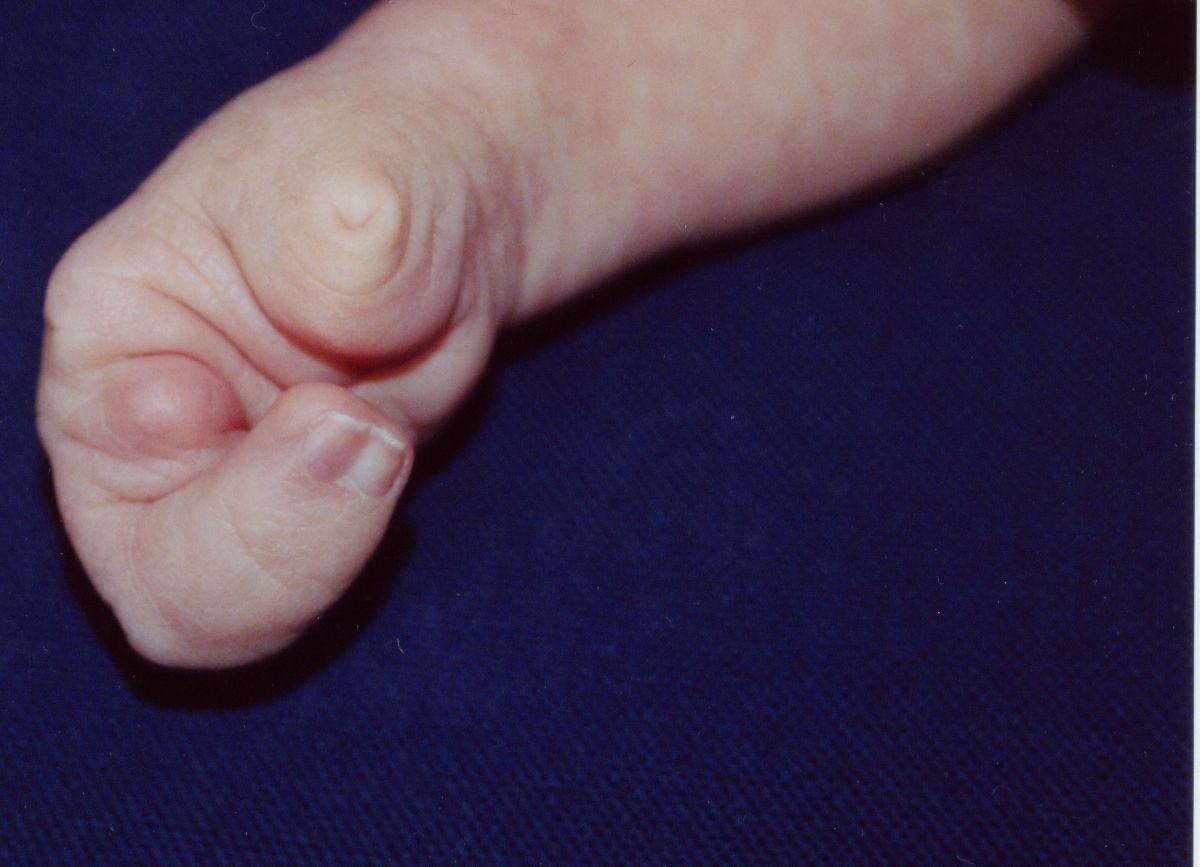
- Symbrachydactyly is distinguished from true transverse arrest by the presence of ectodermal remnants — nubbins, nail vestiges and small skin tags — at the end of the deficient ray.
- Symbrachydactyly is almost always unilateral and sporadic; longitudinal deficiencies, particularly radial, carry a high syndromic burden and must be screened.
- Assessment is function-first: what the child can do with the hand matters more than how many digits it has, and the commonest correct answer is that no surgery is needed.
- “Nubbins mean symbrachydactyly. A clean, smooth, well-padded stump with no ectodermal remnants means true transverse arrest.
- “Non-vascularised toe phalanx transfer must be done before about 18 months of age to retain physeal growth; performed later, the transferred phalanx does not grow and may resorb. Know what the window actually buys: even when transferred at a mean age of 1.5 years, 78 per cent of physes had closed by 10 years and the phalanx finished at 71 per cent of expected length, with growth falling from 0.83 to 0.22 mm per year after the fifth year (Kawabata 2018).
- “Poland syndrome is symbrachydactyly with ipsilateral absence of the sternocostal head of pectoralis major — always examine the chest wall.
- “Any child with radial longitudinal deficiency needs full dysmorphology and haematology assessment, and chromosomal breakage testing (diepoxybutane or mitomycin C) for Fanconi anaemia before elective surgery — a normal blood count does not exclude it, because marrow failure appears later in the first decade. Note the live controversy: one tertiary series found no hand feature predictive of Fanconi anaemia and no positive child with an ISOLATED limb anomaly, and argues against testing isolated hand abnormalities — but it is small and the cost of a miss is a child given marrow-toxic treatment. Follow local guidance; escalate on any additional feature.
Overview
Oligodactyly means a hand with fewer than five digits. It is a description, not a diagnosis, and the first task in the clinic and in the viva is to work out which developmental process produced it, because that determines the natural history, the syndromic risk, the reconstructive options and the counselling.
Congenital upper limb differences occur in approximately 1 in 500 to 1 in 600 live births when all types are counted; the great majority are minor. Deficiencies producing a hand with fewer than five digits are considerably rarer, and most are unilateral, sporadic and non-syndromic.
Two entirely different biological events produce a short hand, and telling them apart takes seconds at the cot side.
1. The hand never formed properly (malformation).
- Symbrachydactyly: the hand plate forms but the digits fail to develop fully. The tell-tale sign is ectodermal remnants at the end of the deficient ray — nubbins, nail vestiges and small skin tags. Soft tissue is present but skeleton is absent or rudimentary. Almost always unilateral and sporadic.
- True transverse arrest (transverse deficiency): development stops cleanly at a level, producing a smooth, well-padded stump with no distal remnants at all, most commonly at the proximal forearm. The classic level is the proximal third of the forearm.
- Longitudinal deficiency: a specific axis fails — radial (thumb and radius), ulnar (ulnar digits and ulna) or central (cleft hand). The hand is deficient along a defined ray rather than at a level.
2. A normally formed hand was subsequently damaged (deformation).
- Constriction ring (amniotic band) sequence: the hand formed normally and was later constricted or amputated in utero. Look for constriction rings on other digits or limbs, acrosyndactyly with proximal fenestrations, and distal lymphoedema. Nothing is genetically wrong with the child.
Why the distinction matters clinically, not just academically:
- Symbrachydactyly has soft tissue but no skeleton — the reconstructive problem is skeletal, so toe phalanx transfer works well.
- Transverse arrest has neither, and there is usually nothing distal to build on — prosthetic provision is the mainstay.
- Constriction ring sequence has normal proximal anatomy and normal tissue quality, and the deficiency is often asymmetrical and unpredictable — reconstruction is highly individualised.
- Longitudinal deficiency carries the syndromic burden. Radial deficiency in particular mandates screening for Fanconi anaemia, VACTERL, Holt-Oram and thrombocytopenia-absent radius.
VACTERL plus THFScreening a Radial Longitudinal Deficiency
Hook:Fanconi is the one that kills. Test for it before you operate, every time.
Aetiology and Embryology
The three signalling axes
Upper limb development occurs between approximately the fourth and eighth weeks of gestation, from the limb bud, and is organised along three axes, each with its own signalling centre. The OMT classification is built directly on this scheme.
- Signalling centre
- Apical ectodermal ridge (AER)
- Key molecule
- Fibroblast growth factors (FGF2, FGF4, FGF8)
- Failure produces
- Transverse deficiency, symbrachydactyly, phocomelia
- Signalling centre
- Zone of polarising activity (ZPA)
- Key molecule
- Sonic hedgehog (SHH)
- Failure produces
- Radial and ulnar longitudinal deficiency, mirror hand, preaxial polydactyly
- Signalling centre
- Dorsal ectoderm
- Key molecule
- WNT7A (dorsalising) and EN1 (ventralising), acting via LMX1B
- Failure produces
- Nail-patella syndrome, dorsal-ventral patterning defects
- The AER and the ZPA maintain each other through an FGF-SHH feedback loop. Disturbance of the AER interrupts outgrowth along the proximal-distal axis and is the accepted developmental basis of transverse deficiency and symbrachydactyly.
- Interdigital apoptosis, mediated by bone morphogenetic proteins, sculpts the separate digits from the hand plate. Failure of apoptosis produces syndactyly; excessive or aberrant apoptosis contributes to the digital loss seen in symbrachydactyly.
The subclavian artery supply disruption sequence
An influential vascular hypothesis proposes that interruption of the early embryonic blood supply in the subclavian arteries, the vertebral arteries or their branches — occurring during or around the sixth week — produces a spectrum of defects depending on the site of occlusion. Bavinck and Weaver grouped Poland, Klippel-Feil and Möbius anomalies, isolated absence of pectoralis major with breast hypoplasia, isolated terminal transverse limb defects and the Sprengel anomaly under the term subclavian artery supply disruption sequence (SASDS). It explains the characteristic unilaterality and sporadic occurrence of symbrachydactyly, and the association with Poland syndrome, better than any genetic model.
Say it explicitly and early.
- Symbrachydactyly, transverse deficiency and constriction ring sequence are sporadic events occurring in the fourth to eighth week, usually before the pregnancy was even confirmed.
- Nothing the mother did or failed to do caused it. Parents almost universally believe otherwise and will not raise it unless you do.
- Recurrence risk in a future pregnancy is very low for sporadic symbrachydactyly and transverse deficiency, and effectively zero for constriction ring sequence.
Set the frame at the first visit.
- Children with unilateral deficiencies adapt remarkably; the developing brain builds function around what is present.
- The commonest correct management decision is no operation. Say so, and mean it.
- Show families, with consent, what other children with the same difference achieve. Peer contact and parent support organisations do more in the first year than any surgeon does.
- Reconstruction, if any, is planned over years, not weeks. There is no urgency.
Classification
The Oberg-Manske-Tonkin (OMT) classification
Proposed in 2010 and subsequently approved by the IFSSH Scientific Committee for Congenital Conditions as a replacement for the Swanson classification. It is organised on developmental biology and dysmorphology terminology rather than on morphology alone, placing each condition in one of three main groups — malformations, deformations and dysplasias — with recognised syndromes catalogued alongside.
- Subdivision
- A. Failure of axis formation or differentiation — whole limb
- Examples
- Proximal-distal: transverse deficiency, symbrachydactyly, brachydactyly. Radial-ulnar: radial and ulnar longitudinal deficiency. Dorsal-ventral: nail-patella syndrome
- Subdivision
- B. Failure of axis formation or differentiation — hand plate only
- Examples
- Cleft hand, syndactyly, polydactyly, camptodactyly, clinodactyly
- Subdivision
- Extrinsic forces acting on an already formed limb
- Examples
- Constriction ring sequence, trigger digit, arthrogryposis-related deformity
- Subdivision
- Abnormal tissue growth
- Examples
- Macrodactyly, tumorous conditions, multiple hereditary exostoses, Ollier disease
- Subdivision
- Catalogued alongside the three groups rather than forming a fourth axis
- Examples
- Poland, Apert, Holt-Oram, VACTERL, Fanconi anaemia, TAR
Where oligodactyly sits
- Most commonly: OMT I.A, failure of formation along the proximal-distal axis — symbrachydactyly or transverse deficiency.
- Also: OMT I.A radial-ulnar — radial or ulnar longitudinal deficiency with absent digits.
- Also: OMT I.B — cleft hand (central deficiency) affecting the hand plate only.
- Also: OMT II — constriction ring sequence with intrauterine amputation.
The examination point: oligodactyly is not a category in OMT. Name the process, then place it.
Assessment
Function first — the principle that governs everything
A child does not present complaining of having four fingers. They present because a parent is worried. Your assessment is of function and of family expectation, in that order.
- 1Step 1 — Watch before you touch
Put age-appropriate toys within reach and observe. How does the child approach, grasp, transfer and release? Is the affected hand used as a dominant hand, a helper, or ignored? Is there any pinch, and between what and what?
Observation gives more information than measurement in a small child
- 2Step 2 — Define the deficiency
Count the rays. Look for nubbins and nail remnants — symbrachydactyly versus transverse arrest. Look for constriction rings elsewhere. Determine whether the deficiency is transverse, longitudinal or central.
Names the process and sets the syndromic risk
- 3Step 3 — Examine what is present
For each existing digit: active and passive range at every joint, stability, sensation, nail and pulp quality, and the presence of extrinsic and intrinsic motors. Assess web space depth and first web width.
Determines what can be built on
- 4Step 4 — Examine the whole limb and the child
Shoulder, elbow, forearm rotation, wrist stability. Chest wall for Poland syndrome. Spine, feet, cardiac and abdominal examination. Growth parameters and development.
The hand is rarely the only finding
- 5Step 5 — Assess the donor sites
Examine the feet: toe length, alignment, web spaces, and whether the second toe is long enough to be a donor. Assess for foot anomalies, which frequently coexist.
No reconstruction plan is complete without a donor assessment
- 6Step 6 — Establish the family's goal
Ask what specifically they hope for: function, appearance, or reassurance. Most want reassurance. Document what the child cannot do that they wish they could.
Operating on parental anxiety is the commonest error in this field
- 7Step 7 — Plan over years
Set a longitudinal plan with review points, not a single decision. Involve the hand therapist, prosthetist and paediatrician from the outset.
There is no urgency; there is a developmental timetable
What to look for on examination
- How to elicit it
- Inspect the end of each deficient ray under good light
- What it means
- Symbrachydactyly rather than true transverse arrest — soft tissue sleeve is present and a toe phalanx can be inserted
- How to elicit it
- Inspect and palpate the terminal segment
- What it means
- True transverse arrest — nothing to build on; prosthetics led
- How to elicit it
- Examine all four limbs
- What it means
- Constriction ring sequence — a deformation, no genetic risk, no recurrence risk
- How to elicit it
- Inspect and stress the wrist
- What it means
- Radial longitudinal deficiency — mandatory syndromic screening
- How to elicit it
- Assess elbow range and ulnar rays
- What it means
- Ulnar longitudinal deficiency — check for proximal femoral focal deficiency and fibular deficiency
- How to elicit it
- Ask the child to push their hands together, or feel the anterior axillary fold
- What it means
- Poland syndrome
- How to elicit it
- Inspect the palm
- What it means
- Cleft hand — classify by first web space (Manske-Halikis)
- How to elicit it
- Offer a small object and observe
- What it means
- The single most important functional determinant; a hand with any pinch is a working hand
- How to elicit it
- Age-appropriate testing; observe whether the child looks at the hand to use it
- What it means
- An insensate part will not be incorporated regardless of how it looks
Outcome measures
- Patient-reported: PODCI (Pediatric Outcomes Data Collection Instrument), PROMIS paediatric upper extremity measures, and the Prosthetic Upper Extremity Functional Index (PUFI) where a prosthesis is in use.
- Observational: the Assisting Hand Assessment (AHA) is validated in children with unilateral upper limb differences and measures how effectively the affected hand is used as a helper in bimanual tasks. It is the most informative single measure in this population.
- Objective: grip and pinch dynamometry once the child is old enough (usually from about 5-6 years), digit length, and range of motion.
- Record the child's own view from around school age. Children and parents frequently disagree about what matters, and the child's view should prevail.
Imaging
Plain radiographs
- Posteroanterior and lateral of both hands and both wrists, and of the forearms where a longitudinal deficiency is suspected.
- Bilateral, always — the unaffected side is the template for reconstruction and reveals bilateral involvement that is not clinically obvious.
- What to look for: which metacarpals and phalanges are present, whether the carpus is formed, physeal presence at each level, and the state of the trapeziometacarpal joint in thumb hypoplasia.
The neonatal caveat
The carpus is largely cartilaginous in infancy. The capitate and hamate ossify at around 1-3 months, and the remaining carpal bones sequentially through childhood, with the pisiform last at around 8-12 years. A radiograph in a neonate massively underestimates what is present. Do not tell a family a bone is absent on the basis of a neonatal film; repeat imaging as ossification proceeds, or use ultrasound or MRI.
Feet
- Radiographs of both feet are required before any toe transfer, to assess donor toe phalangeal length and to identify coexisting foot anomalies, which are common in symbrachydactyly and in cleft hand.
- Chromosomal breakage testing (diepoxybutane or mitomycin C) for Fanconi anaemia in any radial longitudinal deficiency or thumb hypoplasia. Fanconi anaemia presents with progressive marrow failure typically in the first decade, carries a high malignancy risk, and is life-limiting. Diagnosis changes the entire management plan and may make elective hand surgery inappropriate. This is the single most important investigation on the page.
- Full blood count — thrombocytopenia identifies TAR syndrome. Remember the counter-intuitive rule: in TAR the thumb is present despite absent radii, which is the reverse of every other radial deficiency.
- Echocardiography — Holt-Oram syndrome (TBX5) and VACTERL.
- Renal ultrasound — VACTERL; renal anomalies are the most frequently missed association.
- Spinal imaging — vertebral anomalies and tethered cord.
- Clinical genetics referral for any bilateral, familial or syndromic pattern.
- Symbrachydactyly and constriction ring sequence do not require this panel unless there are other findings — but examine the chest wall for Poland syndrome in every symbrachydactyly.
Management
The default position
The commonest correct management decision in oligodactyly is no operation. Children with unilateral differences develop excellent bimanual function, and the developing brain incorporates whatever is present. Surgery must be justified by a specific functional gain that the family and, where old enough, the child can articulate.
The developmental timetable
Timing of Intervention
Examine the whole child. Complete syndromic screening where indicated. Refer to clinical genetics if the pattern is bilateral, familial or syndromic. Counsel the parents explicitly that this is not their fault and that recurrence risk is low. Connect them with a parent support organisation — this is the single most valuable intervention in the first year. No surgery.
Observe developing hand preference and grasp patterns. A passive prosthesis may be introduced from around 6 months, principally to aid sitting balance and bimanual crawling and to normalise the appearance of the limb — evidence for functional benefit at this age is weak, and rejection is common. Involve the hand therapist. Nubbin excision, if the family requests it for appearance, can be done at any time but is not urgent.
Non-vascularised toe phalanx transfer should be performed before about 18 months if it is to be performed at all, because transfer within this window retains physeal growth potential. This is the tightest timing constraint in the whole topic.
First web deepening, syndactyly release (border digits first), constriction ring release. Syndactyly involving digits of unequal length is released earlier, from around 6 months, to prevent progressive deformity.
The usual window, balancing vessel calibre (which improves with age) against cortical plasticity and the need to establish grasp before school. Some centres transfer earlier, from around 18 months to 2 years.
Formal prosthetic fitting where a prosthesis is wanted. Reassess function before school entry. Ask the child, not only the parents.
Distraction lengthening, on-top plasty, secondary tenolysis, revision of appearance. Chest wall reconstruction in Poland syndrome. From this age the child's own preference should determine whether anything further is done.
Surgical Options
Non-vascularised toe phalanx transfer (NVTPT)
in symbrachydactyly the digit has a soft tissue sleeve with a nail remnant but no skeleton. Inserting a free (non-vascularised) toe phalanx into that sleeve creates a skeletal strut that lengthens and stabilises the digit and provides a post against which the remaining digits can pinch.
the proximal phalanx of the second, third or fourth toe, harvested through a dorsal approach.
The critical technical points
- Detail
- Before approximately 18 months; some series argue for before 12-15 months
- Why it matters
- Transfer within this window preserves physeal growth potential; transferred later, the phalanx does not grow and may resorb. Counsel on the ceiling: even inside the window (mean age 1.5 years), Kawabata found the physis closed in 23 per cent by 5 years and 78 per cent by 10, length falling from 87 to 71 per cent of expected, and growth from 0.83 to 0.22 mm per year - so the graft grows well for about five years and then largely stops
- Detail
- Harvest with an intact periosteal sleeve and preserve the collateral ligament attachments where possible
- Why it matters
- The periosteum carries the blood supply that revascularises the graft and is essential for physeal survival
- Detail
- Including both physis and epiphysis
- Why it matters
- A phalanx without its physis cannot grow
- Detail
- Insert into the existing skin sleeve without tension; do not close a tight pocket over the graft
- Why it matters
- Tension causes skin necrosis and graft extrusion
- Detail
- Kawabata graded active metacarpophalangeal motion good in 24, fair in 7 and POOR IN 16 of the transfers assessed
- Why it matters
- The operation lengthens a digit reliably and mobilises it unreliably. Promise length and stability - stability and alignment were good in 37 and 33 respectively - and be cautious about promising movement
- Detail
- Secure the transferred phalanx to the metacarpal or metacarpal remnant, ideally reconstructing a joint or a stable pseudarthrosis
- Why it matters
- An unstable strut gives length without function
- Detail
- Fill the defect and stabilise the toe; syndactylise the donor toe to its neighbour to prevent deviation
- Why it matters
- Prevents progressive toe deviation and floppiness
- Detail
- Multiple phalanges can be transferred at one sitting
- Why it matters
- Efficient use of a single anaesthetic in a small child
Expected outcome
- Series performing transfer before 18 months report continued growth of the transferred phalanx in the great majority of cases, with growth rates approaching, though generally below, those of the native phalanx.
- Transfer after 18-24 months is associated with markedly reduced growth and a significant rate of resorption.
- The transferred digit is stiff — it provides length and a stable post, not motion. Counsel the family that this is a post, not a finger.
Donor morbidity
- Shortening of the donor toe and a tendency to deviation or floppiness. Syndactylising the donor toe to its neighbour mitigates this.
- Foot function is generally unaffected; gait and footwear are not usually a problem. Long-term donor foot outcomes are good in reported series, but parents deserve to see photographs of a donor foot before consenting.
- Excising nubbins before planning reconstruction. The soft tissue sleeve with its nail remnant is the recipient bed for a non-vascularised toe phalanx. Once excised, it cannot be recreated.
- Missing the 18-month window for non-vascularised toe phalanx transfer. Referral delay is the commonest reason this opportunity is lost — refer early, even if no surgery is planned.
- Proceeding to microvascular toe transfer without angiography. The symbrachydactylous hand frequently has an absent or abnormal superficial palmar arch, and discovering that on the table is a disaster.
- Operating without excluding Fanconi anaemia in a child with radial longitudinal deficiency.
- Creating a single mobile digit with nothing to oppose. Plan the pinch pair, not the digit.
- Operating on parental distress rather than on functional deficit. Ask what the child cannot do; if the answer is nothing, do not operate.
- Promising motion after toe transfer in symbrachydactyly. The intrinsics are absent; the transferred toe provides a growing, sensate, stable post with limited independent motion. Say so before, not after.
Outcomes
What the evidence supports
- Reported outcome
- Excellent bimanual adaptation; most children function at or near age norms in daily tasks
- Caveats
- Psychosocial impact is real and is not measured by function scores
- Reported outcome
- Consistent functional gain in grasp span with low complication rates
- Caveats
- Simple, underused, and should precede complex reconstruction
- Reported outcome
- Continued growth of the transferred phalanx in the great majority; stable digit length gain
- Caveats
- The digit is stiff — it is a post, not a moving finger
- Reported outcome
- Markedly reduced growth and a significant rate of resorption
- Caveats
- The reason the timing window matters
- Reported outcome
- Flap survival typically above 90-95% in experienced centres; the toe grows and recovers useful sensation
- Caveats
- Motion is limited where intrinsics are absent; requires a specialist centre and a multi-year commitment
- Reported outcome
- Achievable lengthening of roughly 30-50% of segment length
- Caveats
- High complication rate: pin site infection, regenerate non-union, stiffness
- Reported outcome
- Reliable, durable improvement in grasp and pinch; high parent and patient satisfaction in long-term follow-up
- Caveats
- Technically demanding; results depend heavily on correct positioning and intrinsic reassignment
- Reported outcome
- High rejection rates increasing with age, particularly for heavy or high-maintenance devices
- Caveats
- Activity-specific devices are often the most used
Long-term perspective
- Function scores in unilateral congenital deficiency are consistently better than clinicians predict and often better than parents fear. The affected hand becomes an effective assisting hand in most children.
- Bilateral deficiency is a different problem. Here, reconstruction to create any pinch is transformative, the threshold for surgery is far lower, and the priority is to create two opposing posts on at least one side.
- Psychosocial outcome is the domain most often neglected. Teasing, self-consciousness in adolescence and choices about prosthesis use for appearance rather than function are real and should be addressed explicitly, with access to psychology and peer support.
- The child's own view diverges from the parents' view from school age onward, and follow-up should be structured so the child is asked directly and, where appropriate, seen without the parent.
Counselling the family — the content of the conversation
- This is not your fault; it happened in the first eight weeks, sporadically, and nothing caused it.
- Recurrence risk is very low for sporadic symbrachydactyly, transverse deficiency and constriction ring sequence. Genetics referral if bilateral, familial or syndromic.
- There is no urgency. Nothing needs to be decided in the first months.
- Your child will do more than you expect. Offer peer contact and a parent support organisation.
- Explain the screening you are doing and why, especially the Fanconi test where relevant.
- What specific function will this gain? If neither you nor the family can name it, do not operate.
- What is the donor cost? Show photographs of a donor foot before consenting to any toe transfer.
- How many operations and over how many years?
- What is the failure rate, and what happens if it fails?
- A transferred toe phalanx is a post, not a finger. A transferred toe grows and feels but will not move like a finger if the intrinsics are absent.
- The child's preference governs from school age. Say this to the parents early so it is expected later.
Guidelines, Registries & Global Practice
Classification and registry practice
- The Oberg-Manske-Tonkin classification has been approved by the IFSSH as the replacement for the Swanson scheme and is the international standard for reporting congenital upper limb differences; iterative revision since 2010 has improved comparability between registries.
- Congenital anomaly registries — EUROCAT, the National Birth Defects Prevention Network and comparable national registries — supply the population prevalence figures used in counselling. Reported prevalence varies between registries partly for genuine reasons and partly through differing ascertainment and coding. Prevalence of all congenital upper limb differences is commonly quoted at roughly 1 in 500 to 1 in 600 live births, the great majority minor; deficiencies producing fewer than five digits are considerably rarer.
Areas of genuine international divergence
- Wrist centralisation in radial longitudinal deficiency is the most contested area in paediatric hand surgery. Concerns about recurrence, ulnar growth arrest and loss of motion have shifted many units toward soft tissue distraction alone, ulnar lengthening, or acceptance of the deformity — particularly where elbow flexion is limited and the radial deviation is functionally useful. There is no consensus and no randomised evidence.
- Timing of microvascular toe transfer varies from around 18 months in some specialist centres to 4 years or later in others, reflecting different weightings of technical feasibility against cortical plasticity.
- Prosthetic provision policy differs markedly between health systems — some fund early passive prostheses routinely, others only on demonstrated functional need — and reported rejection rates make both positions defensible. Nubbin excision likewise varies: routine in some units for appearance, deferred in others to preserve the reconstructive option. The safe teaching is to defer until a reconstructive plan is agreed.
Access, equity and multidisciplinary standards
- Microvascular toe-to-hand transfer is concentrated in a small number of high-volume centres worldwide and outcomes are volume-dependent, so referral to a specialist paediatric hand service is the single biggest determinant of what options a child is offered. By contrast non-vascularised toe phalanx transfer needs no microsurgery, implants or specialist equipment and is achievable far more widely; its time-limited window makes early referral the critical variable everywhere. Web space deepening is likewise low-cost, single-stage and high-yield, and is probably the most underused operation in this field.
- Prosthetic access is highly inequitable. Low-cost 3D-printed partial hand devices have widened access; they do not match definitive devices for durability or grip force, but children frequently prefer and actually use them, which is the metric that matters.
- BSSH, ASSH and EFORT paediatric hand guidance all recommend management within a multidisciplinary paediatric hand service spanning hand surgery, therapy, prosthetics, paediatrics, clinical genetics and psychology. The consistent message is that assessment is function-first, most children need no surgery, and referral should be early even where none is anticipated — precisely because some options are time-limited.
MCQ Practice Points
Q: What does the presence of nubbins and nail remnants at the end of a deficient ray indicate? A: Symbrachydactyly, not true transverse arrest. Soft tissue formed but skeleton did not.
Q: Under which OMT category does symbrachydactyly fall? A: I — Malformations, failure of axis formation along the proximal-distal axis (apical ectodermal ridge).
Q: By what age must a non-vascularised toe phalanx transfer be performed to retain growth? A: Approximately 18 months. Later transfer shows markedly reduced growth and a significant resorption rate.
Q: What is the most important technical point in harvesting a toe phalanx for non-vascularised transfer? A: Preserve the periosteal sleeve — it carries the blood supply that revascularises the graft and permits physeal survival.
Q: What defines Poland syndrome? A: Symbrachydactyly with ipsilateral absence or hypoplasia of the sternocostal head of pectoralis major.
Q: In thrombocytopenia-absent radius syndrome, is the thumb present or absent? A: Present. This is the exception to the rule that radial deficiency means an absent or hypoplastic thumb.
Q: Which test must be performed before elective surgery in radial longitudinal deficiency? A: Chromosomal breakage testing (diepoxybutane or mitomycin C) for Fanconi anaemia. A normal blood count does not exclude it.
Q: Which signalling centre governs the proximal-distal axis, and which molecule? A: The apical ectodermal ridge, via fibroblast growth factors. The zone of polarising activity governs the radial-ulnar axis via sonic hedgehog.
Q: What investigation is mandatory before microvascular toe-to-hand transfer in symbrachydactyly? A: Angiography (CT or MR). The superficial palmar arch is frequently absent or abnormal.
Q: What distinguishes Blauth IIIA from IIIB thumb hypoplasia? A: Trapeziometacarpal joint stability. IIIA is stable and reconstructable; IIIB is unstable and requires pollicisation.
Q: What is the recurrence risk of constriction ring sequence in a future pregnancy? A: Effectively zero. It is a deformation, not a genetic malformation.
Q: What is the commonest correct management decision in a child with unilateral oligodactyly? A: No operation. Function-first assessment, therapy, counselling and observation.
Exam Viva Scenarios
Practise clinical reasoning and management decisions out loud
“You are asked to see a newborn with a short right hand. There are two digits — a thumb and a small ulnar digit — and several soft skin nubbins with tiny nail remnants where the other digits should be. The left hand is normal. Take me through your assessment.”
“A 3-year-old has a right hand ending at the metacarpal level with nubbins and no digits at all. The left hand is normal. The parents are asking about toe transfer. How do you counsel and plan?”
“A 9-month-old has a right hand with four digits and no thumb. The forearm is short and the wrist is radially deviated. What is the diagnosis, what must you do before any surgery, and what is the plan?”
Classify
- OMT: 3 groups — malformations, deformations, dysplasias (+ syndromes)
- Oligodactyly usually OMT I.A — failure of formation
- Transverse (symbrachydactyly or arrest) vs longitudinal vs deformation
- Nubbins present equals symbrachydactyly; smooth stump equals transverse arrest
Screen
- Symbrachydactyly: examine the chest wall for Poland syndrome
- Radial deficiency: Fanconi (chromosomal breakage), FBC, echo, renal ultrasound, spine
- TAR syndrome: the thumb IS present
- Constriction ring sequence: no genetic risk, no recurrence
Assess
- Function first — watch the child play before touching
- Is there any pinch, and between what and what?
- Sensation determines whether a part will be used
- Assisting Hand Assessment; ask the child from school age
Treat
- Default is no operation in unilateral deficiency
- Web deepening first — highest value, lowest risk
- Non-vascularised toe phalanx BEFORE 18 months; preserve periosteum
- Microvascular toe transfer 2-4 years; angiography mandatory
- Pollicisation for Blauth IIIB, IV and V
- Never excise nubbins before agreeing a reconstructive plan
Evidence Base
Developmental Biology and Classification of Congenital Anomalies of the Hand and Upper Extremity
- Argues that classification should follow the MOLECULAR pathways of limb development rather than surface morphology, and uses formal dysmorphology terminology (malformation, deformation, dysplasia)
- Maps clinically recognised anomalies onto the three limb axes and their signalling centres - the apical ectodermal ridge for proximal-distal outgrowth, the zone of polarising activity for anteroposterior, and Wnt signalling for dorsoventral
- This is the basis of the OMT classification adopted by the IFSSH, which replaced the older Swanson scheme
- The pathogenetic framing is what places symbrachydactyly with the failures of AER-driven outgrowth rather than with the cleft hands
Symbrachydactyly - the Blauth and Gekeler Types and What Each One Needs
- Blauth and Gekeler described FOUR types, running from short middle phalanges to complete absence of the digital rays
- TYPE I (short finger): function is near normal and syndactyly release is usually the only procedure required
- TYPE II (cleft hand type): a thumb and at least one ulnar finger are present, so pinch already exists - surgery is web separation, resection of non-functional stumps, or finger translocation
- TYPE III (monodactyly): all long fingers absent. Pinch must be CREATED, between the thumb and a toe transfer placed ulnarly - or by bone lengthening as the alternative
- TYPE IV (peromelia): surgery is not always indicated, because function can only be restored if active motion already exists at the wrist or carpometacarpal level