The Dynamic Stabiliser of the Posteromedial Corner
- Semimembranosus arises from the superolateral facet of the ischial tuberosity, anterolateral to the conjoint tendon of semitendinosus and biceps long head.
- The sciatic nerve lies on average 1.2 cm lateral to the ischial tuberosity — the single most important number for proximal hamstring repair.
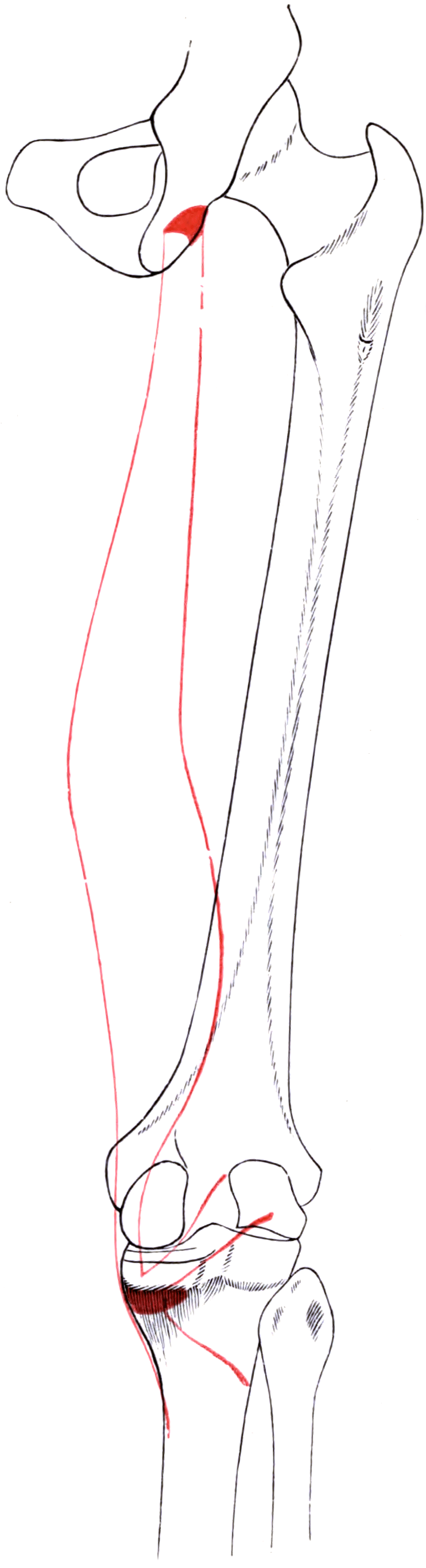
- There are five distal expansions: anterior (deep) arm, direct arm, inferior (distal tibial) arm, the oblique popliteal ligament and the capsular arm to the posterior oblique ligament.
- The oblique popliteal ligament is the largest structure on the posterior knee and is a direct lateral reflection of the semimembranosus tendon.
- It is the principal dynamic restraint of the posteromedial corner; loss of the static posteromedial structures produces anteromedial rotatory instability.
- “Semimembranosus is deep and lateral to semitendinosus proximally, then becomes deep and medial distally — the two cross.
- “A Baker cyst arises through a valvular slit between the semimembranosus tendon and the medial head of gastrocnemius.
- “Semimembranosus bursitis is posteromedial and deep; pes anserine bursitis is anteromedial, 4-5 cm distal to the joint line.
- “The muscle is membranous proximally and fleshy distally — the exact reverse of semitendinosus, which is fleshy proximally and tendinous distally.
Overview
Semimembranosus is the deepest and most powerful of the medial hamstrings. Its name describes its proximal half: a broad, flat, glistening membranous tendon that occupies the upper third of the muscle before giving way to a fleshy belly. Distally the belly reconstitutes into a short, thick tendon that fans out into five separate expansions around the posteromedial knee.
Two facts make this muscle disproportionately important in fellowship examinations. First, it is the only hamstring that inserts as a multi-limbed complex rather than a single tendon — and one of those limbs, the oblique popliteal ligament (OPL), is the largest structure on the back of the knee. Second, it is the dynamic engine of the posteromedial corner (PMC): the static structures of the PMC (the posterior oblique ligament, the posteromedial capsule, the deep medial collateral ligament) are all tensioned by semimembranosus contraction.
Examiners will push on why an isolated superficial medial collateral ligament (sMCL) tear behaves so differently from an sMCL tear that also involves the posteromedial corner.
- In extension, the posterior oblique ligament (POL) and posteromedial capsule are taut and act as the primary static restraint to valgus and to tibial internal rotation. The sMCL is a secondary restraint in this position.
- In flexion, the POL and posteromedial capsule slacken. Their tension is now actively restored by semimembranosus, which pulls the POL and the posteromedial capsule posteriorly and distally through its capsular arm.
- The couple: the static structures hold the corner in extension; semimembranosus holds it through flexion. Divide the muscle's expansions and the corner is only competent at full extension.
- Failure mode: with the PMC incompetent, an applied valgus and external rotation force allows the medial tibial plateau to rotate anteriorly around an intact posterolateral pivot — this is anteromedial rotatory instability (AMRI), and it is the reason a grade III medial injury with a positive anteromedial drawer will not settle with a brace alone.
- Corollary for the ACL: the PMC is a secondary restraint to anterior translation. An unrecognised PMC injury is a recognised cause of high graft force and early ACL reconstruction failure.
DAI-OCThe Five Distal Arms of Semimembranosus
Hook:Direct And Inferior go to bone; Oblique and Capsular go to soft tissue.
The five-arm description is the working scheme every textbook and viva uses, and it is the one to recite. Know that it is a simplification: LaPrade's quantitative dissection of 20 fresh-frozen knees found EIGHT attachments distal to the main common tendon, of which the paper names the three main components — the lateral expansion to the oblique popliteal ligament, the direct arm to the tibia, and the anterior arm — plus a distal tibial expansion forming a posterior fascial layer over popliteus. The extra expansions are small and are not individually named in that report.
Why it matters beyond pedantry: the smaller expansions are what a posteromedial reconstruction is trying to recreate, and they are why a repair that reattaches only the direct arm restores less than the anatomy suggests. Say "at least five, described as eight in quantitative dissection" and you have shown you know where the number comes from.
Attachments, Innervation and Relations
Proximal Origin
- Site: the superolateral facet of the ischial tuberosity, on its lateral and anterior aspect.
- Relationship to the conjoint tendon: semimembranosus is anterolateral; the conjoint tendon of semitendinosus and the long head of biceps femoris is posteromedial. The two footprints are contiguous but distinct, and the semimembranosus footprint extends further anteriorly onto the tuberosity.
- Footprint dimensions: cadaveric work places the semimembranosus footprint at roughly 2.5-3 cm in length, crescentic, lying on the lateral half of the tuberosity. The conjoint footprint is of similar length but sits posteromedially.
- Practical consequence: the semimembranosus footprint is the deep, anterior, lateral part of the origin — the part hardest to see and hardest to re-anchor in a retracted three-tendon avulsion, and the part closest to the sciatic nerve.
Distal Insertion — the Five Arms
The tendon reaches the posteromedial tibia at the level of the joint line and immediately fans out.
1. Anterior (deep) arm. Turns anteriorly and runs deep to the superficial MCL, inserting on the medial tibia just distal to the joint line, beneath the sMCL. It is the arm most often mistaken for the direct arm at surgery.
2. Direct arm. The strongest and most posterior; inserts into a discrete tubercle on the posteromedial aspect of the medial tibial condyle, just distal and posterior to the joint line. This is the palpable, blunt structure the surgeon finds first through a posteromedial approach.
3. Inferior (distal tibial) expansion. A thin band running distally and anteriorly deep to the sMCL toward the posteromedial tibial crest and blending with the fascia over popliteus.
4. Oblique popliteal ligament (OPL). A broad, strong lateral reflection of the tendon that runs superolaterally across the posterior capsule to blend with the lateral posterior capsule, the plantaris origin and the lateral gastrocnemius. This is the largest structure on the posterior aspect of the knee and forms the floor of the popliteal fossa.
5. Capsular arm. Runs proximally and medially into the posterior oblique ligament (POL), the posteromedial capsule, and reaches the posterior horn of the medial meniscus.
The Membranous Character
- Proximal third: flat, wide, membranous tendon (its name).
- Middle third: muscular belly, deep to semitendinosus.
- Distal third: short, thick, rounded tendon.
- Contrast with semitendinosus: fleshy proximally, long round tendon distally, with a tendinous raphe across its mid-belly.
In cadaveric mapping of the proximal hamstring origin the sciatic nerve lies on average 1.2 cm lateral to the ischial tuberosity, running distally and slightly laterally.
- The semimembranosus footprint is the lateral one — so the deep, lateral limit of your dissection is also the nerve's territory.
- In a chronic retracted avulsion the nerve is frequently adherent to the scarred tendon stump and must be identified and neurolysed before the stump is mobilised.
- Retraction on the nerve, not transection, is the usual mechanism of postoperative neuropraxia.
- Work from medial to lateral, identify the nerve early, and place all lateral retractors under direct vision.
Action and Biomechanics
Actions in Each Plane
- Hip: extension. As a biarticular extensor it is most efficient when the knee is extended and the hip flexed — the terminal swing phase of running.
- Knee: flexion, and — critically — internal rotation of the tibia on the femur when the knee is flexed.
- Posteromedial corner: contraction pulls the POL and posteromedial capsule posteromedially and distally, tensioning them through the arc of flexion where they would otherwise be slack.
- Medial meniscus: the capsular arm retracts the posterior horn of the medial meniscus during flexion, preventing entrapment between the femoral condyle and tibial plateau.
- Popliteal fossa floor: the OPL, tensioned by semimembranosus, resists hyperextension and prevents posterior capsular redundancy.
Length-Tension and Moment Arm
- A biarticular muscle: it cannot shorten maximally at both joints simultaneously. Hip flexion with knee extension places it at maximal length — the position of injury in the sprinter.
- The knee flexion moment arm increases from full extension to about 30-50 degrees of flexion, then plateaus. The internal rotation moment arm rises steeply with flexion, which is why the tibial internal rotation contribution is negligible near extension.
- Eccentric demand: the highest muscle-tendon strain during sprinting occurs in late swing, when the hamstrings decelerate the extending knee. This is when the muscle is both long and maximally activated — the classical injury window.
Synergists and Antagonists
- Synergists: semitendinosus, biceps femoris long head, gracilis, sartorius, popliteus (for tibial internal rotation), medial gastrocnemius (knee flexion).
- Antagonists: quadriceps (knee extension), iliopsoas and rectus femoris (hip flexion), biceps femoris (tibial external rotation).
What Happens When It Fails
- Acute complete proximal avulsion: loss of hip extension power, inability to sprint, a "cramping" sitting pain from the ischial stump, and, if the sciatic nerve is tethered, a radiating posterior thigh pain.
- Loss of the distal expansions (surgical or traumatic): the posteromedial corner is competent in extension but slack in flexion — anteromedial rotatory instability.
- Selective medial hamstring weakness: the biceps femoris becomes relatively dominant, producing a subtle tibial external rotation bias, of interest in ACL-deficient and PMC-deficient knees.
The medial hamstrings pull the tibia posteriorly relative to the femur — the same direction as the ACL's restraint of anterior tibial translation.
- Hamstring co-contraction reduces ACL strain, and this is the biomechanical justification for hamstring-dominant rehabilitation after ACL reconstruction and for neuromuscular ACL injury-prevention programmes.
- It is also why quadriceps-dominant landing patterns (small knee flexion angle, high quadriceps activation, low hamstring activation) are a recognised ACL injury risk profile.
- The corollary that examiners like: harvesting semitendinosus and gracilis for an ACL graft removes part of this protective mechanism, with measurable deep-flexion knee flexion strength deficits persisting for years.
Surface Anatomy and Examination
Palpation
- Proximal: the ischial tuberosity is palpable with the hip flexed, deep to the gluteal fold. In a proximal avulsion the tuberosity feels bare and there is a palpable defect and a step in the hamstring mass.
- Distal: with the patient prone and the knee flexed to 90 degrees against resistance, the medial hamstring tendons stand out. The most posterior and most prominent cord is semitendinosus; the semimembranosus is the broader, flatter, deeper structure felt just anterior and lateral to it, filling the space between semitendinosus and the medial gastrocnemius.
- Distal tendon: palpate at the posteromedial joint line, aiming for the tubercle on the posteromedial tibial condyle. Tenderness here in a runner with posteromedial knee pain is distal semimembranosus tendinopathy until proved otherwise.
Isolation Testing
- Prone knee flexion with tibial internal rotation against resistance loads semimembranosus and semitendinosus preferentially.
- Prone knee flexion with tibial external rotation shifts load to biceps femoris. A pain or power difference between the two positions localises the injury medially or laterally.
- Grading: standard Medical Research Council 0-5 scale, but functional grading matters more: the ability to perform a single-leg bridge and, at higher level, a prone hamstring curl at 90 degrees through full range without a lag.
Named Provocative Tests
For proximal hamstring tendinopathy.
- Standing, hip flexed roughly 90 degrees with the knee extended and the foot on a support — an active hamstring stretch.
- Positive: reproduction of deep buttock pain at the ischial tuberosity.
- False positive: lumbar radiculopathy and posterior thigh referred pain from the hip.
For proximal hamstring tendinopathy.
- Supine; the examiner maximally flexes the hip and knee, then slowly straightens the knee.
- Positive: ischial pain as the knee approaches extension.
- More sensitive than a straight-leg raise for tendinopathy, and can be modified to a rapid "modified bent-knee stretch" for higher sensitivity.
For posteromedial corner insufficiency.
- Knee flexed 80-90 degrees, foot externally rotated 10-15 degrees, then an anterior drawer force.
- Positive: the medial tibial plateau rotates anteriorly relative to the femur — anteromedial rotatory instability.
- Trap: in an ACL-deficient knee the whole tibia translates; look specifically at the medial plateau rotating forward.
Separating isolated sMCL from a combined injury.
- Laxity at 30 degrees only — isolated superficial MCL.
- Laxity at 0 degrees as well — the posteromedial corner and posterior oblique ligament are torn, and often the cruciate.
- This one manoeuvre distinguishes an injury that will brace successfully from one that frequently needs repair or reconstruction.
False Positives and Pitfalls
- Ischial pain is produced by ischiofemoral impingement, ischial bursitis, lumbar radiculopathy, deep gluteal syndrome and, in adolescents, apophysitis — not only by hamstring tendinopathy.
- Posteromedial knee pain in a middle-aged patient is far more often a medial meniscus posterior horn tear or a Baker cyst than semimembranosus tendinopathy.
- Pain on resisted knee flexion is not specific: it is positive in gastrocnemius, popliteus and biceps pathology as well.
Complications
Iatrogenic Nerve Injury
- Sciatic neuropraxia after proximal hamstring repair is the commonest neurological complication. Mechanism is retraction, not transection. Avoid by early identification, keeping the knee flexed to relax the nerve, and limiting continuous lateral retraction.
- Posterior femoral cutaneous nerve injury gives a posterior thigh sensory deficit and occasionally a painful neuroma at the gluteal crease incision. Preserve it during the approach.
- Complete sciatic palsy is rare and should prompt immediate re-exploration if it is noted after a repair performed under tension.
Vascular Injury
- Popliteal artery injury during posteromedial exposure or posterior capsulotomy is catastrophic and preventable: keep the medial gastrocnemius interposed and never work lateral to the semimembranosus tendon without visualising the vessels.
- The artery is closest to bone at the level of the posterior joint line and the proximal tibia, and is tethered at the adductor hiatus and at the soleal arch — the anatomical reason knee dislocation threatens it.
Repair-Related Problems
- Re-rupture of a proximal repair: roughly related to delay to surgery and to non-compliance with the post-operative brace protocol. Repairs performed acutely have consistently lower re-rupture rates and better strength recovery than chronic repairs.
- Sitting pain from anchors or knots at the ischial tuberosity — bury the knots and countersink the anchors.
- Heterotopic ossification and adhesions around the tuberosity leading to secondary sciatic tethering.
Functional Morbidity
- Persistent hamstring weakness in deep flexion after chronic repair or after prolonged immobilisation.
- Posteromedial instability after over-aggressive release in arthroplasty or in a posteromedial approach — the medial flexion gap opens and the knee feels unstable on stairs.
- Stiffness after over-tensioned POL reconstruction — the corner is retensioned in the wrong position (flexion rather than extension).
Missed Pathology
- Failing to recognise the PMC component of a medial-sided injury leads to persistent rotatory instability after an otherwise well-performed ACL reconstruction.
- Treating a Baker cyst in isolation without addressing the intra-articular driver results in recurrence rates that are high enough to make isolated excision indefensible in most adults.
Clinical Relevance
Mechanism and Pattern
- Classic mechanism: forced hip flexion with the knee extended — water-skiing, hurdling, a slip with the leg going forward, or an awkward split.
- Immediate features: an audible pop, posterior thigh pain, inability to continue, and within 24-72 hours a large posterior thigh ecchymosis tracking distally.
- Chronic features: buttock pain on sitting, inability to sprint, and a "dead leg" sensation with sciatic tethering.
Anatomical Patterns
- Structures involved
- Bony ischial apophysis with all three tendons attached
- Typical management
- Non-operative if displaced less than 1.5-2 cm; fixation if more
- Structures involved
- Usually the conjoint tendon or semimembranosus alone
- Typical management
- Non-operative; graded rehabilitation
- Structures involved
- Conjoint tendon plus semimembranosus, partial
- Typical management
- Individualised; operative in high-demand athletes
- Structures involved
- All three tendons off the tuberosity
- Typical management
- Operative repair, ideally within 4 weeks
- Structures involved
- Scarred stump encasing the nerve
- Typical management
- Neurolysis plus repair, occasionally with allograft augmentation
Imaging
- Radiographs: essential in adolescents to identify a bony apophyseal avulsion, which changes the whole management.
- MRI (the investigation of choice): coronal and axial oblique sequences along the hamstring origin. Report the number of tendons avulsed, the degree of retraction in cm, and the relationship of the stump to the sciatic nerve.
- Ultrasound: useful for dynamic assessment and for guided injection, but poor at quantifying retraction.
Surgical Repair Principles
- Position: prone, hip extended and knee flexed 30-45 degrees to relax the hamstrings and reduce repair tension.
- Approach: transverse incision in the gluteal crease for acute repairs; a longitudinal or L-shaped extension for retracted chronic cases.
- Sequence: identify the inferior border of gluteus maximus, retract it superiorly, identify the posterior femoral cutaneous nerve, then the sciatic nerve laterally before any lateral mobilisation.
- Fixation: suture anchors into the decorticated tuberosity, restoring the semimembranosus footprint anterolaterally and the conjoint footprint posteromedially. Anatomical restoration of both footprints separately is the goal, not a single mass repair.
They are clinically indistinguishable.
- Both give calf pain, swelling and a positive Homans sign.
- Never diagnose a ruptured cyst without a duplex ultrasound excluding deep vein thrombosis.
- A cyst and a DVT can coexist — a large cyst can compress the popliteal vein and cause one.
Do not mistake healing callus for a tumour.
- An exuberant healing apophyseal avulsion produces a heterogeneous mineralised mass on radiographs and marrow oedema on MRI.
- This has been biopsied as a suspected osteosarcoma or Ewing sarcoma.
- The history of an acute sprinting or hurdling injury and the classic location resolve it — correlate before biopsy.
Surgical Relevance
The Posteromedial Approach to the Knee
- Position: supine with the knee flexed 60-90 degrees and the hip externally rotated, or prone for a direct posterior approach.
- Incision: longitudinal, centred over the posteromedial joint line, just posterior to the palpable sMCL.
- Interval: develop the plane between the medial head of gastrocnemius (retracted posterolaterally, protecting the neurovascular bundle) and the semimembranosus and posteromedial capsule (retracted anteromedially).
- Uses: posterior horn medial meniscal root repair, PCL tibial inlay (via a direct posterior approach), fixation of posteromedial tibial plateau fractures, and excision of a recalcitrant Baker cyst.
- Key protection: the medial head of gastrocnemius is the retractor of choice for the popliteal vessels. Never place a retractor deep to the vessels.
Posteromedial Tibial Plateau Fractures
- The posteromedial fragment is defined in part by the semimembranosus insertion: the direct arm attaches to the posteromedial tibial condyle and the fragment carries that attachment.
- A posteromedial fragment is a shear fragment and will not be controlled by lateral plating — it needs a posteromedial buttress plate applied through this interval.
- Semimembranosus tension is a deforming force on the fragment, drawing it proximally and posteriorly; this must be neutralised, not resisted, by the implant.
Structures at Risk with Distances
- Location relative to a landmark
- Approximately 1.2 cm lateral to the ischial tuberosity
- How to protect it
- Identify it before any lateral dissection in proximal hamstring repair
- Location relative to a landmark
- Posterior and superficial to the sciatic nerve at the gluteal fold
- How to protect it
- Preserve when splitting the gluteal fascia; injury gives posterior thigh numbness
- Location relative to a landmark
- Immediately lateral to the semimembranosus tendon at the joint line
- How to protect it
- Retract the medial head of gastrocnemius laterally to shield them
- Location relative to a landmark
- Crossing the sartorial fascia anteromedially, 1-2 cm distal to the joint line
- How to protect it
- Keep the posteromedial incision behind the sMCL
- Location relative to a landmark
- Runs deep to the sMCL along the medial tibial condyle
- How to protect it
- Encountered during deep exposure of the posteromedial tibia
- Location relative to a landmark
- Its capsular attachment blends with the semimembranosus capsular arm
- How to protect it
- Do not detach the capsular arm during posteromedial capsulotomy
Semimembranosus as a Surgical Resource
- Not a graft donor. Unlike semitendinosus and gracilis, semimembranosus is not harvested as a free tendon graft: its tendon is short, broad and multi-limbed, and harvesting it destroys the posteromedial corner. If an examiner asks which medial hamstring you would never harvest, the answer is semimembranosus.
- As a dynamic tensioner: the classical Hughston posteromedial capsular reconstruction advances the posterior oblique ligament and the semimembranosus insertion anteriorly and proximally to retension the corner.
- In posteromedial corner reconstruction: the semimembranosus tendon is a useful anatomical reference — the direct arm marks the level of the joint line posteromedially and separates the POL (proximal) from the posteromedial capsule (distal).
- Distal release: the anterior (deep) arm can be released to improve posteromedial exposure in a stiff knee or a complex plateau fracture; the direct arm and OPL should be preserved wherever possible.
Total Knee Arthroplasty
- In a fixed varus knee, the sequential medial release proceeds from the deep MCL and osteophytes, to the posteromedial capsule, and — in the tightest knees — to the semimembranosus insertion.
- Semimembranosus release corrects flexion-space tightness medially because the tendon is a flexion-space structure. Releasing it in a knee that is tight in extension only is a technical error and produces medial flexion instability.
- Sequence discipline: osteophytes first, then deep MCL, then posteromedial capsule, then semimembranosus, and only then consider the superficial MCL. Reassess the gaps after each step.
Guidelines, Registries & Global Practice
Anatomical Variation Across Populations
- The number and prominence of the distal semimembranosus expansions is consistent across cadaveric series from European, North American and East Asian populations; the anatomy is reliable and the five-arm description is reproducible.
- The oblique popliteal ligament is present in essentially all knees, but its width and the contribution it receives from the plantaris and the lateral capsule vary. This variability partly explains inconsistent reported contributions to hyperextension restraint.
- Popliteal cyst prevalence varies with the population studied: it is far more common in cohorts with a high prevalence of knee osteoarthritis and inflammatory arthropathy, and is a common incidental MRI finding in asymptomatic adults.
Differences in Described Technique
- Position on the posteromedial corner
- Anatomical repair and advancement of the posterior oblique ligament with the semimembranosus insertion; the historical foundation of posteromedial surgery.
- Position on the posteromedial corner
- Quantitative cadaveric mapping of the medial and posteromedial attachments, with separate anatomical reconstruction of the superficial MCL and posterior oblique ligament.
- Position on the posteromedial corner
- Emphasises the posteromedial approach between semimembranosus and medial gastrocnemius for posteromedial tibial plateau shear fragments, with buttress plating.
- Position on the posteromedial corner
- Non-operative management for the majority of isolated medial collateral injuries, with early specialist referral where there is laxity in full extension or a combined cruciate injury.
- Position on the posteromedial corner
- Supports early operative repair of complete proximal hamstring avulsions in active patients and anatomical repair of acute grade III posteromedial injuries.
Practice Signals
- Registry-level data specific to posteromedial corner surgery is limited; most evidence is cohort and cadaveric. National ligament registries consistently identify untreated concomitant collateral and rotatory instability as a risk factor for ACL graft failure, which is the strongest practice signal for addressing the posteromedial corner.
- Proximal hamstring avulsion repair shows a consistent time-dependent effect across series: acute repair outperforms chronic repair for strength, return to sport and re-rupture, and this has driven a lower threshold for early referral in sports medicine services internationally.
High- and Limited-Resource Practice
- Well-resourced settings: routine MRI for suspected proximal hamstring avulsion and for medial-sided knee injury; anatomical posteromedial reconstruction with allograft or autograft; ultrasound-guided injection for bursitis.
- Limited-resource settings: clinical examination — the palpable defect and the pattern of ecchymosis proximally, and valgus stress at 0 and 30 degrees at the knee — carries most of the diagnostic load. Ultrasound is a highly effective substitute for MRI in experienced hands for both proximal avulsion and popliteal cysts. Acute anatomical repair with suture anchors remains achievable and is the highest-value intervention.
MCQ Practice Points
Q: What is the innervation of semimembranosus? A: The tibial division of the sciatic nerve, L5, S1, S2. Every hamstring except the short head of biceps femoris takes the tibial division; the short head alone takes the common peroneal division.
Q: What is the oblique popliteal ligament and where does it come from? A: It is the broad superolateral reflection of the semimembranosus tendon across the posterior capsule, and it is the largest structure on the back of the knee. It forms the floor of the popliteal fossa and resists hyperextension.
Q: How far is the sciatic nerve from the ischial tuberosity? A: Approximately 1.2 cm lateral. Since the semimembranosus footprint is the lateral one, the nerve is closest to the part of the repair the surgeon reaches last.
Q: A Baker cyst is a distension of which bursa? A: The gastrocnemio-semimembranosus bursa, communicating with the joint through a valvular slit between the semimembranosus tendon and the medial head of gastrocnemius.
Q: What does valgus laxity at full extension tell you? A: The posteromedial corner has failed — the posterior oblique ligament and posteromedial capsule, not just the superficial MCL. This is the finding that turns a braceable injury into a surgical one.
Q: Which medial hamstring is more superficial at the knee? A: Semitendinosus. Semimembranosus is deep and lateral proximally; distally semitendinosus crosses superficial to it to reach the pes anserinus, leaving semimembranosus deep at the posteromedial joint line.
Q: How do you distinguish semimembranosus bursitis from pes anserine bursitis? A: By site. Semimembranosus bursitis is posteromedial and at or just distal to the joint line, deep to the tendon. Pes anserine bursitis is anteromedial, about 4-5 cm distal to the joint line, deep to the pes tendons and superficial to the superficial MCL.
Q: Which arm of semimembranosus reaches the medial meniscus? A: The capsular arm, which blends with the posterior oblique ligament and reaches the posterior horn of the medial meniscus, retracting it during flexion.
Q: Why is semimembranosus never used as a free tendon graft? A: Its distal tendon is short, broad and multi-limbed, and those limbs are the posteromedial corner. Harvesting it destabilises the very structure it supports. Semitendinosus and gracilis are the donors.
Q: A varus knee at total knee arthroplasty is tight medially in flexion but balanced in extension. Which structure do you release? A: The semimembranosus insertion, a flexion-space structure. Releasing the posteromedial capsule addresses extension tightness; releasing semimembranosus addresses flexion tightness.
Exam Viva Scenarios
Practise clinical reasoning and management decisions out loud
“A 34-year-old water-skier falls with the leg forced forward. He has severe buttock pain, cannot weight-bear comfortably and three days later has extensive posterior thigh bruising. Examination shows a palpable defect below the gluteal fold. How do you proceed?”
“A 24-year-old footballer sustains a valgus and external rotation injury. Six weeks on he has valgus laxity at 30 degrees and also at 0 degrees, a positive Lachman, and on the anteromedial drawer the medial plateau rotates forward. What is the pathoanatomy and how would you manage him?”
“A 58-year-old woman with known medial compartment osteoarthritis presents with sudden calf pain and swelling after squatting. There is bruising around the medial malleolus. What is your differential and your management?”
Core Anatomy
- Origin: superolateral ischial tuberosity, anterolateral to conjoint tendon
- Insertion: five arms — direct, anterior (deep), inferior, OPL, capsular
- Nerve: tibial division of sciatic, L5-S2
- Membranous proximally, fleshy distally (reverse of semitendinosus)
Biomechanics
- Hip extension, knee flexion, tibial internal rotation
- Dynamic tensioner of the posteromedial corner in flexion
- Retracts the posterior horn of the medial meniscus
- OPL resists hyperextension and forms the popliteal fossa floor
Numbers
- Sciatic nerve 1.2 cm lateral to ischial tuberosity
- Retraction greater than 2 cm plus three tendons — repair
- Apophyseal avulsion: fix if displaced more than 1.5-2 cm
- Pes anserine bursa 4-5 cm distal to the medial joint line
Clinical Traps
- Valgus laxity at 0 degrees = posteromedial corner failure
- Baker cyst = symptom; find the intra-articular driver
- Ruptured cyst versus DVT — duplex first, always
- Never harvest semimembranosus as a graft
Evidence Base
The Anatomy of the Posterior Aspect of the Knee: An Anatomic Study
- Detailed quantitative dissection of 20 non-paired fresh-frozen knees
- The semimembranosus tendon had eight attachments distal to the main common tendon, the principal ones being the lateral expansion to the oblique popliteal ligament, the direct arm to the tibia and the anterior arm
- The oblique popliteal ligament was the largest posterior knee structure, averaging 48.0 mm long and 9.5 mm wide medially, widening to 16.4 mm at its lateral attachment
- A distal tibial expansion of semimembranosus formed a posterior fascial layer over the popliteus muscle
- The plantaris, popliteofibular ligament, fabellofibular ligament and semimembranosus bursa were present in all specimens
The Posteromedial Corner of the Knee: Medial-Sided Injury Patterns Revisited
- Retrospective review of 93 knees with operatively treated isolated and combined medial-sided injuries
- The posterior oblique ligament was injured in 99 per cent of knees
- The semimembranosus capsular attachment was injured in 70 per cent
- Complete peripheral detachment of the medial meniscus was present in 30 per cent
The Role of the Posterior Oblique Ligament in Repairs of Acute Medial (Collateral) Ligament Tears of the Knee
- The originating clinical description of the posterior oblique ligament as a structure to be addressed in repair of acute medial-sided knee injury
- Reported a series of acute medial collateral ligament tears treated surgically, with follow-up
- Established the posterior oblique ligament in orthopaedic practice as distinct from the superficial medial collateral ligament
- No structured abstract is indexed for this 1973 paper; the findings above are those stated in its title and indexing
The Proximal Origin of the Hamstrings and Surrounding Anatomy Encountered During Repair
- Fourteen fresh-frozen cadaveric specimens dissected prone to map the proximal hamstring origin
- The semitendinosus and biceps femoris share a common tendinous origin on the ischium; the semimembranosus originates just laterally
- The sciatic nerve lay an average of 1.2 plus or minus 0.2 cm from the most lateral aspect of the ischial tuberosity
- The conjoint origin measured 2.7 by 1.8 cm; the semimembranosus origin was crescentic at 3.1 by 1.1 cm
- The inferior gluteal nerve and artery lay an average of 5.0 cm from the inferior border of gluteus maximus at the lateral border of the ischium
Complete Proximal Hamstring Avulsions: A Series of 41 Patients with Operative Treatment
- 41 patients with complete proximal hamstring avulsion treated operatively, mean follow-up 37 months
- 19 results were excellent and 10 good; 5 were moderate and 7 poor
- Mean delay from injury to surgery was 2.4 months in the excellent or good group versus 11.7 months in the moderate or poor group
- The difference in delay between outcome groups was statistically significant
Avulsion of the Proximal Hamstring Origin
- 72 consecutive reconstructions in 71 patients, mean age 40.2 years, mean follow-up 24 months
- Water-skiing was the most frequent mechanism, in 21 cases
- Complete avulsion of the proximal hamstring origin was present in 63 cases (87.5 per cent), with a mean retraction of 7 cm (range 0 to 20 cm)
- Mean postoperative isotonic hamstring strength was 84 per cent and endurance 89 per cent of the contralateral side
- Delayed repair was more technically demanding, increased the likelihood of sciatic nerve involvement and reduced postoperative strength and endurance