The Dynamic Stabiliser of the Medial Longitudinal Arch
- Deepest muscle of the deep posterior compartment, arising from the interosseous membrane and the adjacent tibia and fibula.
- Innervated by the tibial nerve, roots L4 and L5, with motor branches entering the proximal third.
- Primary insertion on the navicular tuberosity, with slips to all three cuneiforms, the cuboid, the sustentaculum tali and the bases of the second to fourth metatarsals.
- The principal invertor of the foot and the dynamic locker of the transverse tarsal joint, allowing the gastrocsoleus to work against a rigid lever.
- Working excursion is only about 1 to 1.5 cm, far less than flexor digitorum longus - the key limitation in transfer surgery.
- “Too-many-toes sign is hindfoot valgus plus forefoot abduction viewed from behind - it is a positional sign, not a strength test.
- “The single heel-raise test is the functional test: failure to invert the heel on rising means the tendon cannot lock the transverse tarsal joint.
- “Tarsal tunnel order from anterior to posterior: Tibialis posterior, flexor Digitorum longus, posterior tibial Artery, tibial Nerve, flexor Hallucis longus.
- “The tendon runs directly behind the medial malleolus in a fibro-osseous groove and has a hypovascular zone just distal to it.
Overview
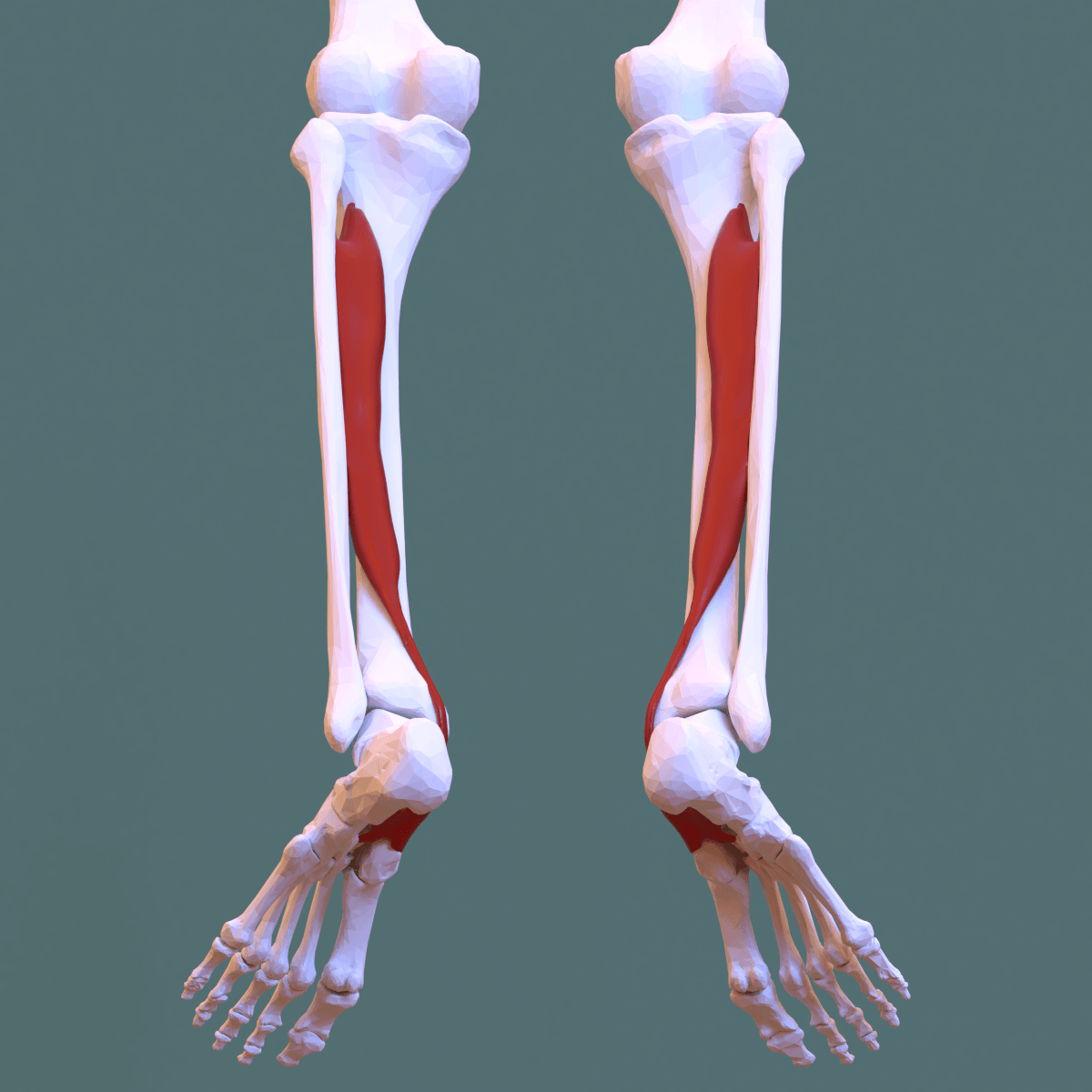
Tibialis posterior is the deepest and most central muscle of the deep posterior compartment of the leg. It arises from the interosseous membrane and the adjoining posterior surfaces of both the tibia and the fibula, so it is the only muscle in the leg that takes origin from both bones and from the membrane between them. Its tendon runs behind the medial malleolus, turns sharply forward beneath the sustentaculum tali, and fans out onto almost every bone of the midfoot.
Functionally it is far more than an invertor. During stance it is the dynamic locker of the transverse tarsal joint: by inverting the hindfoot it brings the talonavicular and calcaneocuboid axes out of parallel, converting the flexible midfoot into a rigid lever against which the gastrocsoleus can push. Its loss produces the single most common acquired deformity of the adult foot.
The transverse tarsal (Chopart) joint comprises the talonavicular and calcaneocuboid joints. Their axes are parallel when the hindfoot is in valgus and divergent when the hindfoot is in varus.
- Hindfoot valgus: axes parallel, the midfoot is unlocked and flexible - useful at heel strike for shock absorption and accommodating uneven ground.
- Hindfoot varus: axes divergent, the midfoot is locked and rigid - essential at heel rise so the triceps surae can transmit force to the forefoot through a stiff lever.
- Tibialis posterior is the muscle that converts one state into the other. It inverts the calcaneus at the start of heel rise, locks the midfoot, and only then can the gastrocsoleus generate an efficient push-off.
- When it fails: the heel stays in valgus, the transverse tarsal joint never locks, the gastrocsoleus pushes against a flexible midfoot, and the arch collapses through the talonavicular joint. The forefoot abducts, the talus plantarflexes and adducts, and the deformity becomes self-perpetuating because the Achilles vector now lies lateral to the subtalar axis and itself becomes an evertor.
That final point is the reason a contracted gastrocnemius must be lengthened during flatfoot reconstruction: in a valgus hindfoot the Achilles is a deforming force, not merely a tight one.
Tom Dick And Very Nervous HarryTarsal Tunnel Contents (Anterior to Posterior)
Hook:Order runs anterior to posterior behind the medial malleolus, deep to the flexor retinaculum (laciniate ligament).
Attachments, Innervation and Relations
Origin
- Interosseous membrane (upper two-thirds) - the dominant origin.
- Posterior surface of the tibia, lateral to the vertical line, below the soleal line.
- Posterior surface of the fibula, medial to the medial crest.
- It is therefore the only leg muscle arising from tibia, fibula and interosseous membrane together, and it lies deep to and between flexor digitorum longus (medial) and flexor hallucis longus (lateral).
Tendon course
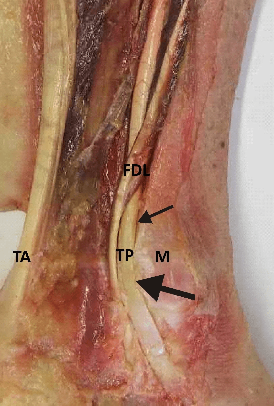
- The tendon forms at about the junction of the middle and distal thirds of the leg and passes deep to flexor digitorum longus at the ankle, crossing from lateral to medial so that at the medial malleolus it becomes the most anterior structure of the tarsal tunnel.
- It runs in a fibro-osseous groove on the posterior aspect of the medial malleolus, held by the flexor retinaculum, then turns sharply forward - roughly 90 degrees - beneath the sustentaculum tali, which acts as its pulley.
- It has a true synovial sheath extending from about 4 cm proximal to the malleolus to near the navicular insertion.
Insertion - the fan
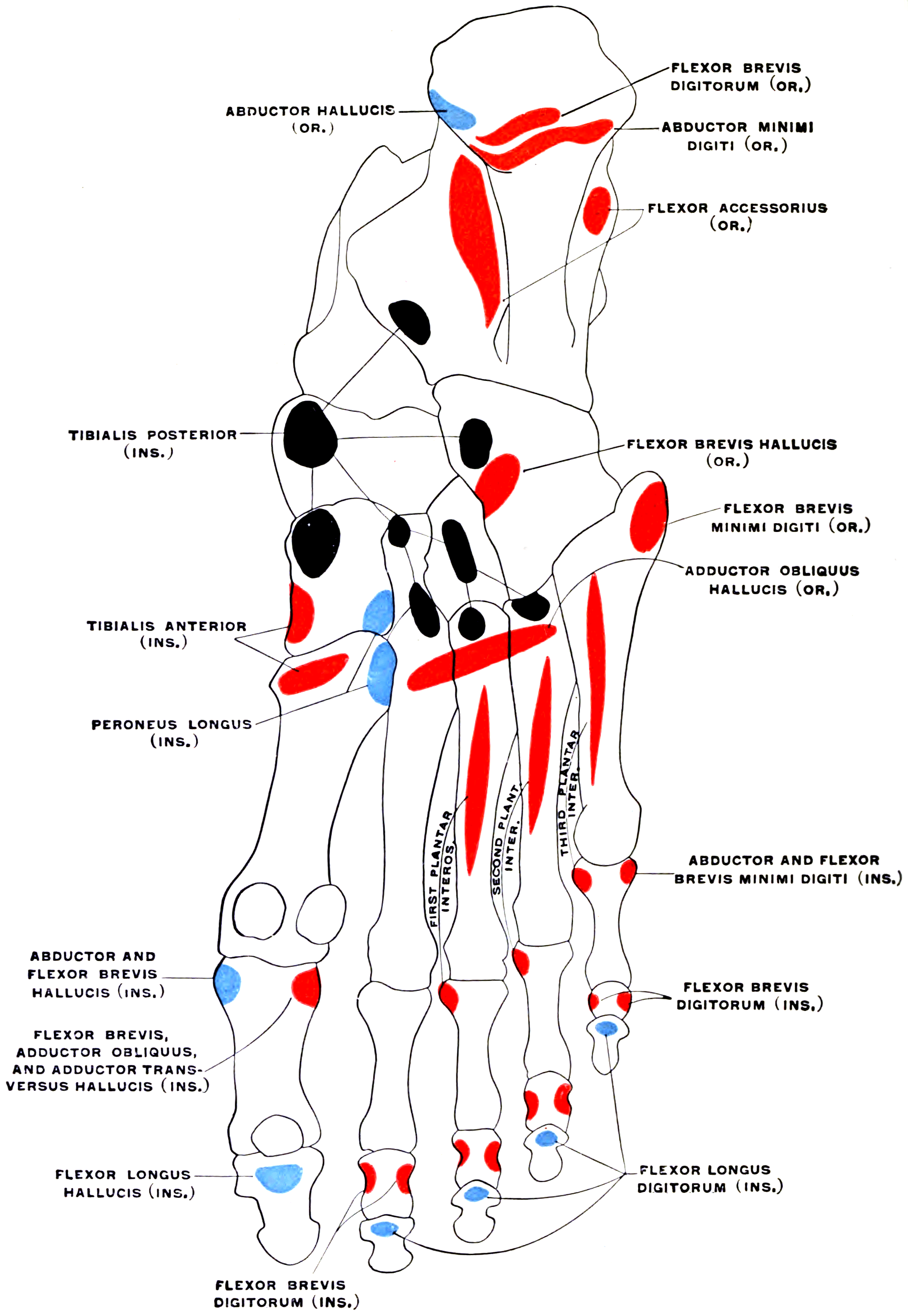
- Navicular tuberosity - the main and strongest slip, and the one that must be preserved or re-attached.
- Plantar slips to the medial, intermediate and lateral cuneiforms, the cuboid, and the bases of the second, third and fourth metatarsals.
- A recurrent slip to the sustentaculum tali.
- A slip blending with the plantar calcaneonavicular (spring) ligament complex.
- This broad, plantarly directed fan is why the muscle supports the arch across the whole midfoot rather than acting at a single point, and why complete detachment is functionally catastrophic while partial detachment is often tolerated.
Accessory navicular
- Present in roughly 10 to 15 per cent of feet. Type I is a sesamoid within the tendon; Type II is a synchondrosis with the navicular (the symptomatic type, from shear across the cartilaginous bridge); Type III is a fused cornuate navicular. A symptomatic type II is treated by excision with tendon advancement (Kidner procedure) when non-operative care fails.
In the medial hindfoot the two tendons look alike. Tibialis posterior is anterior, thicker, and inserts on the navicular tuberosity; flexor digitorum longus is posterior, thinner, and divides into four slips distally. Trace each tendon distally before dividing anything. Harvesting the wrong tendon during a flatfoot reconstruction - taking the posterior tibial as the donor - converts a reconstructable stage II deformity into a disaster.
Action and Biomechanics
Actions by plane
- Frontal: inversion (supination) of the subtalar and transverse tarsal joints - its dominant and defining action. It is the strongest invertor of the foot, roughly twice as strong as its direct antagonist peroneus brevis.
- Sagittal: weak ankle plantarflexion; the tendon passes posterior to the ankle axis but its moment arm is small.
- Transverse: adduction of the forefoot, resisting the abduction that characterises the collapsing flatfoot.
- Arch: elevation and dynamic support of the medial longitudinal arch through its broad plantar insertion, acting as a dynamic sling under the talonavicular joint and augmenting the static spring ligament.
Timing within gait
- Active from just after heel strike through to heel rise - an early and mid-stance muscle.
- Eccentric early: it decelerates the physiological pronation that occurs after heel strike, controlling the rate of arch flattening.
- Concentric at heel rise: it inverts the calcaneus, locks the transverse tarsal joint, and hands the foot over to the gastrocsoleus as a rigid lever.
- It is essentially silent in swing, which is why it is a poor donor for a swing-phase role such as dorsiflexion without phase conversion.
Excursion and strength - the numbers that matter for transfers
- Excursion approximately 1 to 1.5 cm in its working range - the shortest useful excursion of any of the major tendons about the hindfoot.
- Flexor digitorum longus excursion is roughly 2 to 3 cm and its strength is only around one-third that of tibialis posterior.
- Consequence: flexor digitorum longus is not a strength-matched substitute for tibialis posterior. The FDL transfer works not by replacing lost inversion power but by acting as a tenodesis and a proprioceptive checkrein, and by supplementing a bony realignment. This is exactly why the transfer must be combined with a medial displacement calcaneal osteotomy - the osteotomy does the mechanical work; the tendon transfer holds the correction and provides some dynamic support.
Static versus dynamic support of the arch
- Type
- Static (windlass)
- Role
- Tensions on great toe dorsiflexion, raises the arch at push-off
- Consequence of failure
- Arch collapse, loss of windlass, forefoot overload
- Type
- Static
- Role
- Primary static support of the talar head
- Consequence of failure
- Talar head plantar and medial migration - the key lesion in PCFD
- Type
- Static
- Role
- Resists hindfoot valgus and talar tilt
- Consequence of failure
- Stage IV valgus talar tilt in the mortise
- Type
- Static
- Role
- Wedge-shaped arch, Lisfranc complex
- Consequence of failure
- Midfoot break, dorsolateral peritalar subluxation
- Type
- Dynamic
- Role
- Locks the transverse tarsal joint, inverts hindfoot, elevates arch
- Consequence of failure
- Progressive collapsing foot deformity
- Type
- Dynamic
- Role
- Secondary arch support and toe stabilisation
- Consequence of failure
- Clawing, less efficient push-off
Tibialis posterior is a dynamic stabiliser. A dynamic stabiliser cannot fail in isolation and produce a fixed deformity - the fixed deformity requires the static restraints to fail as well. In progressive collapsing foot deformity the sequence is:
- Tendon tenosynovitis and elongation (dynamic failure) - the foot still looks normal.
- Repetitive overload of the spring ligament and the talonavicular capsule (static failure) - the talar head drops medially and plantarwards, the forefoot abducts, and the deformity becomes visible.
- Attenuation of the deltoid ligament - valgus talar tilt in the mortise (stage IV).
This explains why simply repairing or reconstructing the tendon does not correct an established deformity, and why modern reconstruction always includes a bony realignment (medial displacement calcaneal osteotomy, lateral column lengthening, or a medial column procedure) and often a spring ligament repair.
Surface Anatomy and Examination
Palpation
- The tendon is palpable immediately behind and inferior to the medial malleolus, running forward to the prominent navicular tuberosity, which sits roughly 2.5 to 3 cm distal and anterior to the malleolar tip.
- Ask for resisted inversion with the foot in plantarflexion (which relaxes tibialis anterior) - this isolates tibialis posterior better than any other manoeuvre.
- Swelling and tenderness along the tendon course, with a boggy fullness behind the malleolus, is the earliest physical sign of stage I disease.
Named tests and signs
- How to perform
- Stand behind the standing patient and look at the heels
- Positive finding
- More toes visible lateral to the leg on the affected side
- What it means
- Hindfoot valgus with forefoot abduction
- False positives
- Normal variation, external tibial torsion, unilateral comparison essential
- How to perform
- Patient stands on one leg and rises onto the toes, fingertip support only for balance
- Positive finding
- Cannot rise, or rises without the heel inverting
- What it means
- Tibialis posterior cannot lock the transverse tarsal joint
- False positives
- Calf weakness, pain inhibition, poor balance, ankle arthritis - allow finger support and repeat
- How to perform
- Both feet, rise onto toes
- Positive finding
- Heels fail to invert
- What it means
- Screening test only, less sensitive than single heel raise
- False positives
- The unaffected side compensates and masks the deficit
- How to perform
- Foot plantarflexed and inverted against resistance
- Positive finding
- Weakness or pain
- What it means
- Tendon dysfunction or tear
- False positives
- Tibialis anterior substitution if the foot is not plantarflexed
- How to perform
- Passively externally rotate the leg or invert the hindfoot with the patient standing
- Positive finding
- The first metatarsal head lifts off the floor
- What it means
- Loss of medial column stability from tibialis posterior insufficiency
- False positives
- Fixed forefoot supination, midfoot arthritis
- How to perform
- Passive dorsiflexion with knee extended then flexed, hindfoot held neutral
- Positive finding
- Improvement with the knee flexed
- What it means
- Isolated gastrocnemius contracture - needs recession at reconstruction
- False positives
- Allowing midfoot dorsiflexion to substitute
- How to perform
- Passively invert the heel with the patient sitting; observe correction on tiptoe standing
- Positive finding
- Correctable to neutral or beyond
- What it means
- Stage II (flexible) rather than stage III (fixed)
- False positives
- Examiner force overcoming a stiff but not fixed joint
Interpreting the single heel-raise properly
- The test has two components. First, can the patient rise? Second, does the heel invert as they rise?
- A patient may rise onto the toes using the gastrocsoleus alone yet the heel remains in valgus - that is a positive test and indicates tibialis posterior insufficiency.
- Fatigue variant: ask for ten repetitions. Early dysfunction shows as progressive loss of height and loss of heel inversion with repetition, when a single attempt looked normal.
- Fingertip balance support is permitted; without it, balance failure produces false positives.
Additional assessment required before surgery
- Flexibility of the hindfoot - stage II versus stage III. This is the single most important determinant of treatment.
- Forefoot varus (supination) - after correcting the hindfoot to neutral, look at the plane of the forefoot. A fixed forefoot varus of more than about 10 to 15 degrees needs a medial column procedure (Cotton opening wedge medial cuneiform osteotomy or a naviculocuneiform or first tarsometatarsal fusion), otherwise the corrected hindfoot will simply fall back into valgus to get the first ray to the ground.
- Forefoot abduction - assess talonavicular uncoverage on a weightbearing anteroposterior radiograph. Uncoverage of more than about 40 per cent suggests the need for lateral column lengthening.
- Gastrocnemius contracture - a Silfverskiold test in every case.
- Ankle valgus - a weightbearing ankle radiograph or weightbearing CT to exclude stage IV.
Radiographic parameters
- Weightbearing lateral: Meary line (talus-first metatarsal angle) breaks plantarwards at the talonavicular or naviculocuneiform level; the calcaneal pitch falls below about 18 degrees; the lateral talocalcaneal angle increases.
- Weightbearing anteroposterior: increased talonavicular uncoverage, increased talus-first metatarsal angle, forefoot abduction.
- Hindfoot alignment (Saltzman) view: quantifies valgus of the calcaneus relative to the tibial axis.
- Weightbearing CT increasingly demonstrates peritalar subluxation, sinus tarsi and subfibular impingement, and middle facet subluxation, which correlates strongly with the clinical deformity.
- MRI: tenosynovial fluid, tendon thickening or attenuation, longitudinal split tears, and integrity of the spring ligament and deltoid.
Complications
- Mechanism
- Nerve crosses the lateral hindfoot about 1.5 to 2 cm posteroinferior to the fibular tip
- Prevention
- Full-thickness incision to bone, subperiosteal retractors, know the course
- Management
- Observe; neuroma excision and burial if a painful neuroma develops
- Mechanism
- Medial translation of more than 10 mm tents the bundle over the medial calcaneal wall
- Prevention
- Limit translation to 8 to 12 mm; protect the medial cortex from the saw
- Management
- Early recognition; reduce the translation or release the tarsal tunnel
- Mechanism
- Failure to address forefoot abduction, forefoot varus or gastrocnemius contracture
- Prevention
- Full deformity assessment including Silfverskiold and forefoot plane; add lateral column lengthening or Cotton osteotomy as indicated
- Management
- Revision with the omitted procedure; arthrodesis if the deformity is now fixed
- Mechanism
- Excessive lateral column lengthening or excessive medial translation
- Prevention
- Intra-operative assessment with the foot loaded against a flat plate
- Management
- Revision osteotomy; a varus hindfoot is poorly tolerated and causes lateral overload and instability
- Mechanism
- The osteotomy stiffens and lengthens the lateral column
- Prevention
- Restrained graft size (8 to 10 mm), avoid over-lengthening
- Management
- Orthotic management; graft downsizing or calcaneocuboid fusion in refractory cases
- Mechanism
- Loss of long flexor drive to toes 2 to 5
- Prevention
- Divide FDL just distal to the knot of Henry so FHL interconnections continue to drive it; consider tenodesing the stump to FHL
- Management
- Usually asymptomatic; toe flexor tenodesis if a symptomatic lesser toe deformity develops
- Mechanism
- Poor bone quality, smoking, inadequate fixation
- Prevention
- Two-screw fixation, avoid over-translation, stop smoking
- Management
- Revision fixation with bone graft
- Mechanism
- Thin skin, oedematous foot, tension from correction
- Prevention
- Full-thickness flaps, avoid undermining, delay surgery until swelling settles
- Management
- Local wound care, negative pressure dressing
- Mechanism
- Harvesting tibialis posterior removes the dynamic arch stabiliser
- Prevention
- Assess pre-existing planovalgus; consider an alternative donor or a bony stabilisation
- Management
- Orthotic support; arthrodesis in severe collapse
- Mechanism
- Injection into an already degenerate watershed tendon
- Prevention
- Avoid steroid injection; use immobilisation, orthoses and eccentric loading
- Management
- Reconstruct as for stage II disease
Clinical Relevance
Posterior tibial tendon dysfunction and progressive collapsing foot deformity
The condition is now widely termed progressive collapsing foot deformity (PCFD) to acknowledge that the tendon is one component of a multiplanar deformity, but the Johnson and Strom staging with the Myerson modification remains the language of the exam.
- Tendon
- Tenosynovitis, normal length
- Deformity
- None
- Hindfoot
- Normal, heel inverts on rise
- Typical management
- Immobilisation, orthosis, physiotherapy with eccentric loading, tenosynovectomy if refractory
- Tendon
- Elongated, degenerate or torn
- Deformity
- Planovalgus, forefoot abduction
- Hindfoot
- Flexible - corrects passively
- Typical management
- FDL transfer plus medial displacement calcaneal osteotomy, with lateral column lengthening and/or medial column procedure as required, plus gastrocnemius recession
- Tendon
- Non-functional
- Deformity
- Fixed planovalgus
- Hindfoot
- Rigid, subtalar arthritis
- Typical management
- Subtalar, double or triple arthrodesis
- Tendon
- Non-functional
- Deformity
- Fixed deformity plus valgus talar tilt
- Hindfoot
- Rigid, deltoid incompetent
- Typical management
- Deltoid reconstruction with hindfoot fusion; total ankle replacement or tibiotalocalcaneal fusion in end-stage disease
- Stage II is subdivided in common practice into IIA (minimal forefoot abduction, treated with medial displacement calcaneal osteotomy and FDL transfer) and IIB (significant forefoot abduction with more than about 40 per cent talonavicular uncoverage, requiring lateral column lengthening in addition).
- Risk factors: female sex, age over 40, obesity, hypertension, diabetes, seronegative and inflammatory arthropathy, prior local steroid injection, and pre-existing pes planus.
- Presentation: medial hindfoot pain and swelling early; as the deformity progresses the pain often migrates laterally to the sinus tarsi and the subfibular region as the calcaneus impinges beneath the fibula. A patient describing lateral hindfoot pain with a flat foot has advanced disease, not a new problem.
Acute rupture and traumatic injury
- Acute traumatic rupture is uncommon and follows a forced eversion injury or a medial malleolar fracture with entrapment. It is often missed in the trauma setting.
- Tendon entrapment or incarceration within a medial malleolar fracture or a subtalar dislocation prevents reduction; the tibialis posterior tendon is the classic block to reduction of a lateral subtalar dislocation, while the peroneals block a medial dislocation.
- Look specifically for the tendon in any medial malleolar fracture that will not reduce, and in any open reduction of a pilon or plafond injury approached medially.
Tarsal tunnel syndrome
- Compression of the tibial nerve beneath the flexor retinaculum (laciniate ligament) between the medial malleolus and the calcaneus.
- Causes: space-occupying lesions (ganglion, lipoma, varicosities, accessory muscle such as an accessory flexor digitorum accessorius longus), post-traumatic scarring and malunion, tenosynovitis of the adjacent flexor tendons, and hindfoot valgus, which stretches the nerve.
- Findings: burning plantar pain, positive Tinel over the tunnel, a positive dorsiflexion-eversion test (sustained maximal dorsiflexion and eversion with toe extension for 5 to 10 seconds reproduces symptoms), and sensory disturbance in the medial or lateral plantar territory. Heel sensation is often spared because the medial calcaneal branch may arise proximal to the retinaculum.
- Important link: a valgus hindfoot from tibialis posterior dysfunction can itself produce a traction tarsal tunnel syndrome, and correcting the deformity may relieve the neuropathy.
Deep posterior compartment syndrome
- The deep posterior compartment contains tibialis posterior, flexor digitorum longus, flexor hallucis longus, the tibial nerve and the posterior tibial and peroneal vessels.
- Clinical signs: pain on passive toe extension and passive foot eversion, deep calf tenderness that is hard to appreciate because the compartment is covered by soleus, and plantar sensory disturbance.
- It is the compartment most often missed because the deep transverse fascia is not opened. The late consequence is a fibrotic, contracted deep compartment with claw toes, a cavovarus foot and plantar sensory loss.
Accessory navicular and the Kidner procedure
- A symptomatic type II accessory navicular causes medial midfoot pain over a prominent bony bump, worse in stiff footwear and with activity, typically in an adolescent.
- Non-operative management with activity modification, orthoses and immobilisation succeeds in the majority.
- Kidner procedure: excision of the accessory ossicle with advancement and re-attachment of the tibialis posterior tendon plantarwards onto the navicular. Modern practice usually re-attaches with a suture anchor. Debate persists as to whether simple excision without formal advancement gives equivalent results.
Injecting corticosteroid into the tibialis posterior tendon sheath in a patient with tenosynovitis is a recognised precipitant of tendon rupture, because the tendon is already degenerate and lies in a hypovascular watershed segment against a bony pulley. If injection is contemplated at all it must be image-guided, strictly intrasheath, and limited - and in most units it is avoided altogether in favour of immobilisation, orthoses and eccentric loading.
Surgical Relevance
The medial hindfoot approach
- Incision: a curved incision centred 1 cm posterior to the medial malleolus, curving distally toward the navicular tuberosity, roughly 8 to 10 cm long. This single exposure gives access to the tibialis posterior tendon, the flexor digitorum longus, the spring ligament and the navicular.
- Structures at risk: the saphenous vein and nerve anteriorly, the posterior tibial neurovascular bundle posteriorly (it lies immediately deep and posterior to flexor digitorum longus), and the medial calcaneal branches of the tibial nerve crossing the distal part of the wound.
- Key relation: the flexor digitorum longus lies directly posterior to tibialis posterior and anterior to the neurovascular bundle. The safe plane in which to harvest FDL is therefore between the two tendons, staying strictly anterior to the bundle.
FDL transfer with medial displacement calcaneal osteotomy - the operation to know
Why this combination
- The medial displacement calcaneal osteotomy (MDCO) moves the tuberosity medially by 8 to 12 mm, bringing the insertion of the Achilles tendon medial to the subtalar joint axis. This converts the Achilles from a deforming evertor back into an invertor, and it shifts the weightbearing axis of the hindfoot medially. It does the mechanical work.
- The flexor digitorum longus transfer re-establishes a dynamic medial sling and a proprioceptive checkrein. It cannot restore native strength (FDL is roughly one-third as strong as tibialis posterior) but combined with a realigned hindfoot it does not need to.
- Gastrocnemius recession or tendo-Achilles lengthening is added whenever the Silfverskiold test is positive - and it usually is.
- Spring ligament repair or reconstruction is increasingly added when the ligament is grossly attenuated at surgery.
- Additional bony procedures are chosen from the deformity: lateral column lengthening for significant forefoot abduction, and a Cotton osteotomy or medial column fusion for fixed forefoot varus.
Tibialis posterior as a tendon transfer donor
- Transfer for foot drop is the classic use: the tibialis posterior tendon is transferred anteriorly through the interosseous membrane to the dorsum of the foot (usually the lateral cuneiform, or split between the cuneiforms) to restore dorsiflexion in a common peroneal nerve palsy, in leprosy, or in post-polio paralysis.
- Circumtibial routing (around the medial border of the tibia subcutaneously) is an alternative but gives a less direct line of pull and a poorer vector.
- The interosseous route requires a generous window in the membrane - at least 3 to 4 cm - to prevent the tendon binding, and the anterior tibial neurovascular bundle must be protected.
- Limitations: excursion of only 1 to 1.5 cm against a required 3 cm, and the muscle is out of phase (a stance-phase muscle asked to do a swing-phase job). Results are best regarded as producing a dynamic tenodesis rather than true active dorsiflexion, and most patients are converted from a flail foot to a plantigrade one rather than to normal.
- Consequence to warn about: harvesting tibialis posterior in a foot with any pre-existing planovalgus tendency will accelerate arch collapse.
- Split posterior tibial tendon transfer (SPOTT) is used for spastic equinovarus in which the tibialis posterior is the deforming muscle - varus present through stance rather than only in swing. Half the tendon is passed behind the tibia and fibula and attached to peroneus brevis.
- Varus that is worst in swing phase with the foot supinating as it is lifted - the deforming muscle is tibialis anterior; perform a split anterior tibial tendon transfer (SPLATT).
- Varus present throughout stance, with the patient loading the lateral border from foot-flat onwards - the deforming muscle is tibialis posterior; perform a split posterior tibial tendon transfer or a posterior tibial intramuscular lengthening.
- Where dynamic EMG is available it settles the question. Where it is not, watching the phase of the deformity is a legitimate and defensible clinical basis for the decision.
- Both operations require a passively correctable deformity and simultaneous correction of any equinus.
Guidelines, Registries & Global Practice
Terminology and consensus
- An international consensus group has recommended replacing the term adult-acquired flatfoot deformity with progressive collapsing foot deformity (PCFD), and has proposed a classification describing the deformity by its components - hindfoot valgus, midfoot and forefoot abduction, forefoot varus, peritalar subluxation and ankle instability - rather than by a single linear stage. Both systems are examinable; the Johnson and Strom stages with the Myerson modification remain the common clinical language, and a candidate should be able to speak both.
- Weightbearing CT has changed the assessment of peritalar and middle facet subluxation, and is increasingly regarded as the reference standard for quantifying three-dimensional deformity where available.
Anatomical variation across populations
- Accessory navicular is present in roughly 10 to 15 per cent of feet across most populations, with reported prevalence varying by imaging modality and by population, and is bilateral in the majority.
- The number and distribution of the plantar insertional slips varies considerably; the navicular slip is universal, the cuneiform and metatarsal slips vary.
- Flexible flatfoot is far more common than symptomatic PCFD and, in the absence of a tight Achilles or a tarsal coalition, is a normal variant in children that requires reassurance, not treatment.
Side-by-side guidance
- Position relevant to tibialis posterior
- Support a substantial trial of non-operative management (orthoses, immobilisation, eccentric strengthening, weight management) before reconstruction in stage I and early stage II; stage-directed surgery thereafter.
- Position relevant to tibialis posterior
- Emphasise deformity-specific reconstruction combining tendon transfer with bony realignment, and correction of gastrocnemius contracture; arthrodesis reserved for rigid deformity.
- Position relevant to tibialis posterior
- Recommends describing the deformity by its components rather than a single stage, and highlights peritalar subluxation and the spring ligament as central to the pathoanatomy.
- Position relevant to tibialis posterior
- In medial malleolar fixation, emphasises inspection for interposed deltoid, periosteum and tibialis posterior tendon in any irreducible fragment.
- Position relevant to tibialis posterior
- Tibialis posterior transfer through the interosseous membrane is the standard drop-foot reconstruction, taught as a dynamic tenodesis with careful patient counselling.
Resource-dependent practice
- Well-resourced settings: weightbearing CT, MRI, custom orthoses, arthroereisis implants and total ankle replacement for stage IV.
- Limited-resource settings: the diagnosis of PCFD is entirely clinical - too-many-toes, single heel raise, passive correctability - and weightbearing radiographs suffice for planning. Medial displacement calcaneal osteotomy with FDL transfer requires only a saw, two screws and basic instruments, and is entirely deliverable at district hospital level. Where implants are limited, a well-executed triple arthrodesis remains a reliable and durable operation for rigid deformity.
- Tibialis posterior transfer for drop foot is one of the highest-value operations in leprosy and post-polio programmes worldwide, and is performed in enormous numbers in South Asia and sub-Saharan Africa.
Registry and outcome signals
- There is no dedicated international registry for flatfoot reconstruction. Institutional cohorts consistently show durable improvement after combined tendon transfer and bony realignment for flexible stage II deformity, with the principal causes of failure being under-correction, an unaddressed gastrocnemius contracture, and unrecognised fixed forefoot varus.
- Total ankle replacement registries show that pre-operative coronal-plane deformity greater than about 10 to 15 degrees is associated with higher failure rates, which is why hindfoot realignment must precede or accompany arthroplasty in stage IV disease.
MCQ Practice Points
Q: What is unique about the origin of tibialis posterior? A: It arises from the interosseous membrane and from both the tibia and the fibula - the only leg muscle to take origin from all three.
Q: What is the nerve supply and root value? A: The tibial nerve, roots L4 and L5, with motor branches entering the proximal third of the muscle.
Q: What is the anterior-to-posterior order of the tarsal tunnel contents? A: Tibialis posterior, flexor digitorum longus, posterior tibial artery and veins, tibial nerve, flexor hallucis longus - Tom, Dick And Very Nervous Harry.
Q: Where is the hypovascular zone of the tendon? A: Approximately 1 to 4 cm distal to the medial malleolus, where the tendon turns around the malleolus and the sustentaculum tali. This is where degenerative tears occur.
Q: What is the working excursion of tibialis posterior, and why does it matter? A: Only about 1 to 1.5 cm. Dorsiflexion requires around 3 cm, so a transfer for drop foot functions largely as a dynamic tenodesis rather than as a true active dorsiflexor.
Q: What distinguishes stage II from stage III dysfunction? A: Flexibility. In stage II the hindfoot corrects passively to neutral; in stage III the deformity is fixed and arthrodesis is required.
Q: How far is the calcaneal tuberosity translated in a medial displacement calcaneal osteotomy? A: 8 to 12 mm. Translation greater than about 10 mm increases the risk of traction on the tibial nerve on the medial calcaneal wall.
Q: At what level is flexor digitorum longus divided during a transfer, and why is "just distal to the knot of Henry" the wrong answer?
A: Divide PROXIMAL to the point where the flexor hallucis longus slip joins FDL. The slip is what keeps the lesser toes flexing after the harvest, so the cut must leave it attached to the distal tendon - which is O'Sullivan's own conclusion from 16 dissections. The familiar rule fails because the interconnection is not at the knot: in the largest series (166 legs) it lay a mean 9 to 11 mm distal to the knot in about half of feet, so a cut placed "just distal to the knot" takes the slip away with the graft in roughly half of cases. Expose the slip, pull FHL, watch the toes, then cut proximal to what you have seen - and in the one foot in seven with no interconnection at all, tenodese the distal stump to FHL.
Q: Which nerve is most at risk during a calcaneal osteotomy and where is it? A: The sural nerve, crossing the lateral hindfoot approximately 1.5 to 2 cm posterior and inferior to the tip of the lateral malleolus.
Q: Why does hindfoot inversion stiffen the midfoot? A: It makes the talonavicular and calcaneocuboid axes divergent. Parallel axes (hindfoot valgus) unlock the transverse tarsal joint; divergent axes (hindfoot varus) lock it into a rigid lever for push-off.
Q: Which type of accessory navicular is typically symptomatic and how is it treated? A: Type II, a synchondrosis with the navicular subjected to shear. When non-operative treatment fails, excision with advancement and re-attachment of the tibialis posterior tendon - the Kidner procedure.
Q: How much subtalar motion is lost after an isolated talonavicular fusion? A: Approximately 90 per cent. Because of this coupling, an isolated talonavicular fusion behaves close to a hindfoot fusion.
Exam Viva Scenarios
Practise clinical reasoning and management decisions out loud
“A 54-year-old woman presents with an 18-month history of medial ankle pain and a foot that she says has become flatter. More recently the pain has moved to the outside of her heel. Examination shows a valgus hindfoot with a too-many-toes sign on the right, she cannot perform a single heel raise on that side, and the hindfoot corrects passively to neutral. Ankle dorsiflexion is minus 5 degrees with the knee extended and plus 10 degrees with the knee flexed. How do you assess and manage her?”
“A 30-year-old man has a complete common peroneal nerve palsy 18 months after a knee dislocation. There has been no recovery, EMG shows no reinnervation of tibialis anterior, and he wears an ankle-foot orthosis he dislikes. Sensation on the dorsum of the foot is absent. The foot is supple and plantigrade. What are his options?”
“You are reducing a bimalleolar ankle fracture. The fibula has been plated anatomically but the medial malleolar fragment will not reduce despite adequate exposure and the medial clear space remains widened. What is going on and what do you do?”
Anatomy
- Origin: interosseous membrane plus posterior tibia and fibula - all three
- Insertion: navicular tuberosity plus slips to all cuneiforms, cuboid, sustentaculum, MT bases 2-4
- Nerve: tibial nerve, L4-L5, entering the proximal third
- Deepest muscle of the deep posterior compartment
- Watershed 1-4 cm distal to the medial malleolus
Function
- Principal invertor; locks the transverse tarsal joint at heel rise
- Dynamic support of the medial longitudinal arch
- Active from after heel strike to heel rise; silent in swing
- Excursion only 1-1.5 cm; roughly three times stronger than FDL
Examination
- Too-many-toes sign from behind
- Single heel raise: can they rise, and does the heel invert?
- First metatarsal rise sign
- Passive correctability separates stage II from stage III
- Silfverskiold test in every case
Staging
- I: tenosynovitis, no deformity
- II: flexible planovalgus (IIA minimal, IIB abduction over 40% uncoverage)
- III: fixed deformity, subtalar arthritis
- IV: valgus talar tilt in the mortise
Surgery
- Stage II: FDL transfer plus MDCO 8-12 mm plus gastrocnemius recession
- Add Evans lateral column lengthening for abduction; Cotton for forefoot varus
- Divide FDL distal to the knot of Henry
- Sural nerve about 1.5-2 cm posteroinferior to the fibular tip
- Transfer for drop foot: through the interosseous membrane to the lateral cuneiform
Evidence Base
Anatomy of the Sural Nerve with Emphasis on the Incision for Medial Displacement Calcaneal Osteotomy
- 20 cadaveric specimens dissected specifically to map the sural nerve within the operative field of a medial displacement calcaneal osteotomy
- Distances were expressed as RATIOS between fixed bony landmarks rather than as absolute centimetre measurements, because absolute distances scale with foot size
- Median ratio of the nerve position from the lateral malleolar tip toward the posteroinferior calcaneal margin was 0.26 (range 0.19 to 0.32) along the diagonal
- The same nerve sat at a median 0.36 of the VERTICAL distance (range 0.20 to 0.47) and 0.43 of the HORIZONTAL distance toward the Achilles border (range 0.34 to 0.52), so it is closest to the malleolus along the diagonal - the direction the incision actually runs
- Concluded it is relatively safe to make the oblique incision through a point no less than one third of the distance from the tip of the lateral malleolus to the posteroinferior margin of the calcaneus
Tibialis Posterior Tendon Dysfunction - The Original Staging
- Established that dysfunction of the tibialis posterior tendon evolves through a series of stages, each with characteristic pain, clinical signs and radiographic changes
- The staging system permits clarification and individualisation of the dysfunction, the expected pathological changes, and the surgical treatment
- The three stages are tenosynovitis with a tendon of normal length, tendon elongation with a flexible deformity, and fixed hindfoot deformity
- Restored attention to the importance of the tibialis posterior tendon in normal hindfoot function
FDL Transfer and Calcaneal Osteotomy for Stage II Deficiency
- Retrospective review of 129 patients operated between 1990 and 1997 for stage II posterior tibial tendon deficiency
- All had a painful flexible flatfoot without fixed forefoot supination deformity
- Treated with medial translational calcaneal osteotomy plus flexor digitorum longus transfer to the navicular
- Mean AOFAS hindfoot score 79 at a mean 5.2 years (range 54 to 93); 118 patients entirely satisfied, 7 partially, 4 dissatisfied; 125 (97%) had pain relief
- THE PRICE OF THE OSTEOTOMY IS SUBTALAR MOTION, AND THE PAGE SHOULD SAY SO: subtalar movement was normal in only 56 of 129 (44%), slightly decreased in 66 (51%) and moderately decreased in 7 (5%) - so roughly half the feet lost some hindfoot motion
- Inversion and plantarflexion power remained symmetrical in 95 patients but was mildly weak in 18 and moderately weak in 8; there were 7 significant complications in 6 patients
- Established the combination of tendon transfer with bony realignment as the standard for flexible stage II deformity
Epidemiological Factors Associated with Rupture of the Posterior Tibial Tendon
- Review of 67 patients with posterior tibial tendon rupture, average age 57 years
- 45 of 67 (60%) had hypertension, obesity, diabetes mellitus, previous medial foot surgery or trauma, or steroid exposure
- 35 (52%) had hypertension, diabetes or obesity; statistical correlation was strongest with obesity
- Supports a degenerative rather than a purely traumatic aetiology in most cases
The First Metatarsal Rise Sign
- Described a previously unreported clinical sign, tested with the patient standing and fully weightbearing on both feet
- Externally rotating the shank of the affected side, or passively bringing the heel into varus, raises the head of the first metatarsal when the tibialis posterior is dysfunctional, whereas it stays on the ground with normal function
- A prospective study designed to validate the sign against surgical exploration
- The comparison is the paper's actual argument: in these same feet the too-many-toes, single heel-rise and double heel-rise signs were NEGATIVE in 20% to 35%, while the first metatarsal rise sign was positive in all of them
- Requires no equipment and complements the too-many-toes and single heel-raise tests
Adult-Acquired Flatfoot Deformity - The Role of the Spring Ligament
- Review establishing that adult-acquired flatfoot deformity, formerly termed posterior tibial tendon dysfunction, encompasses a wide range of deformities varying in location, severity and rate of progression
- Emphasised that establishing the diagnosis as early as possible is among the most important factors in treatment
- Prompt, aggressive non-surgical management first; surgical correction should be strongly considered when it fails, to avoid worsening of the deformity
- In all four stages the surgical goal is proper alignment with retention of as much flexibility as possible, and management of severe flexible deformity remains controversial
The Myth of Muscle Balance - Relative Strengths and Excursions of Normal Muscles About the Foot and Ankle
- Muscle fibre lengths and muscle weights below the knee were measured in the lower limbs of five cadavers to derive the relative strength and excursion of each muscle
- The plantarflexors of the ankle were found to be SIX times as strong as the dorsiflexors
- The authors discarded the concept of muscle balance in tendon transfer surgery and proposed that task appropriateness should be the guide
- Because muscle fibre length and excursion are constantly related, contracture is accompanied by decreased excursion - and tendon lengthening improves the deformity but does NOT improve the decreased active range of movement
Isolated Gastrocnemius Tightness
- Prospective case-control study: 34 consecutive patients with metatarsalgia or related midfoot and forefoot symptoms against 34 age, weight and sex-matched asymptomatic controls, measured with an electrogoniometer
- Knee EXTENDED, mean maximal dorsiflexion was 4.5 degrees in patients versus 13.1 degrees in controls (p less than 0.001); knee FLEXED to 90 degrees it was 17.9 versus 22.3 degrees (p = 0.09, NOT significant) - which is the entire logic of the Silfverskiold test
- THE PREVALENCE DEPENDS ON THE THRESHOLD: at 5 degrees or less, contracture was present in 65% of patients and 24% of controls; at 10 degrees or less, in 88% of patients but ALSO in 44% of controls
- The hindfoot must be held in neutral during measurement to prevent midfoot substitution