The Extensor Mechanism of the Elbow
- Long head arises from the infraglenoid tubercle of the scapula; lateral head from the posterior humerus proximal and lateral to the spiral groove; medial head from the posterior humerus distal and medial to the groove, extending to the medial intermuscular septum.
- All three are radial nerve (C6, C7, C8) but by SEPARATE branches taken off at different levels — the long and lateral heads in the axilla and spiral groove, the medial head by a long branch that arises proximally and runs distally alongside the ulnar nerve.
- The radial nerve crosses the posterior humerus from a mean of 20.7 cm proximal to the medial epicondyle to 14.2 cm proximal to the LATERAL epicondyle; a standard triceps-splitting approach exposes about 15.4 cm of humerus from the lateral epicondyle before reaching it.
- Exposure of the distal humeral articular surface: triceps-splitting 35 per cent, triceps-reflecting (Bryan-Morrey) 46 per cent, olecranon osteotomy 57 per cent — and even the osteotomy shows less than 60 per cent.
- The distal triceps footprint is broader than the area most repairs restore, but its measured size depends on definition - 466 mm squared for the bony attachment (Yeh, 27 elbows) against about 134 mm squared for discrete tendinous insertions (Athwal, 15 elbows). Anatomic footprint repair produces less repair-site motion under cyclic loading than cruciate or simple anchor techniques, with no difference in ultimate strength.
- “The medial head branch arises high and runs distally WITH the ulnar nerve, which is why the medial head can be spared in a lesion at the spiral groove and why ulnar nerve dissection endangers it.
- “In a spiral groove radial nerve palsy the triceps is typically SPARED, because the branches to the long and lateral heads arise proximal to or within the groove — preserved triceps with wrist and finger drop localises the lesion to the groove or below.
- “Bryan and Morrey's triceps-reflecting approach reflects the extensor mechanism medial to lateral in continuity with the forearm fascia and ulnar periosteum — the whole point is maintaining CONTINUITY, so the triceps is never detached.
- “Triceps insufficiency after total elbow arthroplasty is reconstructed by direct suture, anconeus rotation or Achilles tendon allograft, selected on tissue quality, retraction and the state of the olecranon.
Overview
Triceps brachii occupies the entire posterior compartment of the arm and provides essentially all of the elbow extension power a human being has (the anconeus contributes a small amount). Everything a surgeon needs to know about it follows from three anatomical facts:
- Its three heads have separate radial nerve branches taken at different levels, so the pattern of triceps weakness localises a radial nerve lesion.
- The radial nerve runs in the spiral groove between the lateral and medial heads, and the exposure of the humerus a posterior approach can deliver is defined by where that nerve crosses.
- Its distal insertion is the extensor mechanism of the elbow, and any operation that divides, reflects or detaches it risks an extensor lag that is difficult to salvage.
Wilkinson and Stanley performed all three standard posterior approaches on cadaveric elbows, painted the visible articular surface with methylene blue, then disarticulated and measured. The median percentage of the distal humeral articular surface exposed was:
- Triceps splitting: 35 per cent
- Triceps reflecting (Bryan-Morrey): 46 per cent
- Olecranon osteotomy: 57 per cent
Olecranon osteotomy exposed significantly more than triceps splitting; the difference from triceps reflecting was not statistically significant. Even the olecranon osteotomy approach failed to provide visualisation of more than 40 per cent of the distal humeral articular surface — the authors' point being that none of these approaches lets you see the whole distal humeral joint surface.
How to use this in a viva. The choice is not "which gives the best view" in the abstract; it is a trade-off:
- Extra-articular or simple intra-articular fracture, or the need to convert to arthroplasty intra-operatively: avoid the osteotomy. A triceps-reflecting or para-tricipital (Alonso-Llames) approach keeps every option open.
- Complex intra-articular (AO type C) distal humeral fracture in a young patient requiring anatomic reduction of the articular surface: olecranon osteotomy — chevron, apex distal, intra-articular, at the bare area of the sigmoid notch, pre-drilled before the cut.
- Total elbow arthroplasty: Bryan-Morrey triceps-reflecting or a triceps-on (para-tricipital) approach — never an osteotomy, because the implant will occupy the space and the olecranon must be preserved.
- Elderly, comminuted, likely arthroplasty: para-tricipital, working on either side of an intact triceps, with the ability to convert.
LLM: Axilla, Groove, LongTriceps Heads and Their Nerve Levels
Hook:The medial head branch travels furthest and keeps company with the ulnar nerve — which is why the medial head is spared in a groove lesion and endangered by ulnar nerve dissection.
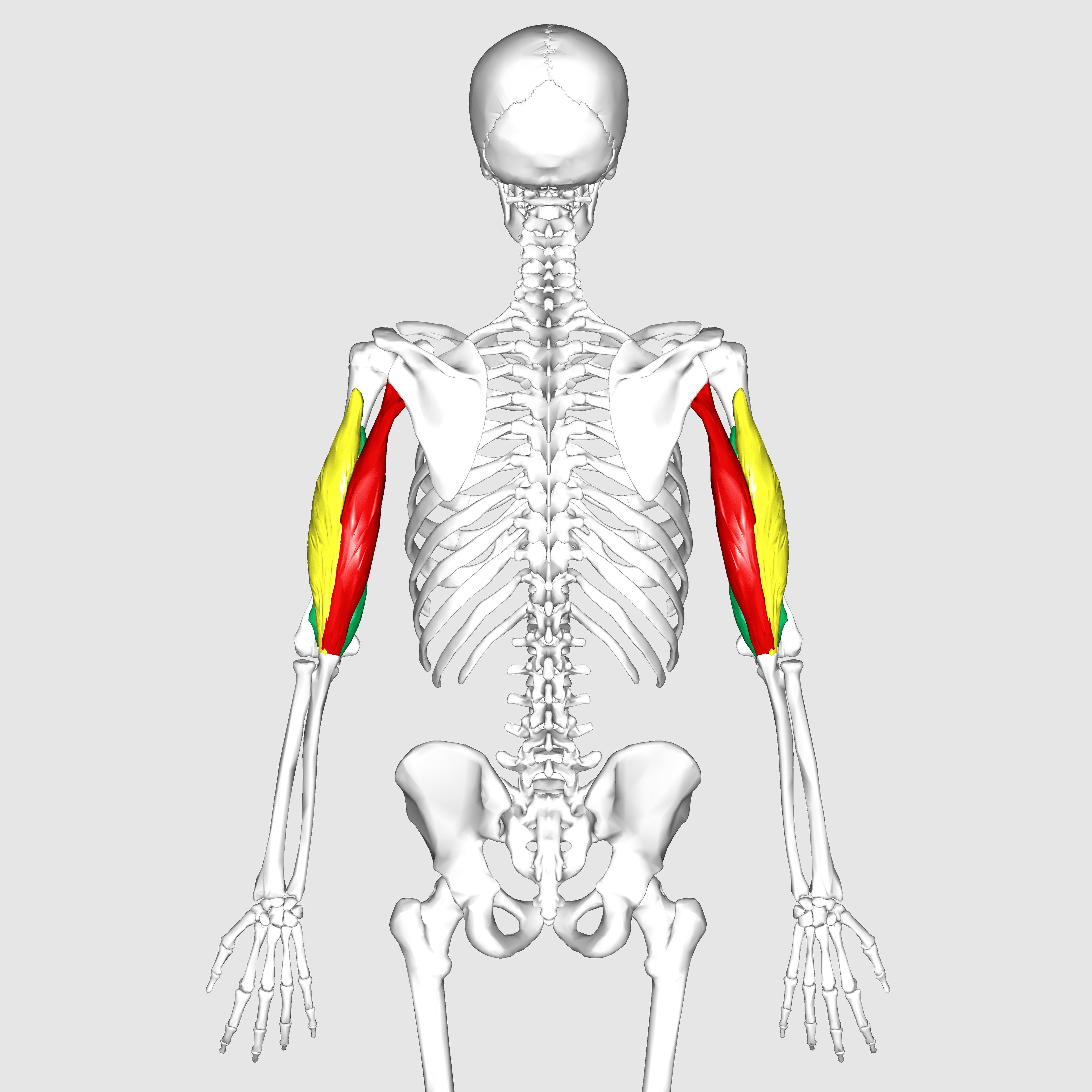
Attachments, Innervation and Relations
Origins
- Long head: the infraglenoid tubercle of the scapula, immediately inferior to the glenoid rim. It is the only head that crosses the shoulder joint, and therefore the only head with a shoulder action (extension and adduction). It forms the medial border of the quadrangular space and the lateral border of the triangular space, and divides the two.
- Lateral head: a linear origin on the posterior surface of the humerus, proximal and lateral to the spiral groove, running obliquely from just below the surgical neck down toward the groove, and from the lateral intermuscular septum.
- Medial (deep) head: a broad origin on the posterior surface of the humerus, distal and medial to the spiral groove, extending from the groove down to within 2 cm of the trochlea, and from the medial and lateral intermuscular septa. It lies deep to the other two heads throughout.
The Spiral Groove Divides the Origins
The single most useful way to remember the origins: draw the spiral groove on the posterior humerus. The lateral head arises above and lateral to it; the medial head arises below and medial to it; the radial nerve and profunda brachii run in the groove between them.
Insertion
- A common tendon onto the posterior and superior surface of the olecranon, with a superficial expansion continuing into the deep antebrachial (forearm) fascia and blending laterally with the anconeus fascia.
- Footprint - and two published figures that differ threefold. Yeh and colleagues measured the average bony footprint at 466 mm squared in 27 elbows. Athwal and colleagues, below, measured 44 mm squared for the medial head and 115 mm squared for the combined long and lateral heads in 15 elbows - about 134 mm squared where all three insert together. Both are quoted verbatim from their papers, and neither defines the boundary it measured to, so they are almost certainly not measuring the same thing: a broad osseous attachment area including the fascial expansion, against discrete tendinous insertions. Do not quote a single number as settled. The point that survives either measurement is the clinically useful one - the attachment is broader than the region a suture-anchor repair reconstitutes.
- Layered structure. Athwal and colleagues, dissecting 15 cadaveric limbs, found:
- In 8 of 15 (53 per cent) the medial head had a SEPARATE insertion onto the olecranon, lying deep to the combined long and lateral head insertion. Mean area of the medial head insertion 44 mm squared; mean area of the combined long and lateral head insertion 115 mm squared.
- In 7 of 15 (47 per cent) all three heads inserted together, mean area 134 mm squared — but even then the medial head fibres were oriented deep to the long and lateral heads.
- Clinical consequence: an isolated avulsion of the deep medial head can occur with the superficial tendon intact, presenting as pain and subtle extension weakness with an apparently continuous triceps. It has been treated by arthroscopic repair.
Bony Landmarks to Quote
- Long head origin: infraglenoid tubercle, immediately inferior to the glenoid.
- Lateral head origin: begins approximately 3 to 5 cm distal to the greater tuberosity on the posterolateral humerus.
- Medial head origin: extends from the spiral groove to within about 2 cm of the olecranon fossa.
- Insertion: posterior and superior olecranon; bony footprint measured at 466 mm squared, tendinous insertions at about 134 mm squared - see the reconciliation above.
Action and Biomechanics
Primary Action
Extension of the elbow. The triceps provides the overwhelming majority of elbow extension torque; anconeus contributes a small amount and acts principally as a stabiliser.
Long Head: The Shoulder Contribution
- Because it arises from the infraglenoid tubercle of the scapula, the long head also produces extension and adduction of the shoulder, and contributes to inferior stabilisation of the glenohumeral joint by resisting inferior translation, particularly with the arm at the side carrying a load.
- Two-joint muscle consequence: the long head is at a mechanical disadvantage when the shoulder is flexed (it is shortened at the shoulder while being asked to extend the elbow). This is the basis for the observation that triceps extension is strongest with the shoulder in extension or neutral, and is exploited in overhead triceps strengthening, which loads the long head at length.
Regional Function of the Three Heads
- Medial (deep) head is the workhorse for low-load, everyday extension — it is active through the whole arc and is recruited first.
- Lateral and long heads are recruited progressively with increasing load, particularly the lateral head, which has the largest cross-sectional area of the three.
- This is why triceps weakness after surgery may be evident only against resistance or overhead, and why a patient can extend against gravity while being functionally weak.
Comparative Extension Strength
- Nerve
- Radial C6-C8
- Origin
- Scapula and posterior humerus
- Role
- Essentially all elbow extension torque
- Consequence of loss
- Cannot extend against gravity; cannot rise from a chair or use crutches
- Nerve
- Radial (proximal branch)
- Origin
- Posterior humerus below the groove
- Role
- Low-load extension through the arc
- Consequence of loss
- Subtle weakness; may extend against gravity but fatigues
- Nerve
- Radial (terminal branch of medial head nerve)
- Origin
- Posterior lateral epicondyle
- Role
- Weak extension; dynamic lateral stabiliser
- Consequence of loss
- Negligible extension loss; possible PLRI contribution
- Nerve
- -
- Origin
- -
- Role
- Substitutes for extension with the arm dependent
- Consequence of loss
- Masks triceps loss — always test against gravity
What Happens When It Fails
- Triceps insufficiency — loss of active extension against gravity. The patient cannot push up out of a chair, cannot use a walking stick or crutches, cannot reach overhead to a shelf, and cannot stabilise the arm for fine work.
- The functional threshold is the ability to extend against gravity. A patient who can do that will manage most activities of daily living; a patient who cannot is significantly disabled.
- Compensation: patients learn to position the shoulder so gravity extends the elbow, and to lock the elbow by using the anterior deltoid to drive the arm forward. This is why triceps insufficiency is frequently missed unless specifically tested against gravity with the shoulder flexed to 90 degrees.
Surface Anatomy and Examination
Palpation
- The long head is palpable as a distinct rounded belly in the medial posterior arm, best appreciated with the shoulder abducted and the elbow extended against resistance.
- The lateral head forms the visible lateral posterior bulge of the upper arm below the deltoid.
- The common tendon is palpable as a broad flat band above the olecranon; a palpable defect 2 to 3 cm proximal to the olecranon is the hallmark of a distal triceps rupture.
- The olecranon and the posterior border of the ulna are subcutaneous and are the fixed landmarks.
Clinical Tests and What They Mean
- How to perform
- Shoulder flexed to 90 degrees, elbow flexed; ask the patient to straighten the elbow with the arm forward
- Positive finding
- Cannot achieve full active extension
- What it means
- Triceps insufficiency — the functional threshold
- False positives
- Testing with the arm dependent lets gravity extend the elbow and hides the deficit
- How to perform
- Support the arm; passively extend fully, then ask the patient to hold it
- Positive finding
- The arm drops — a measurable lag in degrees
- What it means
- Partial insufficiency or attenuated repair; quantify and document at each review
- False positives
- Pain inhibition; fixed flexion deformity gives an apparent but passive-limited lag
- How to perform
- Prone or with the arm over the couch edge and the forearm hanging; squeeze the triceps muscle belly
- Positive finding
- No passive elbow extension on squeezing
- What it means
- Complete distal triceps tendon rupture
- False positives
- Partial tear may still produce some extension; an intact lateral expansion can preserve the response
- How to perform
- Tap the triceps tendon just above the olecranon with the elbow flexed and supported
- Positive finding
- Absent or reduced
- What it means
- C7 root or radial nerve lesion proximal to the triceps branches
- False positives
- Poor relaxation; reinforce
- How to perform
- Palpate 2 to 3 cm proximal to the olecranon with the elbow flexed
- Positive finding
- A step or gap in the tendon
- What it means
- Distal triceps rupture, often with an avulsed flake of olecranon bone on the lateral radiograph
- False positives
- Olecranon bursitis obscures the palpation; a partial deep medial head avulsion may have NO palpable defect
Imaging
- Lateral radiograph: the flake sign — a small avulsed fleck of bone pulled off the olecranon by the tendon — is present in a majority of complete distal triceps ruptures and is nearly diagnostic.
- Ultrasound: dynamic, cheap and good for continuity, but operator dependent; it can distinguish a partial from a complete tear.
- MRI: best for characterising partial tears, for distinguishing a superficial (long and lateral head) tear with an intact deep medial head from a complete rupture, and for identifying the rare isolated deep medial head avulsion where the superficial tendon is intact.
The Diagnostic Trap
A patient with a partial distal triceps tear in which the lateral expansion into the forearm fascia remains intact can still extend the elbow against gravity. Extension strength is reduced but present, the squeeze test may be equivocal, and the injury is frequently dismissed as a strain. Persistent posterior elbow pain with weakness after a fall on the outstretched hand or a forced eccentric extension warrants imaging.
Complications
Denervation and Donor Morbidity
- Transferring a single triceps head branch to the axillary nerve leaves the other two heads intact and does not produce a functional extension deficit — the anatomical basis of the transfer.
- Splitting the medial head in a posterior approach transiently denervates nothing, since the branch enters proximally, but a split carried too far distally can injure the terminal branch continuing to the anconeus.
- Harvesting the lateral head as a flap costs some extension power in high-demand patients; the anconeus is preferred where its arc will reach.
Post-Operative Stiffness and Heterotopic Ossification
- Posterior elbow surgery carries a high risk of both. Early active motion, avoidance of forced passive stretching, and prophylaxis in high-risk cases (associated head injury or burns, or after excision of established heterotopic bone) are the mitigations.
Wound Problems
- The posterior elbow has thin, poorly vascularised skin under tension. Wound breakdown over the olecranon exposes hardware and joint, and is a genuine risk in rheumatoid and revision surgery.
- Mitigation: full-thickness flaps raised on the fascia, avoid multiple parallel incisions, offset the skin incision laterally around the olecranon tip to avoid a scar directly over the bony prominence, and use the anconeus rotation flap for cover if a defect arises.
Clinical Relevance
Epidemiology and Mechanism
- The least common of all tendon ruptures. Male predominance, typically in the fourth to fifth decades.
- Mechanism: eccentric loading of a contracting triceps — a fall on the outstretched hand with the elbow forced into flexion, or a failed bench press or push.
- Risk factors: anabolic steroid use, local corticosteroid injection, chronic renal failure and secondary hyperparathyroidism, hyperparathyroidism, olecranon bursitis, and previous olecranon fracture surgery.
Classification by Extent
- Partial superficial — long and lateral heads torn, deep medial head insertion intact. Extension preserved but weak.
- Complete — all heads avulsed; extension against gravity lost.
- Isolated deep medial head avulsion — rare; superficial tendon intact, subtle presentation, arthroscopically repairable.
Repair
- Indication: complete ruptures and partial ruptures with greater than about 50 per cent involvement or functional weakness, in patients with reasonable demand.
- Technique matters - but read what was actually measured. Yeh and colleagues biomechanically compared three repairs in 27 cadaveric elbows: cruciate transosseous repair, suture anchor repair, and anatomic (footprint) repair. Load at yield and peak load were similar for all three, so the entire difference between techniques rests on one endpoint - cyclic loading over 1500 cycles produced significantly less displacement with the anatomic footprint repair. Gap formation under repetitive load is a plausible surrogate for what fails during early rehabilitation, and it is the right thing to optimise in the absence of better evidence; but this is a time-zero cadaveric measurement with no healing, and no clinical series has since shown a difference in re-rupture. Say "less gapping in the laboratory", not "better outcome". (The same paper's 466 mm squared bony footprint should be quoted with the caveat set out in the Insertion section above.)
- Chronic or retracted ruptures may require Achilles tendon allograft, anconeus rotation, or a triceps turn-down.
Rehabilitation
- Typically splinted in about 30 degrees of flexion for 2 weeks, then progressive passive and active-assisted flexion with extension assisted, avoiding resisted extension for around 6 weeks and heavy loading for 4 to 6 months.
Surgical Relevance
The Four Options at the Distal Humerus
- What is done to the triceps
- Nothing — work in the medial and lateral windows either side of an intact triceps
- Articular exposure
- Limited articular view; excellent extra-articular view
- Best for
- Extra-articular distal humeral fractures; elderly patients where arthroplasty may be needed; keeps every option open
- Main risk
- Inadequate view for a complex articular reconstruction
- What is done to the triceps
- Longitudinal midline split of the tendon and muscle
- Articular exposure
- 35 per cent (median, Wilkinson and Stanley)
- Best for
- Simple patterns; humeral shaft exposure up to 15.4 cm from the lateral epicondyle
- Main risk
- Extensor weakness if the split repair fails; radial nerve at the proximal limit
- What is done to the triceps
- Extensor mechanism reflected MEDIAL to LATERAL in CONTINUITY with the forearm fascia and ulnar periosteum
- Articular exposure
- 46 per cent (median)
- Best for
- Total elbow arthroplasty; synovectomy; intra-articular distal humeral fracture where arthroplasty may be needed
- Main risk
- Triceps insufficiency if the reattachment fails; ulnar nerve must be identified
- What is done to the triceps
- Triceps left completely attached to the osteotomised olecranon fragment
- Articular exposure
- 57 per cent (median) — the best, but still under 60 per cent
- Best for
- Complex AO type C intra-articular distal humeral fracture in a young patient
- Main risk
- Osteotomy non-union or symptomatic hardware; PRECLUDES conversion to arthroplasty
Bryan-Morrey Triceps-Reflecting Approach in Detail
- Described in 1982 by Bryan and Morrey, prompted by the problem of triceps avulsion and loss of continuity after total elbow arthroplasty.
- The defining feature is continuity. The triceps mechanism is reflected from medial to lateral in continuity with the forearm fascia, the olecranon periosteum and the ulnar periosteum. The tendon is never divided or detached from the fascial sleeve; a subperiosteal sleeve of tissue is peeled off the olecranon carrying the extensor mechanism with it.
- The ulnar nerve must be identified and mobilised first, since the dissection begins medially.
- The ulnar collateral ligament may be released from the humerus for more exposure, but must then be securely reattached.
- A lateral-to-medial variant exists (the extensor mechanism reflected the other way).
- In their series of 49 consecutive total elbow arthroplasties, Bryan and Morrey reported no loss of triceps function and no significant weakness.
- Reattachment is through cruciate transosseous drill holes in the olecranon with heavy non-absorbable suture.
Olecranon Osteotomy Technique
- Chevron (apex distal) osteotomy, made intra-articularly at the bare area of the sigmoid notch — the transverse non-articular strip roughly 2 to 2.5 cm from the tip of the olecranon, where there is no cartilage to damage.
- Pre-drill and pre-tap the fixation (an intramedullary screw or tension band) before making the cut, so the reduction is anatomic on closure.
- Protect the ulnar nerve medially throughout.
- Never perform an olecranon osteotomy if arthroplasty is a possibility — the osteotomy destroys the bone the implant needs and adds a non-union risk to an already compromised elbow.
Guidelines, Registries & Global Practice
Anatomical Variation
- The medial head has a separate olecranon insertion in about 53 per cent of specimens and a fused but still deeper-oriented insertion in the remainder. This is not a rare variant but a coin-flip, and it explains the existence of the isolated deep medial head avulsion.
- A fourth head of triceps and accessory slips from the latissimus dorsi or the medial intermuscular septum are described. A hypertrophic accessory medial head slip is one cause of snapping triceps over the medial epicondyle.
- The dorsoepitrochlearis (latissimocondyloideus), a slip from latissimus dorsi to the medial epicondyle or triceps, is a well-described variant present in a small percentage of limbs and can compress the ulnar nerve or the neurovascular bundle in the axilla.
Side-by-Side Guidance
- Position on triceps handling and distal humeral exposure
- Describes the para-tricipital, triceps-splitting, triceps-reflecting and olecranon osteotomy approaches, recommending the olecranon osteotomy for complex articular fractures and cautioning explicitly against it when arthroplasty may be required.
- Position on triceps handling and distal humeral exposure
- Originated the triceps-reflecting approach and the triceps insufficiency reconstruction algorithm; increasingly favours triceps-on approaches for arthroplasty where exposure permits.
- Position on triceps handling and distal humeral exposure
- Support total elbow arthroplasty as a reasonable primary treatment for comminuted distal humeral fractures in low-demand elderly patients, with the corollary that the approach must preserve the arthroplasty option.
- Position on triceps handling and distal humeral exposure
- No formal guideline; consensus favours repair for complete ruptures and for partial ruptures with functional weakness, with anatomic footprint techniques preferred where early motion is planned.
Registry Signals
- National joint registries (including the National Joint Registry for England, Wales, Northern Ireland and the Isle of Man, and the Australian Orthopaedic Association National Joint Replacement Registry) capture total elbow arthroplasty, though in far smaller volumes than hip and knee. Reported revision rates are substantially higher than for the large joints, with aseptic loosening, infection and periprosthetic fracture the leading indications. Triceps insufficiency is generally NOT captured as a revision reason in registries, because it is treated by soft-tissue reconstruction rather than component revision — meaning registry data systematically under-represent this complication and institutional series remain the best evidence.
- Total elbow arthroplasty for acute distal humeral fracture in the elderly is increasing in registry-reported volume in several countries, which makes the approach decision — and the preservation of the triceps and olecranon — more, not less, important.
High- versus Limited-Resource Practice
- Well-resourced settings: pre-operative CT with three-dimensional reconstruction for complex distal humeral fractures; the full range of approaches; Achilles allograft available for extensor reconstruction.
- Limited-resource settings: allograft is generally unavailable, so anconeus rotation and triceps turn-down become the reconstruction options of choice for extensor mechanism deficiency — both autologous, both single-stage, both requiring no implants. This is a genuine practical advantage of knowing the local flap anatomy well.
- Where arthroplasty is unavailable, fixation is attempted in a wider range of elderly patients, which raises the value of the para-tricipital approach as a low-morbidity default.
Rehabilitation Consensus
- Protected active extension for 6 weeks after any extensor mechanism repair or reflection is near-universal.
- Early active-assisted flexion with extension assisted or gravity-assisted is preferred over immobilisation, which produces stiffness — the elbow is the joint least forgiving of prolonged immobilisation in the entire skeleton.
MCQ Practice Points
Q: Where do the three heads of triceps arise? A: Long head — infraglenoid tubercle of the scapula. Lateral head — posterior humerus ABOVE and lateral to the spiral groove. Medial head — posterior humerus BELOW and medial to the groove.
Q: Why is the medial head of triceps spared in a spiral groove lesion? A: Its branch arises proximally, in the axilla or upper arm, and runs distally with the ULNAR nerve rather than crossing the posterior humerus.
Q: What does a preserved triceps with wrist and finger drop tell you? A: The radial nerve lesion is at or distal to the spiral groove. Triceps weakness as well implies an axillary or posterior cord lesion.
Q: How much of the distal humeral articular surface does each posterior approach expose? A: Triceps splitting 35 per cent, triceps reflecting 46 per cent, olecranon osteotomy 57 per cent (medians, Wilkinson and Stanley).
Q: How far proximal to the lateral epicondyle does the radial nerve cross the posterior humerus? A: A mean of 14.2 cm (and 20.7 cm proximal to the medial epicondyle).
Q: What defines the Bryan-Morrey approach? A: The extensor mechanism is reflected medial to lateral in CONTINUITY with the forearm fascia and the olecranon and ulnar periosteum — it is never detached.
Q: When must you NOT perform an olecranon osteotomy? A: When conversion to total elbow arthroplasty is a possibility — the osteotomy destroys the bone the implant needs and adds a non-union risk.
Q: What is the area of the distal triceps footprint on the olecranon?
A: Give the range and say why. Yeh measured the bony footprint at 466 mm squared in 27 elbows; Athwal measured discrete tendinous insertions totalling about 134 to 159 mm squared in 15. Neither paper states the boundary it measured to, so the two are not the same quantity. 466 mm squared is the figure most often set as the exam answer because it comes from the repair-biomechanics paper - quote it, but be able to say it is a bony attachment area and that tendinous-insertion measurements are roughly a third of it. The clinical point is independent of which is right: the attachment is broader than the area a single row of anchors restores, and anatomic footprint repair shows less displacement under cyclic loading than cruciate or anchor repairs - though only in cadavers, at time zero, with no difference in ultimate strength.
Q: Which structure separates the quadrangular space from the triangular space? A: The long head of triceps. Quadrangular space transmits the axillary nerve and posterior circumflex humeral vessels; the triangular space transmits the circumflex scapular vessels.
Q: What passes through the triangular interval? A: The radial nerve and profunda brachii artery, between teres major above, the long head of triceps medially and the humerus or lateral head laterally.
Q: What radiographic sign suggests a distal triceps rupture? A: The flake sign — a small avulsed fleck of olecranon bone on the lateral radiograph.
Exam Viva Scenarios
Practise clinical reasoning and management decisions out loud
“A 34-year-old man has a comminuted intra-articular AO type C3 distal humeral fracture. A 78-year-old woman with rheumatoid arthritis has a comminuted low transcondylar distal humeral fracture. What posterior approach do you use for each, and why?”
“A patient presents after a mid-shaft humeral fracture with a wrist drop. On examination the triceps is grade 5, the wrist and fingers cannot be extended, and there is numbness over the dorsal first web space. Where is the lesion and why is the triceps working?”
“Eight months after a total elbow arthroplasty for rheumatoid arthritis, a 56-year-old woman cannot push herself out of a chair with that arm. How do you assess and manage this?”
Origins
- Long head: infraglenoid tubercle of the scapula (crosses the shoulder)
- Lateral head: posterior humerus ABOVE/lateral to the spiral groove
- Medial head: posterior humerus BELOW/medial to the groove
- Insertion: posterior olecranon; bony footprint 466 mm², tendinous ~134 mm² - quote the range
Innervation
- Radial nerve C6, C7, C8 - THREE SEPARATE branches
- Long head: branch in the axilla
- Lateral head: branches within the spiral groove
- Medial head: proximal branch running distally with the ULNAR nerve; continues to anconeus
Radial Nerve Numbers
- Crosses posterior humerus 20.7 cm from medial epicondyle to 14.2 cm from lateral epicondyle
- Crosses the lateral septum about the MIDPOINT of the humerus (17 cm from the head)
- Triceps split exposes 15.4 cm from the lateral epicondyle
- Modified posterior approach (both heads reflected medially) exposes 26.2 cm
Approaches
- Para-tricipital (triceps-on): keeps every option open
- Triceps split: 35 per cent articular exposure
- Bryan-Morrey triceps-reflecting: 46 per cent; CONTINUITY preserved
- Olecranon osteotomy: 57 per cent; NEVER if arthroplasty possible
Insufficiency
- Test extension AGAINST GRAVITY - dependent arm hides it
- Reconstruct: direct repair / anconeus rotation / Achilles allograft
- Mayo: extension restored in 15 of 16; 11 excellent, 3 good
- Prevent by triceps-on approach and cruciate transosseous reattachment
Evidence Base
Posterior Surgical Approaches to the Elbow: A Comparative Anatomic Study
- Triceps splitting, triceps reflecting and olecranon osteotomy each performed on four adult cadaveric elbows
- Visible articular surface painted with methylene blue, the elbow then disarticulated and the exposed percentage measured
- Median exposed articular surface: triceps splitting 35 per cent, triceps reflecting 46 per cent, olecranon osteotomy 57 per cent
- Olecranon osteotomy exposed significantly more than triceps splitting (p equals 0.03), but was not significantly better than triceps reflecting
- Even the olecranon osteotomy approach failed to provide visualisation of more than 40 per cent of the distal humeral articular surface
Extensive Posterior Exposure of the Elbow: A Triceps-Sparing Approach
- Approach developed specifically because of triceps avulsion and loss of continuity after total elbow arthroplasty
- The triceps mechanism is reflected medial to lateral in CONTINUITY with the forearm fascia and the olecranon and ulnar periosteum
- A lateral-to-medial variant of the reflection is also described
- The ulnar collateral ligament may be released from the humerus for additional exposure but must be securely reattached
- Used in 49 consecutive total elbow arthroplasties with no loss of triceps function and no significant weakness
Triceps Insufficiency Following Total Elbow Arthroplasty
- Review of 887 total elbow arthroplasties performed 1982 to 2001; 16 elbows in 14 patients underwent a subsequent triceps procedure (infection-related cases excluded)
- Mean age 54 years; mean follow-up after triceps reconstruction 67 months
- Three techniques used, selected on tissue quality, tendon retraction and the state of the olecranon: direct suture in 7, anconeus rotation in 4, Achilles tendon allograft in 4
- Capacity to extend against gravity was restored in 15 of 16 elbows
- Mayo Elbow Performance Score: 11 excellent, 3 good, 2 clinical failures
Alternative Operative Exposures of the Posterior Humeral Diaphysis with Reference to the Radial Nerve
- Ten cadaveric specimens; three posterior operative approaches performed in each
- The radial nerve crossed the posterior humerus from a mean of 20.7 plus or minus 1.2 cm proximal to the medial epicondyle to 14.2 plus or minus 0.6 cm proximal to the lateral epicondyle
- As it crossed, the nerve gave several branches to the LATERAL head of triceps but NO branches to the medial head were found posteriorly in any specimen
- At the lateral humerus the nerve trifurcated into a branch to the medial head, the lower lateral brachial cutaneous nerve, and the continuation of the radial nerve
- Exposure achieved: triceps splitting 15.4 plus or minus 0.8 cm; plus proximal nerve mobilisation an extra 6 cm; modified posterior approach reflecting both lateral and medial heads medially 26.2 plus or minus 0.4 cm
The Distal Triceps Tendon Footprint and a Biomechanical Analysis of Three Repair Techniques
- Triceps tendon footprint measured in 27 cadaveric elbows; a distal tendon rupture was then created and repaired
- Average bony footprint of the triceps tendon was 466 mm squared — a large area on the olecranon
- Three repairs compared: cruciate transosseous, suture anchor, and anatomic footprint repair
- Load at yield and peak load were similar across all three techniques
- Cyclic loading over 1500 cycles produced significantly less displacement with the anatomic footprint repair
Isolated Avulsion of the Medial Head of the Triceps Tendon: An Anatomic Study and Arthroscopic Repair
- Fifteen cadaveric upper extremities examined to characterise the triceps insertion, with particular attention to the medial head
- In 8 specimens (53 per cent) the medial head had a SEPARATE insertion onto the olecranon, deep to the long and lateral head insertions
- Mean area of the medial head insertion 44 mm squared; mean combined long and lateral head insertion 115 mm squared
- In the remaining 7 (47 per cent) all three heads inserted together with a mean area of 134 mm squared, but medial head fibres were still oriented deep to the others
- Two clinical cases of isolated medial head avulsion were repaired arthroscopically; at two years both were pain-free with better strength and improved DASH and Mayo Elbow Performance Scores