Segmental posterior fixation of the most mobile spinal motion segment — anatomy of the vertebral artery governs every decision
- Preoperative CT angiography of the vertebral arteries is mandatory before any C1-C2 screw fixation — trajectory choice is dictated by VA anatomy, not surgeon preference
- Magerl transarticular screws demand a REDUCED or reducible C1-C2 relationship before drilling; a fixed subluxation is an absolute contraindication
- High-riding VA on one side: unilateral Magerl or C2 pedicle screw is contraindicated on that side — switch to pars, translaminar, or Goel-Harms hybrid
- VA injury during drilling: complete and place the screw for tamponade, abandon the contralateral screw, obtain immediate angiography
- The C2 nerve root and its venous plexus overlie the C1 lateral mass entry — control with bipolar and haemostatic agents; root sacrifice versus retraction remains debated
- Fusion of C1-C2 sacrifices roughly half of cervical rotation — document this in consent
- “Ponticulus posticus (arcuate foramen) mimics a broad C1 posterior arch — placing a 'C1 posterior arch' screw through it transfixes the vertebral artery
- “Internal carotid artery lies directly anterior to the C1 lateral mass — bicortical C1 screws risk ICA injury; keep the tip just short of the anterior cortex
- “Translaminar C2 screws are the VA-independent bailout but are biomechanically weaker and risk ventral laminar breach into the canal
- “Steep Magerl trajectory (starting near C7-T1 skin level) can be blocked by thoracic kyphosis or a barrel chest — check trajectory on sagittal CT preoperatively
Indications and Decision Thresholds
Scope note. This page is the C1-C2 fusion technique guide — indications, vertebral-artery anatomy, and the Magerl-versus-Goel-Harms decision. For the atlantoaxial pathology that drives the operation (rheumatoid involvement, instability, Crowned Dens, the Ranawat classification), see atlantoaxial arthritis.
C1-C2 fusion is offered when the atlantoaxial segment is unstable, painful beyond salvage, or destroyed, and when the pathology is confined to C1-C2. Exclude cranial settling, occipital condyle deficiency and subaxial instability before committing to the segment: extension to the occiput or subaxial spine converts the plan to an occipitocervical or extended posterior construct.
Type II odontoid fracture. In the Anderson-D'Alonzo type II fracture the operative candidates are displacement greater than 5 mm, angulation greater than 11 degrees, comminution, a posterior oblique fracture line, and age greater than 50 years, beyond which nonunion risk rises steeply. An anterior odontoid screw preserves rotation but demands a favourable anterior-oblique fracture line, an intact transverse ligament, a reducible acute fracture and adequate bone density; if any criterion fails, the operation is a posterior C1-C2 fusion.
Odontoid nonunion. An established nonunion is a posterior fusion indication. The anterior screw has no role once the fracture surfaces are sclerotic.
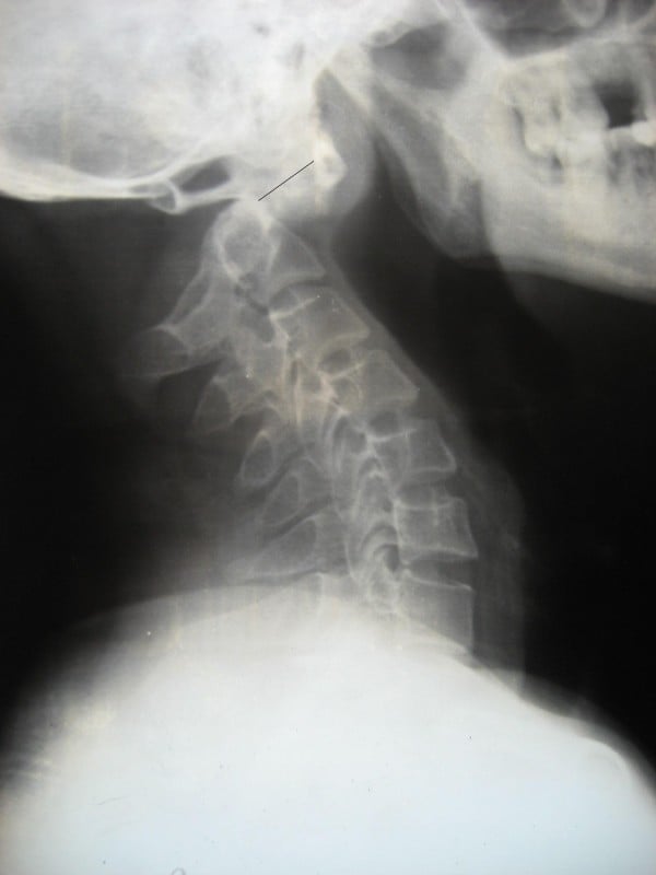
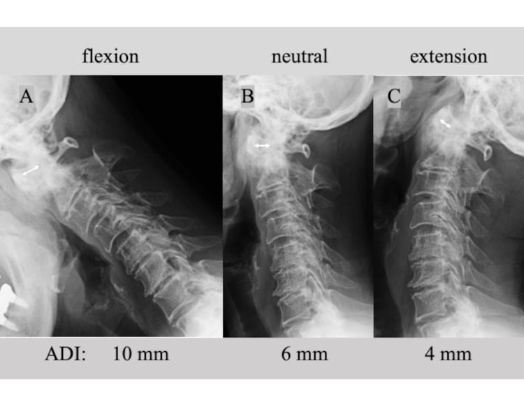
Transverse ligament rupture. The atlantodens interval (ADI) is drawn on the lateral radiograph from the posterior cortex of the anterior C1 arch to the anterior dens cortex at their midpoints. An ADI greater than 3 mm in adults (greater than 5 mm in children) implies transverse ligament incompetence, and greater than 5 mm in an adult implies additional alar and apical failure. An intrasubstance ligament rupture (Dickman type I on MRI) does not heal and is fused; a bony avulsion of the ligament insertion (type II) may heal in a rigid orthosis with close surveillance.
Irreducible or recurrent atlantoaxial rotatory fixation. Paediatric cases failing traction and recurring after reduction are fused.
Os odontoideum. Instability on flexion-extension films, cord signal change, or myelopathy mandates fusion. The asymptomatic stable os remains controversial (see controversies).
Rheumatoid atlantoaxial instability. Fuse for progressive ADI widening, myelopathy, intractable occipitocervical pain, or a posterior atlantodens interval (PADI) less than 14 mm; PADI is the more predictive measurement because it is the space available for the cord. Operate before fixed cranial settling develops, which converts a C1-C2 fusion into an occipitocervical construct.
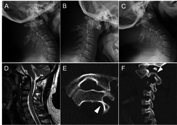
C1-C2 osteoarthritis. Unilateral lateral mass arthrosis causing intractable rotational occipitocervical pain, confirmed by image-guided joint block and refractory to conservative care.
Tumour or infection. Destruction of the dens, C1 lateral mass or C2 body with instability, often combined with anterior debridement and decompression.
Basilar invagination. Where the invagination is reducible, the Goel philosophy is joint distraction with intra-articular spacers and C1-C2 fixation.
Surgical Anatomy — The Vertebral Artery Governs Everything
The V3 segment. The vertebral artery exits the C2 transverse foramen, loops laterally then superiorly through the C1 transverse foramen, and turns medially in the groove on the superior surface of the C1 posterior arch, roughly 1.5 cm from the midline, before piercing the atlanto-occipital membrane and dura. That groove sets the exposure limit: subperiosteal dissection is safe to about 12-15 mm from the midline on the superior surface of the arch and 8 mm on the inferior surface. Dissect strictly subperiosteally and keep cautery off the superolateral arch; straying superolaterally with cautery or a periosteal elevator is the classic mechanism of exposure-phase vertebral artery injury.
Ponticulus posticus. The arcuate foramen is reported in roughly 3 to 15 percent of people depending on the series and the imaging method: an ossified bridge over the vertebral artery groove that mimics a broad posterior arch on lateral fluoroscopy. A "posterior arch" C1 screw started on the bridge drills straight into the artery. Identify it on the preoperative CT and lateral radiographs, and do not judge the entry point from arch thickness on fluoroscopy alone.
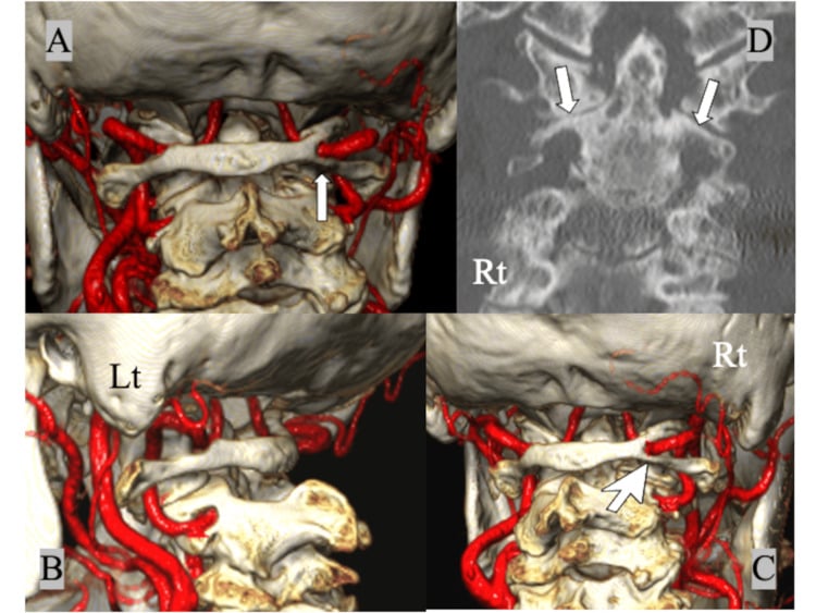
The internal carotid artery. It lies immediately anterior to the C1 lateral mass, within a few millimetres in some patients, often anteromedial and asymmetric. Bicortical C1 screws improve pull-out but risk the carotid and the hypoglossal nerve, so most surgeons stop just short of the anterior cortex, judged on the lateral fluoroscopic view and the preoperative CT angiogram.
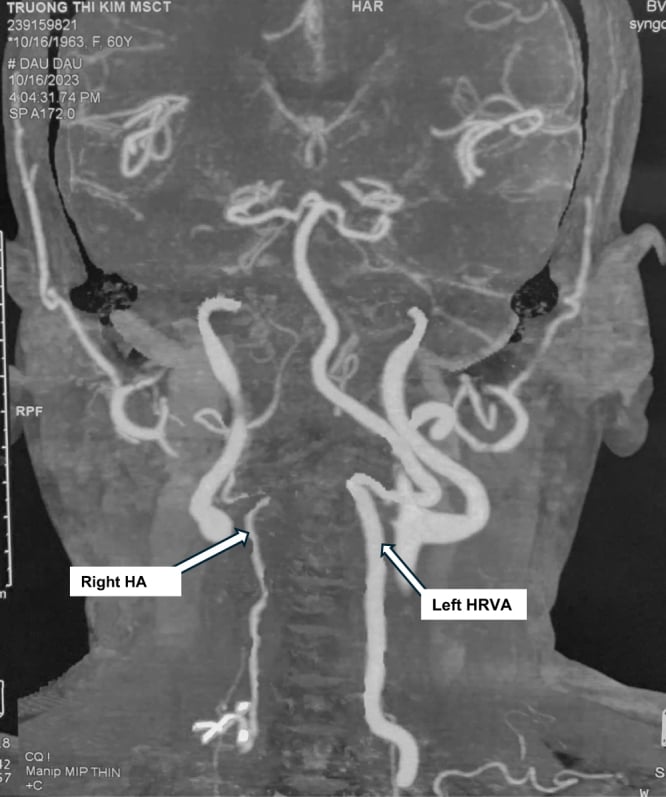
C2 and the high-riding artery. The vertebral artery runs through the C2 transverse foramen beneath the pars and pedicle. A high-riding vertebral artery, a large, cephalad and medially looping vessel that narrows the C2 isthmus (an internal height less than 5 mm or an isthmus width less than 4 mm is a common threshold), is found on at least one side in up to 20 percent of patients and contraindicates a transarticular or pedicle screw on that side. Paramore's 94 CT studies put it at 18 percent prohibited on at least one side, 18 to 23 percent once borderline anatomy is counted, and bilateral in only 3 patients. The anomaly is usually unilateral, which is why the construct is chosen side by side.
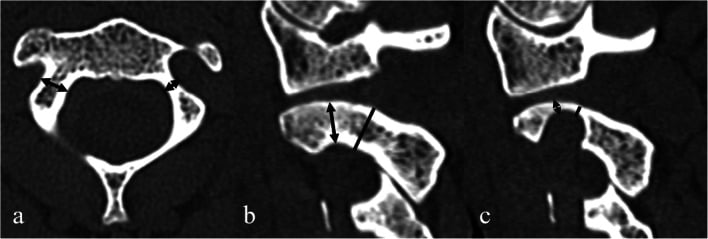
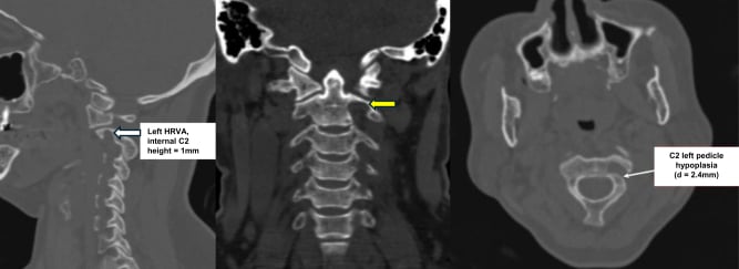
C2 terminology. The pars interarticularis lies between the superior and inferior articular processes; the pedicle connects the posterior elements to the body and sits medial and cephalad to the artery. The pedicle screw trajectory is more medial (about 20-30 degrees) and more cephalad than the pars screw.
The C2 nerve root. It crosses the C1-C2 joint posteriorly, in the greater occipital nerve territory, embedded in a rich venous plexus directly over the C1 lateral mass screw entry. That plexus is the source of the notorious bleeding at that step.
Preoperative imaging. CT angiography is mandatory before any C1-C2 screw construct: map the artery on both sides, measure C2 isthmus height and width on sagittal and axial reconstructions, simulate the Magerl trajectory on the sagittal cut through the isthmus, and note dominance or hypoplasia, anomalous V3 loops, a ponticulus posticus and an anomalous carotid position anterior to C1. Template the screw trajectories and lengths on the CT before incision; the trajectory is chosen to fit the artery, never the reverse. Flexion-extension lateral radiographs quantify reducibility, and MRI assesses cord signal, rheumatoid pannus and transverse ligament integrity by the Dickman classification.
Technique Comparison — The Exam Table
The wiring techniques are now adjuncts or salvage, and the choice today is between Magerl transarticular screws and the Goel-Harms screw-rod construct, made on reducibility and the vertebral artery rather than habit.
- Construct
- Single midline sublaminar wire passed under the C1 posterior arch and looped around the C2 spinous process, compressing one midline corticocancellous graft
- Rigidity / fusion rate
- Poor rotational control; fusion 70 to 85 percent; halo or rigid orthosis mandatory
- Prerequisites
- Intact C1 posterior arch and intact C2 lamina and spinous process
- Key limitations
- Wire tension pulls C1 posteriorly and can worsen posterior subluxation; inadequate as a stand-alone construct today
- Construct
- Bilateral sublaminar wires under both C1 and C2 compressing two wedge-shaped interlaminar bone grafts
- Rigidity / fusion rate
- Better rotational and extension control than Gallie, but still 70 to 85 percent fusion; halo needed
- Prerequisites
- Intact posterior elements at both levels; safe sublaminar wire passage at C1 and C2
- Key limitations
- Sublaminar passage at two levels risks dural tear and cord injury; no reduction capacity
- Construct
- Screws from the C2 inferior articular process across the C1-C2 facet joint into the C1 lateral mass, supplemented by posterior graft and wiring
- Rigidity / fusion rate
- Historically the most rigid construct; fusion greater than 95 percent with supplemental grafting
- Prerequisites
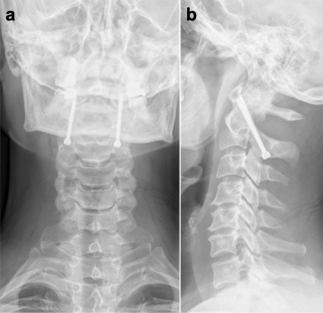
- C1-C2 must be REDUCED before drilling; safe C2 isthmus with no high-riding vertebral artery; trajectory not blocked by thoracic kyphosis or body habitus
- Key limitations
- Vertebral artery at risk in the isthmus; no capacity for intraoperative reduction; steep trajectory often needs a separate percutaneous stab incision
- Construct
- C1 lateral mass screws plus C2 pedicle screws connected by rods, with C2 pars or translaminar screws as anatomy-driven alternatives
- Rigidity / fusion rate
- Segmental and rigid; fusion greater than 95 percent; biomechanically equivalent to transarticular fixation
- Prerequisites
- Adequate C1 lateral mass bone stock; C2 screw option selected to fit vertebral artery anatomy on preoperative CT angiography
- Key limitations
- C2 venous plexus bleeding; C2 (Greater occipital) neuralgia; technically demanding
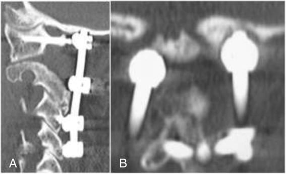
Choosing the construct. Magerl screws are drilled along a fixed trajectory across a joint that must already be reduced. The Goel-Harms construct places each screw independently and reduces at the screw heads before the rods are locked, so it serves a fixed or irreducible subluxation, and it remains feasible when a high-riding artery or a narrow isthmus prohibits a transarticular screw on one or both sides. Biomechanically it is equivalent to transarticular fixation.
When one side is hostile. A high-riding artery or a pars too small for a safe pedicle or transarticular screw takes a C2 translaminar screw on that side (or bilaterally), or the construct becomes a unilateral screw plus contralateral translaminar hybrid. Fixation is achieved without violating the artery; accept lower rotational stiffness and check laminar thickness.
- Joint already reduced and stays reduced
- Normal C2 isthmus on CT angiography with no high-riding vertebral artery
- Slim patient, no fixed kyphosis blocking the low-to-high trajectory
- Fewer implants, lower cost
- Fixed or irreducible subluxation needing intraoperative reduction at the screw heads before rod locking
- High-riding vertebral artery or narrow isthmus on one or both sides
- Each screw placed independently (segmental), so unilateral anatomy does not sink the construct
- Rescue options at C2: pedicle, pars or translaminar tailored to the artery
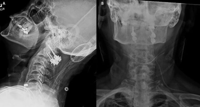
Goel-Harms Construct — Operative Technique
Positioning and preparation. Prone on a radiolucent table with the head in a Mayfield clamp, the neck neutral or slightly flexed at C1-C2 to open the interlaminar interval but with overall alignment set to the intended fused position; reverse Trendelenburg reduces venous bleeding. Awake fibreoptic intubation and an awake positioning check are considered in myelopathic or grossly unstable patients, with neuromonitoring (SSEP and MEP) and a baseline taken before positioning in myelopathy. Lateral fluoroscopy confirms reduction after positioning and before incision, with navigation if available.
In theatre, recheck the CT angiogram, mark the planned C2 screw type for each side, and cross-match blood because the venous plexus can bleed briskly. Have bipolar diathermy, flowable haemostatic matrix and a 3.5 mm polyaxial screw system with smooth-shank C1 options open.
Exposure. A midline posterior incision from occiput to C3 through the avascular median raphe, then subperiosteal exposure of the C1 posterior arch within the 12-15 mm superior and 8 mm inferior lateral limits, followed by the C2 lamina, pars and inferior articular processes. Preserve the C2-C3 facet capsule and the semispinalis cervicis insertion on C2 where possible.
The C2 root and venous plexus. Dissect along the inferior border of the C1 arch onto the lateral mass; the root and its plexus are met here. Control bleeding with bipolar and haemostatic matrix and do not linger: prolonged dissection before screw placement worsens blood loss, whereas placing the C1 screw early tamponades the plexus.
Sacrifice or retract the root? Sectioning the root improves exposure of the joint for decortication and grafting and reliably stops the plexus bleeding, at the cost of occipital numbness and occasional neuralgia or dysaesthesia. Caudal retraction preserves the root but limits joint access. Both are defensible; know the debate.
C1 lateral mass screw. Entry is at the centre of the lateral mass at its junction with the inferior aspect of the posterior arch, with a trajectory 10-15 degrees medial and cephalad, aiming at the anterior arch tubercle on lateral fluoroscopy and stopping just short of the anterior cortex because the internal carotid lies in front. A smooth, unthreaded proximal shank leaves the screw standing proud of the plexus and the C2 root and reduces irritation. Where the arch is thick, notching the inferior arch (Tan modification) or a posterior arch screw are alternatives; beware the ponticulus posticus.
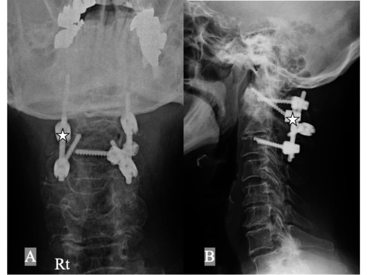
C2 screw, chosen to fit the artery. Three trajectories:
- Pedicle screw, the strongest: entry in the upper-outer quadrant of the isthmus surface, the medial pedicle border palpated with a Penfield in the canal, then 20-30 degrees medial and 20-25 degrees cephalad along the pars into the body. Contraindicated with a high-riding artery on that side.
- Pars screw: the same entry, a steeper cephalad and less medial trajectory, and a shorter screw stopping before the transverse foramen. Safer with moderate arterial risk, but less purchase.
- Translaminar screw (Wright): the artery-independent bailout. Entry at the junction of spinous process and lamina, the screws crossing within the laminae to the contralateral side, offset cephalad and caudad so that they can cross. It risks ventral laminar breach into the canal, is biomechanically weaker, and occupies the laminar graft bed.
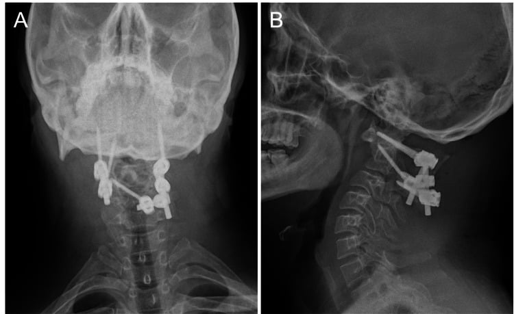
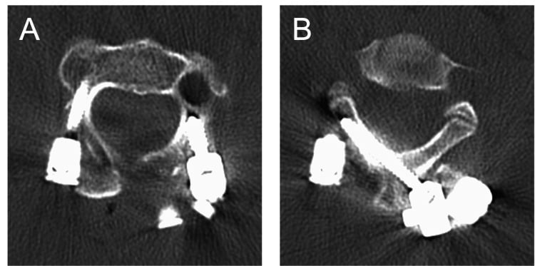
Reduction and rod locking. Connect the rods and, where reduction is needed, lever C1 posteriorly or anteriorly against the fixed C2 screws before final tightening; confirm restoration of the atlantodental interval on lateral fluoroscopy. Never drill Magerl or pedicle trajectories with C1 unreduced.
Fusion bed, grafting and closure. Decorticate the inferior C1 arch, the C2 lamina and, for the highest fusion rates, the C1-C2 joint itself: distract, curette the cartilage and pack cancellous graft or a structural spacer. Goel joint spacers additionally distract and reduce basilar invagination. Lay corticocancellous autograft, structural or morcellised, with iliac crest remaining the gold standard, as an interlaminar strut with or without a supplemental cable, or as onlay graft. Layered closure over the muscle; a drain is optional.
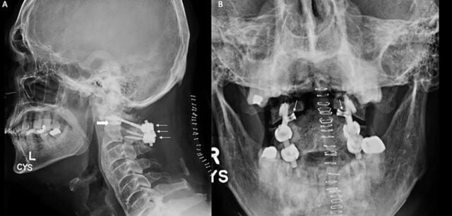
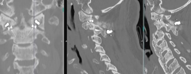
Aftercare. A rigid or semi-rigid collar for 6 to 12 weeks, though rigid segmental fixation permits a soft collar for comfort in many units. Upright lateral radiographs before discharge, radiographs at 6 weeks and 3 months with flexion-extension films to confirm fusion, and CT at 6 to 12 months if fusion is in doubt or symptoms persist.
Salvage. Nonunion or screw failure is revised with the alternative C2 trajectory, joint grafting if not previously done, or extension to occipitocervical fusion if C1 purchase is lost.
Magerl Transarticular Technique — Essentials
Reduction first. Transarticular screws are drilled along a fixed trajectory: if C1 is subluxed on C2 the screw will miss the C1 lateral mass and may enter the vertebral artery or the hypoglossal canal. Confirm reduction on lateral fluoroscopy before drilling, reducing with positioning, Mayfield adjustment or a temporary clamp. If the joint is irreducible, abandon Magerl and use Goel-Harms with intraoperative joint reduction instead.
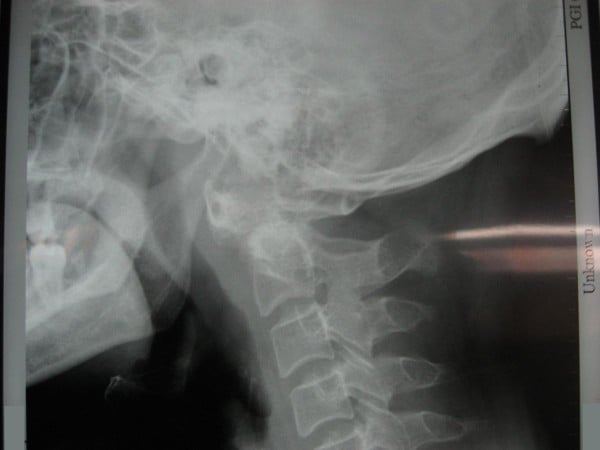
Trajectory. Entry is 2-3 mm superior and 2-3 mm lateral to the medial edge of the C2-C3 facet on the C2 inferior articular process. The trajectory is strictly sagittal (0-10 degrees medial) and steeply cephalad along the C2 isthmus, across the joint and into the C1 lateral mass, aiming at the anterior arch tubercle. That steepness often requires percutaneous stab incisions near the cervicothoracic junction, and a thoracic kyphosis or a barrel chest can make the trajectory unachievable, so simulate it on the preoperative sagittal CT.
The graft is not optional. Always supplement with a posterior interlaminar graft held by wiring or a clamp (a Gallie-type graft): screws alone leave the graft uncompressed and fusion rates fall. A unilateral screw plus posterior wiring is an accepted compromise when only one side has a safe arterial course.
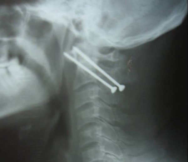
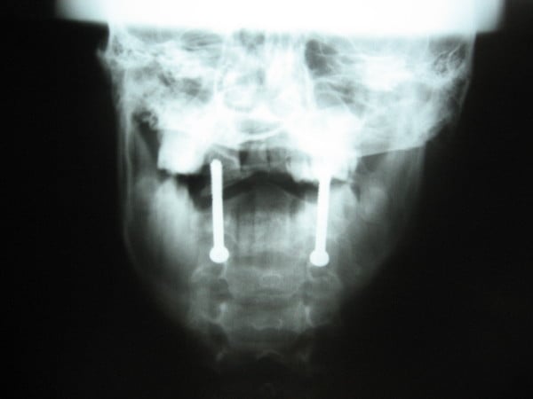
RATMagerl prerequisites
Hook:No RAT, no transarticular screw — otherwise convert to Goel-Harms.
Complications, Prevention and Management
Vertebral artery injury. Roughly 2 to 4 percent of patients with transarticular screws, lower with modern planning. Brisk arterial bleeding during drilling or tapping is met by completing the hole and placing the screw, which tamponades the vessel; the contralateral screw is not attempted, the wound is closed, and immediate angiography characterises occlusion versus pseudoaneurysm. The table below carries the rest of the prevention and management for this and every other structure at risk.
Never attempt the second-side screw after a suspected vertebral artery injury on the first side. Unilateral occlusion is usually tolerated, through collateral flow via the contralateral vertebral artery and the circle of Willis; bilateral injury risks brainstem and cerebellar infarction and death.
Nonunion. Under 5 percent with segmental constructs and joint grafting; inadequate joint preparation leads to it, and salvage is described with the technique above.
Loss of rotation. C1-C2 contributes roughly half of cervical axial rotation, and fusion sacrifices it permanently; counsel drivers and athletes preoperatively and document it. Adjacent segment strain at the occipito-atlantal and subaxial levels can follow over years.
- Where encountered
- In the groove on the superior surface of the C1 posterior arch, roughly 1.5 cm from the midline, and at the C1 lateral mass entry point
- Mechanism
- Lateral subperiosteal dissection beyond the safe zone on the C1 arch, or plunging during C1 lateral mass drilling
- Avoid by
- Stay subperiosteal and dissect no further laterally than needed; identify the arch groove; preoperative CT angiography; navigation or fluoroscopic control of trajectory
- If injured
- Pack and place the screw to tamponade, abandon the contralateral screw to avoid bilateral injury, immediate angiography; occlusion with good collateral flow is observed with neurology input and antiplatelet cover; stent or coil a pseudoaneurysm or dissection; watch for posterior circulation deficit
- Where encountered
- Narrow C2 pars or isthmus with a dominant vertebral groove, present in a substantial minority and often asymmetric
- Mechanism
- Transarticular or C2 pars screw driven through a thin isthmus
- Avoid by
- Measure isthmic height and width on preoperative CT for each side independently; select a side-specific trajectory — translaminar or short pars screw where the isthmus will not accept a transarticular screw
- If injured
- Same as V3 injury; convert the unsafe side to a translaminar screw or a laminar hook rather than repeat the trajectory
- Where encountered
- Bony bridge over the vertebral groove on the C1 arch, mimicking a broad, safe arch on fluoroscopy
- Mechanism
- Mistaking the anomalous bridge for the posterior arch and drilling a C1 screw directly into the artery
- Avoid by
- Recognise the anomaly on preoperative CT and lateral radiographs; do not use arch thickness on fluoroscopy alone to judge the entry point
- If injured
- Abandon that entry point, control bleeding, angiography and management as for V3 injury
- Where encountered
- Immediately anterior to the C1 lateral mass, often lying anteromedial and asymmetric
- Mechanism
- Bicortical C1 lateral mass screw or an over-long transarticular screw breaching the anterior cortex
- Avoid by
- Plan screw length on CT with attention to the carotid position; favour unicortical or carefully measured purchase; check depth on lateral fluoroscopy
- If injured
- Leave the screw in situ, urgent vascular and endovascular assessment before any removal; angiography for pseudoaneurysm or fistula
- Where encountered
- Crossing the C1-C2 joint directly over the C1 lateral mass entry point
- Mechanism
- Retraction, crush, or chronic irritation by a prominent screw head; brisk venous bleeding from the plexus obscuring the field
- Avoid by
- Gentle handling with bipolar control of the plexus, smooth-shank C1 screws seated below the root, deliberate sharp section rather than crush if sacrifice is chosen
- If injured
- Usually self-limiting occipital numbness or C2 neuralgia; treat with neuropathic agents; revise only for a screw head demonstrably impinging on the root
- Where encountered
- Ventral surface of the C1 and C2 laminae during sublaminar wiring, translaminar screw placement, or C2 pedicle drilling
- Mechanism
- Sublaminar passage of wire or cable, ventral translaminar cortical breach, medial C2 pedicle breach, or drill plunge into the canal
- Avoid by
- Palpate the medial pedicle wall and the laminar trough, image the trajectory intraoperatively, direct translaminar screws away from the ventral cortex, avoid sublaminar instrumentation in a stenotic canal
- If injured
- Primary dural repair or sealant with flat bed rest for a cerebrospinal fluid leak; revise any symptomatic malposition; a medial breach threatening the canal is removed and redirected
Guidelines, Registries & Global Practice
- AO Spine upper cervical classification and knowledge forum publications frame odontoid fracture and atlantoaxial injury management; operative thresholds for type II fractures (displacement greater than 5 mm, angulation greater than 11 degrees, age greater than 50, comminution) are widely adopted internationally.
- Congress of Neurological Surgeons guidelines on odontoid fractures acknowledge equipoise between rigid immobilisation and surgery in selected type II fractures but note high nonunion rates with non-operative care in the elderly.
- Global practice variation: modern segmental screw-rod constructs dominate in well-resourced centres; posterior wiring with halo immobilisation remains a legitimate, effective option where fluoroscopy, navigation, or implant availability is limited — examiners expect candidates to justify technique choice by resources as well as anatomy. In parts of Asia, Goel joint-spacer techniques for basilar invagination and congenital craniovertebral anomalies are a major practice stream reflecting regional case mix.
- Epidemiology: odontoid fractures are the commonest cervical fracture in the elderly worldwide, driven by low-energy falls in ageing populations; rheumatoid atlantoaxial instability is declining in incidence in regions with early access to disease-modifying therapy, but remains prevalent where such access is limited.
- No arthroplasty registry applies; fusion outcome data derive from institutional series and multicentre AO Spine cohorts, consistently reporting fusion above 95 percent for segmental constructs.
Controversies & Areas of Uncertainty
- C2 nerve root sacrifice versus preservation: sacrifice improves joint exposure and haemostasis with mostly well-tolerated occipital numbness; preservation avoids sensory loss but limits joint preparation. Comparative series show broadly similar patient satisfaction — both positions are defensible in a viva.
- Asymptomatic stable os odontoideum: prophylactic fusion versus surveillance remains unresolved; most fuse once instability, cord signal change, or symptoms appear.
- Type II odontoid fracture in the very elderly: operative fixation versus accepting a fibrous nonunion in a collar — surgery improves union and possibly survival in fit patients, but perioperative risk is substantial in the frail; shared decision-making is the examinable answer.
- Bicortical versus unicortical C1 screws: bicortical purchase is stronger but risks the ICA; most surgeons accept unicortical fixation within a rigid segmental construct.
- Joint fusion versus posterior onlay only: direct C1-C2 joint decortication and grafting (Goel philosophy) probably maximises fusion, at the cost of more plexus bleeding and root handling.
- Navigation and robotics reduce breach rates in anomalous anatomy in cohort data, but fluoroscopy-guided freehand technique by experienced surgeons remains standard in much of the world.
MCQ Practice Points
A: About 12 to 15 mm from the midline superiorly (8 mm inferiorly) — beyond this the V3 segment of the vertebral artery in its groove is at risk.
A: A high-riding vertebral artery narrowing the C2 isthmus on that side (an irreducible C1-C2 subluxation contraindicates the technique on both sides).
A: The internal carotid artery (with the hypoglossal nerve nearby).
A: Place the screw to tamponade, do not attempt the contralateral VA-risking screw, close, and obtain immediate angiography with neurology observation.
A: Crossing translaminar screws — drawbacks are lower biomechanical strength, risk of ventral laminar breach into the canal, and occupation of the laminar graft bed.
A: Greater than 3 mm in adults and greater than 5 mm in children; greater than 5 mm in an adult suggests additional alar/apical ligament failure. In rheumatoid disease, PADI less than 14 mm better predicts neurological risk.
Exam Viva Scenarios
Practise clinical reasoning and management decisions out loud
“A 60-year-old presents 9 months after a type II odontoid fracture treated in a collar, with persistent neck pain. CT shows a sclerotic nonunion with 4 mm displacement. Flexion-extension views show motion at the fracture. How do you manage this?”
“You plan C1-C2 fusion for os odontoideum with instability. CT angiography shows a dominant right VA with a high-riding course narrowing the right C2 isthmus to 3 mm. The left side is normal. What is your construct?”
“While drilling the right C2 pedicle track you encounter sudden pulsatile arterial bleeding from the hole. Talk me through your immediate and subsequent management.”
“A 58-year-old with longstanding rheumatoid arthritis has neck pain and early hand clumsiness. Flexion lateral radiograph shows an ADI of 9 mm and PADI of 12 mm. MRI shows periodontoid pannus indenting the cord. What is your management?”
Indications
- Type II odontoid fracture (displaced greater than 5 mm, angulated greater than 11 degrees, age greater than 50, comminuted) and any odontoid nonunion
- Os odontoideum with instability; transverse ligament rupture (adult ADI greater than 3 mm; Dickman type I does not heal)
- Rheumatoid instability: PADI less than 14 mm, myelopathy, progressive ADI — exclude cranial settling first
- Irreducible/recurrent rotatory fixation; C1-C2 osteoarthritis after positive joint block; tumour/infection destruction
Anatomy
- C1 arch exposure limits: 12-15 mm lateral to midline superiorly, 8 mm inferiorly (V3 in its groove)
- High-riding VA / narrow C2 isthmus in up to 20 percent — CT angiography mandatory
- Ponticulus posticus mimics a wide posterior arch — screw through it hits the VA
- ICA anterior to C1 lateral mass — avoid bicortical breach; C2 root and venous plexus over the C1 entry
Techniques
- Gallie/Brooks wiring: historic, 70-85 percent fusion, halo needed; Brooks better rotational control
- Magerl transarticular: most rigid, above 95 percent fusion with posterior graft; needs REDUCED C1-C2, safe isthmus, achievable trajectory
- Goel-Harms: modern workhorse — C1 lateral mass (smooth shank, aim anterior tubercle) + C2 pedicle; pars or translaminar as VA-driven alternatives
- Always prepare a fusion bed: joint decortication/grafting plus interlaminar autograft
Complications & Counselling
- VA injury: place the screw (tamponade), never attempt the contralateral side, immediate angiography, neurology observation
- C2 neuralgia/numbness, screw malposition, dural tear, nonunion (under 5 percent with segmental constructs)
- Loss of roughly 50 percent of cervical rotation — mandatory consent point
- Postoperative collar 6 to 12 weeks; radiographic surveillance; CT if fusion doubtful
Evidence
Posterior C1-C2 fusion with polyaxial screw and rod fixation
- Technique description plus the initial 37 patients: 3.5 mm polyaxial screws into both C1 lateral masses and through the C2 pars into the pedicle, then reduction at the screw heads and locking to a 3 mm rod
- No neural or vascular damage related to the technique; solid fusion reported in all patients on EARLY clinical and radiographic follow-up
- Neither structural bone graft nor sublaminar wiring is required - cancellous graft alone suffices, so no wire passes the cord
- It works where transarticular screws cannot: FIXED subluxation of C1 on C2, because reduction is performed at the implants, and an aberrant vertebral artery path
- Because the joint surfaces are left intact, the instrumentation can be REMOVED after temporary fixation in selected young patients and C1-C2 motion regained
Plate and screw fixation for atlanto-axial subluxation
- Thirty cases of atlantoaxial dislocation over 3 years 9 months, fixed with plates and screws in the LATERAL MASSES of atlas and axis - the priority description of segmental C1-C2 fixation, seven years before Harms
- One hundred per cent union, with no morbidity, no mortality and no instrument fatigue or failure; mean follow-up 19 months
- Onlay AND interfacetal bone grafts produced the fusion; the rationale is the thick corticocancellous lateral mass, which gives biomechanically strong purchase
- Immediate rigid segmental fixation permitted early mobilisation with minimal external support - no halo
- The authors note the method can be modified to achieve occipitocervical fusion in selected cases
The anatomical suitability of the C1-2 complex for transarticular screw fixation
- Consecutive series of 94 fine-slice C1-C2 CT studies reviewed on a purpose-designed reconstruction that visualises the potential screw path in the plane of the reconstruction
- SEVENTEEN OF 94 (18 per cent) had a high-riding transverse foramen on at least one side that would PROHIBIT a transarticular screw - left in 9, right in 5, BILATERAL in 3
- A further 5 patients (5 per cent) were judged feasible but risky, giving the paper's headline conclusion: 18 to 23 per cent may be unsuitable on at least one side
- Mean age of the anomalous group (35.9 years) did not differ from the series overall (35.7 years), so age does not predict the anomaly and every patient needs the scan
- The limitation is anatomical and unilateral far more often than bilateral - which is why the decision is made SIDE BY SIDE, not for the patient as a whole
Posterior C2 fixation using bilateral, crossing C2 laminar screws
- Technical note and initial case series of TEN patients using bilateral crossing C2 laminar screws - a trajectory that does not place the vertebral artery at risk at any point
- Indications in the series were trauma, neoplasm, pseudarthrosis and degenerative disease, and included craniocervical and atlantoaxial constructs as well as incorporation of C2 into subaxial fixations
- The author's stated rationale is that anatomical variability of the C2 transverse foramen precludes safe transarticular screws in up to 20 per cent of patients, and that C2 PEDICLE screws still carry significant arterial risk
- The screws are placed under direct vision of the laminar cortices, so the trajectory can be checked by feel and sight rather than by fluoroscopy alone