BMU Concept | Coupling Mechanisms | RANK/RANKL/OPG Axis | Wolff's Law
- Basic Multicellular Unit (BMU) is the functional remodelling team of osteoclasts and osteoblasts
- Coupling links resorption to formation via local factors (TGF-β, IGFs, BMPs) released from bone matrix
- RANK/RANKL/OPG axis is the master regulator of osteoclast formation and bone resorption
- Wolff's Law: bone adapts to mechanical loading via mechanotransduction through osteocytes
- Sclerostin (from osteocytes) inhibits Wnt signaling to suppress bone formation when loading is low
- “Remodelling removes microdamage and adapts bone architecture to mechanical stress
- “Cortical bone remodels at 2-3% per year; trabecular bone at 25% per year
- “Osteocytes sense mechanical strain and orchestrate remodelling via sclerostin regulation
- “Coupling imbalance causes metabolic bone disease (osteoporosis when formation less than resorption)
Overview
Why this page sits at the centre. Remodelling is the process; almost everything else in metabolic bone disease is either a component of it or a failure of it. The two cells that run the cycle are osteoblasts and osteoclasts; the tissue they turn over is described in bone composition and structure, the signalling in bone signalling pathways, and the same machinery redirected after injury is bone healing.
What it is. Bone remodelling is the lifelong process of coordinated bone resorption and formation that maintains skeletal integrity, repairs microdamage and regulates calcium homeostasis. It occurs in discrete anatomical-functional units called Basic Multicellular Units (BMUs), and replaces approximately 10% of the adult skeleton annually.
Why bone remodels. The cycle serves three purposes at once, and every regulatory input described below is pulling on one of them.
- Structural maintenance - remove microcracks and fatigue damage before catastrophic failure, and prevent the accumulation of old bone with reduced mechanical properties
- Metabolic - mobilise calcium from bone to maintain serum calcium homeostasis, responding to PTH and vitamin D for calcium and phosphate regulation
- Mechanical adaptation - Wolff's law: architecture adapts to the pattern of mechanical stress, optimising the strength-to-weight ratio for functional demands
Bone modelling versus remodelling
Remodelling is not the only way bone changes, and examiners frequently test the contrast with modelling.
Remodelling is coupled and stays in one place. Resorption and formation are coupled and occur sequentially at the same surface within a BMU, so existing bone is replaced or repaired without changing the overall size or shape of the bone. It is the dominant process in the adult skeleton and maintains tissue quality and mineral homeostasis.
Modelling is uncoupled and changes the shape. Formation and resorption occur independently at different surfaces and are not coupled to each other, so the size and shape of the bone change - periosteal apposition widening the diaphysis while the endosteal surface resorbs. Modelling dominates during growth but persists in adults: it is how bone obeys Wolff's law, laying new bone on surfaces under high strain.
Why the distinction matters. Periosteal apposition with age is modelling, and by increasing bone diameter and the section modulus it partly offsets the strength lost to endosteal and trabecular remodelling. Modelling-based formation is also a key mechanism of the most potent anabolic drugs: teriparatide and especially romosozumab stimulate formation on previously quiescent surfaces without preceding resorption, so the large, rapid bone-mineral-density gains with romosozumab reflect modelling-based formation rather than merely rebalanced remodelling.
(Bone microstructure and composition are developed in the bone-composition-structure topic; the Wnt and sclerostin signalling driving modelling-based formation is developed in the bone-signaling-pathways and osteocytes-mechanotransduction topics.)
The therapeutic point is that every osteoporosis drug is an intervention on one arm of this cycle, which is why the RANKL:OPG axis is worth knowing precisely: denosumab is an antibody that does what OPG does, bisphosphonates poison the osteoclast that RANKL activated, and teriparatide works on the formation arm instead - so the disease they treat, osteoporosis, is a remodelling imbalance rather than a disease of bone material. Osteopetrosis is the mirror image: the resorption arm fails, and more bone is not better bone.
Anatomy
Remodelling happens everywhere in the skeleton, but where it happens governs what goes wrong. The anatomy that matters is the hierarchy from whole bone down to the cell, and the vascular supply that reaches each remodelling site.
- Structure
- Long bone with cortical shell and trabecular metaphyseal/epiphyseal bone. Example: femur has thick cortical diaphysis (80-90% of cross-section) and porous trabecular ends.
- Remodelling Characteristics
- Trabecular bone: high surface-to-volume ratio (20-30 m²/L vs cortical 2-5 m²/L), remodelling rate 25% per year. Cortical bone: lower surface area, remodelling rate 2-3% per year.
- Clinical Relevance
- Metabolic bone diseases (osteoporosis, hyperparathyroidism) preferentially affect trabecular bone first due to higher remodelling rate. Vertebral compression fractures occur before hip fractures.
- Structure
- Osteons (Haversian systems) in cortical bone, trabeculae (packets) in trabecular bone. Osteon equals concentric lamellae around central canal (100-300 μm diameter). Trabecula equals 100-200 μm thick plate or rod.
- Remodelling Characteristics
- Cortical remodelling creates new osteons (cutting cones tunnel through, fill with lamellar bone). Trabecular remodelling occurs on the surface (hemi-osteons, scalloped resorption cavities filled with new bone).
- Clinical Relevance
- Excessive cortical remodelling increases porosity (type 2 osteoporosis, hyperparathyroidism) weakening bone. Trabecular perforation (when a resorption cavity crosses an entire trabecula) causes irreversible microarchitectural damage.
- Structure
- Basic Multicellular Unit (BMU): anatomical-functional team of osteoclasts, osteoblasts, vasculature and nerves organised in a specific spatial relationship.
- Remodelling Characteristics
- The BMU is the smallest remodelling unit, and the unit in which resorption and formation are coupled in time and space.
- Clinical Relevance
- Disruption of BMU organisation (glucocorticoid excess causing osteoblast and osteocyte apoptosis) uncouples resorption from formation, leading to bone loss.
Cortical and trabecular bone as remodelling surfaces
The building block is the osteon. Cortical bone is built from Haversian systems: concentric lamellae, three to seven layers deep, around a central Haversian canal that carries a capillary and a nerve. Volkmann's canals run transversely and connect adjacent Haversian canals, so the vascular network is continuous across the cortex.
Old cycles leave a record. Interstitial lamellae are the remnants of previous osteons stranded between intact ones, and cement lines are the basophilic scalloped borders marking the edge of an earlier resorption cavity. A cortical section is therefore a history of remodelling as much as a structure.
Porosity is the clinical variable. Excessive cortical remodelling (hyperparathyroidism, Paget disease) increases cortical porosity by creating more Haversian canals, and porosity over 20% significantly weakens bone against a normal of 5-10%. This is why primary hyperparathyroidism causes cortical bone loss at the radius in excess of trabecular bone loss at the spine.
The cells of the remodelling team
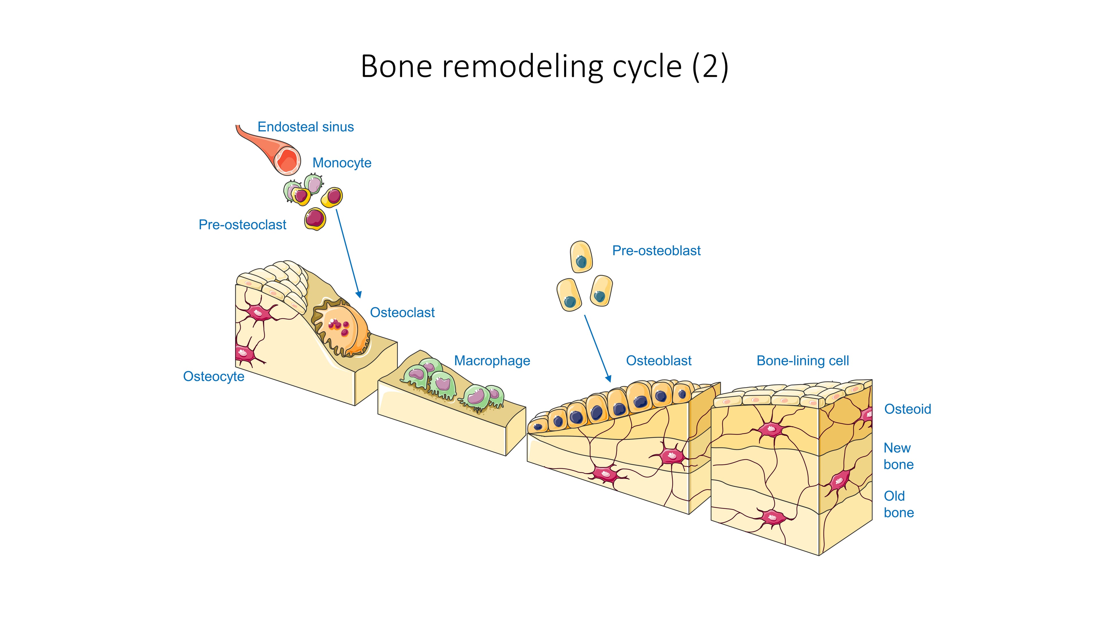
Osteoclasts resorb. Multinucleated giant cells with 10-100 nuclei, derived from haematopoietic stem cells of the monocyte-macrophage lineage, 20-100 μm in diameter. The ruffled border is a deeply folded membrane facing the bone, the clear zone is an actin ring that seals the extracellular space around it (requiring αvβ3 integrin binding to RGD sequences in bone matrix), and the basolateral membrane carries the bicarbonate-chloride exchanger that maintains intracellular pH.
How an osteoclast dissolves bone. A vacuolar H+-ATPase pumps protons into the sealed compartment, acidifying it to pH 4.5 and dissolving the mineral; cathepsin K and matrix metalloproteinases (MMP-9, MMP-13) then degrade collagen in that acid environment. Degraded collagen is then removed by transcytosis - endocytosed, transported across the cell and released at the basolateral membrane into the bloodstream, which is where the CTX fragments measured in serum come from. The cell survives 2-3 weeks on a resorbing surface and then undergoes apoptosis.
Osteoblasts form. Cuboidal cells 20-30 μm across, derived from mesenchymal stem cells and arranged in a single layer on the bone surface during formation. Extensive rough endoplasmic reticulum synthesises type I collagen (90% of bone matrix protein), a prominent Golgi packages the non-collagenous proteins (osteocalcin, osteopontin, bone sialoprotein), and membrane-bound alkaline phosphatase hydrolyses pyrophosphate, removing an inhibitor of mineralisation and generating phosphate for hydroxyapatite.
What the osteoblast produces, and what becomes of it. Osteoid is deposited at 1-2 μm per day and mineralisation begins after a lag of 10-20 days, which allows collagen cross-linking; refilling a resorption cavity takes 3 months. When deposition finishes, the osteoblast becomes an osteocyte (10-20% are embedded in the matrix), becomes a bone lining cell, or undergoes apoptosis.
Osteocytes sense. Stellate cells in lacunae of 10-20 μm throughout mineralised bone, and much the most numerous bone cell (90-95% of all bone cells, at a density of 20,000-30,000 cells/mm³). The cell body has fewer organelles than an osteoblast; 40-100 dendritic processes per cell run through canaliculi of 0.3 μm to neighbouring osteocytes and lining cells, forming a syncytium-like network. A fluid-filled pericellular space of 0.1 μm separates each process from the canalicular wall, and mechanical loading drives fluid through it.
What the osteocyte does with what it senses. Fluid shear stress activates integrins and ion channels such as Piezo1 and generates intracellular calcium signals; loading then decreases sclerostin (SOST) expression, increases Wnt signalling and promotes formation, while unloading does the reverse. Osteocytes live for years to decades, and their apoptosis is itself a signal: a dying osteocyte at a damaged site attracts osteoclasts and triggers targeted remodelling there.
Bone lining cells cover and gate. Flattened inactive osteoblasts, 1-2 μm thick, covering the 95% of adult bone surface that is quiescent. They form the canopy over trabecular remodelling sites, isolating the bone remodelling compartment from the marrow space; they retract and digest the surface osteoid layer when osteoclasts are recruited, exposing mineralised bone for resorption; they are a reservoir that can re-differentiate into active osteoblasts; and they are connected to the underlying osteocytes by gap junctions through the canaliculi.
The vascular supply to a remodelling site
Cortical bone is supplied from within. The nutrient artery enters at the mid-diaphysis and gives endosteal branches to the capillaries of the Haversian canals, while periosteal arteries reach the same system through Volkmann's canals. A cutting cone advances with its own capillary, drawn from the existing Haversian canal, and that vessel becomes the Haversian canal of the new osteon as the tunnel closes behind it.
Trabecular bone is supplied from outside. Marrow sinusoids lie directly against the trabecular surfaces, so a trabecular BMU is nourished across a short diffusion distance of 100-300 μm. There is no vasculature inside a trabecula - it is too thin - and its osteocytes are supplied by diffusion through the canaliculi from the surface.
The vessel is not a bystander. During resorption, a capillary sprout advances with the cutting head, high blood flow delivers osteoclast precursors along with RANKL and M-CSF from the circulation, and degraded matrix (the CTX and NTX fragments) is carried away. During formation, the established capillary delivers amino acids for collagen synthesis and calcium and phosphate for mineralisation, and removes the lactate of osteoblast glycolysis. Endothelial cells also regulate the cells around them, producing VEGF (angiogenesis and osteoblast recruitment), endothelin-1 (osteoblast proliferation) and nitric oxide (osteoclast apoptosis).
Where this shows itself clinically. In avascular necrosis of the femoral head, scaphoid or talus, loss of blood supply kills the osteocytes, mechanosensing stops, and remodelling continues without mechanical feedback until the subchondral bone fractures. Bisphosphonates, conversely, concentrate where blood flow and bone turnover are highest - the vertebrae and proximal femur.
The Basic Multicellular Unit (BMU)
The BMU is the anatomical-functional team of cells that executes remodelling in one discrete location: osteoclasts at the leading edge, osteoblasts behind them, the bone lining cells and osteocytes of the surrounding surface, a blood vessel, the marrow that supplies precursors, and the canopy that encloses the whole thing. Its organisation is what makes formation follow resorption in the right place.
The cortical BMU is a tunnel. 20-50 osteoclasts form a cutting head that bores a longitudinal tunnel of 200 μm diameter, advancing at 20-40 μm per day. 50-100 μm behind it lies the reversal zone, where mononuclear cells clean the surface and prepare it for formation. Osteoblasts then lay concentric lamellae from the outside inwards - the closing cone - filling 80-90% of the resorbed tunnel and leaving a central Haversian canal of 10-20 μm that carries the capillary and nerve supplying the osteocytes of the new osteon. The cutting cone is 1-2 mm long and the result is a new secondary osteon.
The trabecular BMU is a trench. 5-10 osteoclasts excavate a scalloped cavity on the trabecular surface - a Howship's lacuna, 40-60 μm deep and about 1 mm across. At maximum depth the mononuclear reversal cells deposit the basophilic scalloped cement line, and osteoblasts then fill the cavity from the bottom upwards with 4-5 lamellae of about 5 μm each, completely or partly restoring the surface. The result is a hemi-osteon, or trabecular packet, sitting on a concave cement line.
- Cortical BMU
- Cutting cone (longitudinal tunnel)
- Trabecular BMU
- Resorption cavity (surface trench)
- Cortical BMU
- Advances longitudinally through cortex
- Trabecular BMU
- Excavates trabecular surface
- Cortical BMU
- 3-6 months complete cycle
- Trabecular BMU
- 3-6 months complete cycle
- Cortical BMU
- 2-3% per year
- Trabecular BMU
- 25% per year (higher surface area)
- Cortical BMU
- New osteon (Haversian system)
- Trabecular BMU
- New bone packet on trabecular surface
Trabecular perforation is the irreversible event. When cavity depth exceeds the thickness of the trabecula - typically a cavity deeper than 60 μm in a trabecula under 100 μm thick - the osteoclasts tunnel straight through and the strut is disconnected. Osteoblasts cannot rebuild across an empty space, so connectivity is lost permanently, and this is part of why bone in osteoporosis is weaker than bone mineral density alone predicts. Cavity depth can reach 100 μm in high-turnover states, which is exactly when perforation becomes likely.
In the cortex the volume resorbed approximately equals the volume formed, so each cycle produces a new structural unit and remodelling is balanced. On a trabecular surface the cycle produces a packet rather than a structure, and if the cavity is deeper than the strut is thick it produces a hole instead.
That, and the ten-fold difference in surface area, is why metabolic bone disease reaches the spine and hip first and hits them hardest.
The Remodeling Cycle - Phases and Timeline
Bone Remodelling Cycle
Mechanical strain, microdamage, hormonal signals or cytokines activate a quiescent bone surface. Bone lining cells retract, exposing mineralised surface, and osteoclast precursors are recruited from the circulation.
RANKL from osteoblasts and stromal cells, M-CSF for precursor survival, and removal of OPG inhibition.
Multinucleated osteoclasts attach through integrin binding and form the ruffled border and sealing zone. Acidification to pH 4.5 dissolves mineral and cathepsin K degrades collagen.
Approximately 0.05 cubic millimetres of bone is removed, creating a Howship's lacuna in trabecular bone or a resorption tunnel in cortical bone.
2-3 weeks - shorter than formation, which is what creates the transient imbalance.
Osteoclasts undergo apoptosis or migrate away, and mononuclear cells appear in the resorption cavity.
Growth factors released from bone matrix during resorption (TGF-β, IGF-I, IGF-II, BMPs) recruit and activate osteoblast precursors.
A thin layer of hypermineralised matrix marks the junction between old and new bone.
Cuboidal osteoblasts secrete osteoid, which mineralises after the maturation lag.
The resorption cavity is refilled and bone volume restored. In cortical bone concentric lamellae form a new osteon; in trabecular bone a new packet forms on the surface.
Approximately 3 months - formation takes considerably longer than resorption.
Osteoblasts complete matrix synthesis and become flat bone lining cells or embedded osteocytes, and the surface is covered by lining cells until the next activation.
Variable, from weeks to years depending on mechanical and metabolic demand.
Formation takes longer than resorption (3 months against 2-3 weeks), so every active BMU carries a temporary deficit. Raise the remodelling rate - postmenopausal oestrogen deficiency, hyperparathyroidism - and more BMUs sit in the resorption phase at any one moment, producing net bone loss even when each individual BMU is perfectly balanced. This is the remodelling transient.
Primary and secondary mineralisation
The osteoid laid down during the formation phase does not mineralise all at once. Mineralisation proceeds in two kinetically distinct phases, and that distinction underlies both how antiresorptive drugs raise bone mineral density and how over-suppression can make bone brittle.
Primary mineralisation is fast. It begins after the osteoid maturation lag at the mineralisation front, deposits hydroxyapatite quickly, and reaches roughly 70 per cent of the bone packet's final mineral content within days to a few weeks. This is the phase that tetracycline labelling measures as the mineral apposition rate.
Secondary mineralisation is slow. Over months to years the crystals grow and mature, the apatite lattice is perfected and water is displaced, progressively increasing the tissue mineral density and the stiffness of each completed packet. It continues quietly long after the BMU has finished and the surface has gone quiet.
Why this matters at the bedside. By slowing activation frequency, bisphosphonates and denosumab let existing packets complete secondary mineralisation and reduce the proportion of young, under-mineralised packets; this accounts for a substantial part of the measured rise in bone mineral density, beyond simply preventing resorption. Profound, prolonged suppression takes the same mechanism too far, yielding uniformly old, fully mineralised, homogeneous bone that deflects cracks poorly and tolerates microdamage badly - one contributor to the reduced toughness implicated in atypical femoral fracture. A higher and more uniform mineral content raises stiffness but lowers toughness, so bone quality depends on a healthy distribution of packet ages rather than on maximal mineralisation.
(Static bone matrix composition - collagen, hydroxyapatite, mineral-to-matrix ratio - is developed in the bone-composition-structure topic; this section covers the dynamic mineralisation tied to the remodelling cycle.)
Coupling - Linking Resorption to Formation
Coupling is the mechanism that ensures bone formation occurs at sites of prior resorption. Three things achieve it: the growth factors buried in the matrix, direct signalling between the two cell types, and the physical organisation of the BMU itself.
Bone matrix is a store of growth factors, and resorption is what opens it. Osteoclasts release into the local microenvironment factors that were laid down with the matrix, producing a high local concentration exactly where bone has just been removed. That is why formation occurs precisely at the site of prior resorption.
Transforming growth factor-beta (TGF-β) is the most abundant growth factor in bone matrix, at a concentration of 200 micrograms per kilogram. Released during resorption, it is a chemoattractant for osteoblast precursors and draws osteoprogenitors to the resorption site; it stimulates preosteoblast proliferation but inhibits terminal differentiation.
Insulin-like growth factors I and II (IGF-I, IGF-II) are stored in matrix as complexes with IGF-binding proteins, and are released and activated during resorption. They stimulate osteoblast proliferation and collagen synthesis and enhance osteoblast differentiation and function.
The bone morphogenetic proteins BMP-2, BMP-4, BMP-6 and BMP-7 are present in the matrix as potent osteoinductive factors. They promote differentiation of mesenchymal stem cells towards osteoblasts and activate the Runx2 transcription factor.
Uncoupling occurs when formation does not match resorption, and the pattern of failure names the disease: in osteoporosis formation is inadequate and bone is lost; in Paget disease resorption is followed by excessive, disordered formation; in glucocorticoid excess resorption is normal but formation is suppressed; and in myeloma osteoclast-activating factors drive resorption with no coupled formation at all.
Molecular Regulation - RANK/RANKL/OPG System
The RANK/RANKL/OPG axis is the master regulatory system for bone remodelling, controlling osteoclast formation and activity, and it is the axis on which denosumab acts.
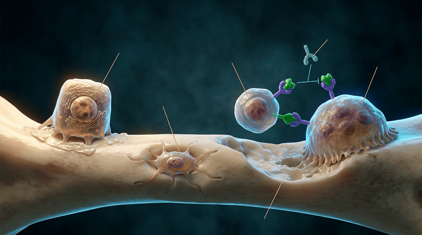
RANKL (receptor activator of nuclear factor kappa-B ligand) is produced by osteoblasts, osteocytes and stromal cells in a dominant membrane-bound form and a soluble form. It binds RANK on osteoclast precursors and activates the NFκB pathway, driving osteoclast differentiation, activation and survival.
RANK is the transmembrane receptor for RANKL, expressed by osteoclast precursors and mature osteoclasts. Ligand binding signals through NFκB, NF-AT, AP-1 and MAPK cascades to switch on the osteoclastogenesis genes.
OPG (osteoprotegerin) is produced by the same osteoblasts and osteocytes but has no transmembrane domain: it is a soluble decoy receptor that binds RANKL before it can reach RANK, and so inhibits osteoclast formation. The bone-forming lineage therefore holds both the accelerator and the brake.
The ratio, not either protein alone, sets the resorption rate.
- Osteoclast Activity
- Increased resorption
- Bone Effect
- Net bone loss
- Clinical Examples
- Postmenopausal osteoporosis, hyperparathyroidism
- Osteoclast Activity
- Physiological turnover
- Bone Effect
- Homeostasis
- Clinical Examples
- Healthy young adult
- Osteoclast Activity
- Decreased resorption
- Bone Effect
- Increased bone mass
- Clinical Examples
- Osteopetrosis (extreme), anabolic therapy
What pushes the ratio up. RANKL is increased by continuous PTH exposure, 1,25-dihydroxyvitamin D3, glucocorticoids, the inflammatory cytokines IL-1, IL-6 and TNF-α, and thyroid hormone. OPG is decreased by oestrogen deficiency and by glucocorticoids - which is why steroids appear on both lists and do double damage.
What pushes it down. OPG is increased by oestrogen, TGF-β, BMP-2 and mechanical loading acting through the osteocyte.
Oestrogen maintains bone mass by increasing OPG production and decreasing RANKL production by osteoblasts, holding the RANKL:OPG ratio low. Menopause reverses both arms at once: RANKL rises, OPG falls, the ratio climbs sharply, and bone is lost at up to 3-5% per year for 5-10 years.
Mechanical Regulation - Wolff's Law and Mechanotransduction
Wolff's law (1892): bone adapts its architecture and mass to the mechanical stresses placed upon it. The cell that makes this happen is the osteocyte, which senses strain and directs the remodelling response.
The stimulus is fluid, not strain directly. Loading deforms the matrix, matrix deformation drives fluid through the lacunar-canalicular network, and the resulting shear stress acts on the osteocyte membrane and its processes. The fluid flow amplifies the underlying strain, which is how a cell buried in mineral becomes an exquisitely sensitive mechanosensor.
Sensing runs through four structures, which operate together:
- Primary cilium - a mechanosensitive organelle detecting fluid flow
- Integrins - connecting cytoskeleton to matrix, sensing deformation
- Gap junctions - connexin 43 channels coordinating the osteocyte network
- Mechanosensitive ion channels - allowing calcium influx
The signal then leaves the cell. Calcium influx triggers intracellular signalling, prostaglandin E2 is produced and nitric oxide released, and gene expression changes - sclerostin, RANKL and OPG.
Hormonal Regulation of Bone Remodeling
Systemic hormones adjust the remodelling rate to meet metabolic demand, and the skeleton pays for calcium homeostasis when the two conflict.
- Source
- Parathyroid glands
- Effect on Remodelling
- Increases resorption
- Mechanism
- Increases RANKL, decreases OPG
- Clinical Relevance
- Hyperparathyroidism causes bone loss
- Source
- Exogenous (teriparatide)
- Effect on Remodelling
- Increases formation
- Mechanism
- Stimulates osteoblasts, anabolic
- Clinical Relevance
- Anabolic therapy for osteoporosis
- Source
- Kidney (1,25-OH2-D3)
- Effect on Remodelling
- Increases resorption
- Mechanism
- Increases RANKL, enhances calcium absorption
- Clinical Relevance
- Deficiency causes osteomalacia
- Source
- Thyroid C-cells
- Effect on Remodelling
- Decreases resorption
- Mechanism
- Direct osteoclast inhibition
- Clinical Relevance
- Used for Paget disease
- Source
- Ovaries
- Effect on Remodelling
- Decreases resorption
- Mechanism
- Decreases RANKL, increases OPG
- Clinical Relevance
- Menopause accelerates bone loss
- Source
- Adrenal cortex
- Effect on Remodelling
- Complex (net bone loss)
- Mechanism
- Decreases osteoblast function, increases apoptosis
- Clinical Relevance
- Steroid-induced osteoporosis
- Source
- Pituitary
- Effect on Remodelling
- Increases formation
- Mechanism
- Stimulates IGF-1 production
- Clinical Relevance
- Acromegaly increases bone turnover
- Source
- Thyroid
- Effect on Remodelling
- Increases turnover
- Mechanism
- Increases both resorption and formation
- Clinical Relevance
- Hyperthyroidism increases fracture risk
Intermittent PTH given as a daily injection (teriparatide) is anabolic: it stimulates osteoblasts, increases formation and improves microarchitecture. Continuous PTH, as in hyperparathyroidism, is catabolic: RANKL rises persistently, osteoclasts are driven, and bone is lost.
The difference is the temporal pattern alone. Intermittent exposure preferentially stimulates osteoblasts without sustained RANKL-mediated resorption; continuous exposure keeps RANKL high and resorption wins.
Classification
Remodelling is classified by how fast it runs and by whether the two arms are still linked. Turnover state is measured by activation frequency and by bone turnover markers; coupling status is inferred from the relationship between the formation and resorption markers. Together they decide whether a patient needs the resorption arm suppressed or the formation arm driven.
Classification by turnover state
- Activation Frequency (Ac.f)
- Ac.f 0.4-1.0 per mm² per year (activation frequency measured by histomorphometry). Represents the number of new remodelling sites initiated per unit bone surface per year.
- Bone Turnover Markers
- CTX 200-600 pg/mL (resorption marker). P1NP 20-80 μg/L (formation marker). Balanced: formation markers proportional to resorption markers.
- Clinical Features
- Skeletal homeostasis maintained. Resorbed bone volume equals formed bone volume. 10% of skeleton replaced annually. Bone mass stable.
- Example Conditions
- Healthy adults age 30-50. Premenopausal women. Eugonadal men. Adequate calcium and vitamin D nutrition. Normal hormonal milieu (PTH, thyroid, oestrogen, testosterone).
- Activation Frequency (Ac.f)
- Ac.f greater than 1.0, often 2-5 times normal. Excessive BMU activation with a shortened remodelling cycle (less than 3 months against a normal 3-6 months).
- Bone Turnover Markers
- CTX elevated (greater than 600 pg/mL, often 800-1500 pg/mL). P1NP elevated (greater than 80 μg/L). BSAP elevated. May be uncoupled, with resorption markers disproportionately higher than formation markers.
- Clinical Features
- Rapid bone loss (2-4% per year against a normal 0-1%). Increased remodelling space (the volume of bone in active remodelling, normally 2-5% of trabecular bone). Microarchitectural deterioration with trabecular perforation. Fragility fracture risk elevated.
- Example Conditions
- Postmenopausal osteoporosis (oestrogen deficiency increases RANKL). Primary hyperparathyroidism (PTH increases RANKL). Hyperthyroidism (thyroid hormone increases osteoclast activity). Paget disease (abnormal excessive remodelling with mosaic pattern). Inflammatory arthritis (cytokines increase RANKL).
- Activation Frequency (Ac.f)
- Ac.f less than 0.4, often less than 0.2 in severe cases. Reduced BMU activation, with a prolonged or arrested remodelling cycle.
- Bone Turnover Markers
- CTX low (less than 200 pg/mL, often less than 100 pg/mL). P1NP low (less than 20 μg/L). BSAP low. May have low vitamin D (25OHD less than 50 nmol/L).
- Clinical Features
- Reduced turnover slows fracture healing. Microdamage accumulates because it is not repaired. Adynamic bone shows no tetracycline labelling and minimal osteoid. Risk of atypical femoral fracture with long-term bisphosphonate use.
- Example Conditions
- Adynamic bone disease (end-stage renal disease with low PTH). Long-term bisphosphonate therapy (greater than 5 years alendronate or 3 years zoledronate). Hypoparathyroidism. Chronic glucocorticoid excess (osteoblast and osteocyte apoptosis).
Classification by coupling status
- Definition
- Resorption and formation tightly linked in time and space. Volume of bone resorbed equals volume formed. Coupling mechanisms (matrix factors, cell-cell signals, canopy) intact.
- Bone Balance
- Neutral bone balance. Bone mass stable. A remodelling transient exists but has minimal impact on bone mass.
- Markers
- CTX and P1NP proportional (P1NP/CTX ratio 0.1-0.2 in pmol units). Example: CTX 400 pg/mL with P1NP 40 μg/L indicates balanced coupling.
- Clinical Conditions
- Healthy premenopausal women. Eugonadal men. Adequate nutrition. Normal hormonal status. Represents skeletal homeostasis.
- Definition
- Resorption and formation dissociated. Cavity depth or number exceeds what formation can replace. Coupling disrupted by oestrogen deficiency (RANKL up, OPG down) or inflammatory cytokines (RANKL up).
- Bone Balance
- Negative bone balance with progressive bone loss. Resorption cavities incompletely filled (wall width less than cavity depth). Trabecular perforation risk.
- Markers
- CTX disproportionately elevated against P1NP. P1NP/CTX ratio low (less than 0.1). Example: CTX 800 pg/mL with P1NP only 50 μg/L.
- Clinical Conditions
- Postmenopausal osteoporosis. Primary hyperparathyroidism. Hyperthyroidism. Rheumatoid arthritis (inflammatory cytokines). Myeloma (RANKL-secreting plasma cells). Immobilisation (disuse osteoporosis).
- Definition
- Formation proceeds without preceding resorption, or exceeds it. Osteoclast function impaired (genetic defect, carbonic anhydrase II deficiency) or osteoblast activity excessive without coupling to resorption (fibrous dysplasia).
- Bone Balance
- Positive bone balance but pathological bone: increased mass with abnormal structure (sclerotic, fragile, obliterated marrow). Dense bone on radiograph.
- Markers
- CTX low or normal. P1NP disproportionately elevated. BSAP elevated. P1NP/CTX ratio high (greater than 0.3). Example: CTX 200 pg/mL with P1NP 100 μg/L.
- Clinical Conditions
- Osteopetrosis (defective osteoclast function, mutations in TCIRG1, CLCN7). Pycnodysostosis (cathepsin K deficiency). Sclerotic phase of Paget disease. Fibrous dysplasia (Gs-alpha mutation, osteoblast overactivity).
The therapeutic goal is to restore coupled, balanced remodelling - not to drive remodelling as low as it will go. Suppression beyond that point accumulates microdamage and brings its own fracture risk, which is the whole story of the atypical femoral fracture.
Classification by disease pattern: Paget disease
Paget Disease - Evolution of Remodelling Pattern
Excessive osteoclast activity with minimal coupled formation. Large multinucleated osteoclasts (100+ nuclei, against a normal 10-20).
Advancing osteolytic front with minimal new bone formation. High CTX, low P1NP initially.
Lytic V-shaped wedge advancing along the bone - the blade of grass sign in long bones, osteoporosis circumscripta in the skull.
Bone pain and pathological fracture risk, because lytic bone is weak.
Excessive resorption followed by excessive formation. Coupling is maintained but both processes are abnormally elevated.
Mosaic pattern - a jigsaw appearance created by repeated remodelling cycles and their cement lines - with woven bone instead of lamellar bone.
Mixed lytic-sclerotic appearance with thickened trabeculae, cortical thickening, bone enlargement and deformity.
Both CTX and P1NP extremely elevated (300-800% above normal). BSAP particularly elevated.
Reduced resorption with continued excessive formation, until eventually both processes slow.
Dense sclerotic bone retaining the mosaic pattern. Woven bone predominates and marrow spaces are obliterated.
Diffuse sclerosis, cortical thickening and bone enlargement. The bone is dense but mechanically weak and prone to insufficiency fracture.
Moderately elevated or normalising. BSAP may remain elevated despite reduced remodelling activity.
Investigations
Investigating remodelling means answering three questions in order: how fast is bone turning over, why, and - rarely - what is the bone actually doing at the cellular level. Biochemical markers answer the first, serum biochemistry and imaging the second, and transiliac biopsy with tetracycline labelling the third.
Bone turnover markers
- Type
- Resorption marker
- Source
- Type I collagen degradation products released during osteoclastic bone resorption. Measured in serum. Cleared by kidneys (falsely elevated in renal impairment).
- Normal Range
- Premenopausal women: 200-600 pg/mL. Postmenopausal: 300-800 pg/mL. Men: 150-500 pg/mL. Diurnal variation: 20-30% higher in the morning (fasting sample preferred, 8 AM).
- Clinical Use
- Monitor antiresorptive response: expect 50-70% reduction within 3-6 months of bisphosphonate or denosumab. Drug holiday decision: resume therapy if CTX increases above 600 pg/mL. Predict fracture risk: CTX greater than 800 pg/mL is associated with 2-fold increased fracture risk independent of BMD.
- Type
- Formation marker
- Source
- Propeptide cleaved from procollagen during collagen synthesis by osteoblasts. Measured in serum. Direct reflection of osteoblast activity.
- Normal Range
- Premenopausal women: 20-80 μg/L. Postmenopausal: 30-100 μg/L. Men: 15-70 μg/L. Less diurnal variation than CTX (10-15%).
- Clinical Use
- Monitor anabolic therapy: expect 100-200% increase within 1-3 months of teriparatide or romosozumab; if absent, check compliance. Assess coupling: P1NP/CTX ratio indicates balance (normal 0.1-0.2). A greater P1NP increase predicts a greater BMD gain with anabolic therapy.
- Type
- Formation marker
- Source
- Isoenzyme of alkaline phosphatase produced by osteoblasts. Measured in serum. Hydrolyses pyrophosphate to facilitate mineralisation.
- Normal Range
- Adults: 5-25 μg/L (varies by assay). Higher in children (growing skeleton) and postmenopausal women. Less affected by liver or kidney disease than total ALP.
- Clinical Use
- Particularly useful in Paget disease (marked elevation, 100-500 μg/L) and for monitoring its treatment: BSAP should normalise within 3-6 months of zoledronate. Less responsive to short-term change than P1NP (half-life 1-2 weeks against 30 minutes for P1NP).
- Type
- Resorption marker
- Source
- Type I collagen degradation product. Measured in urine (second morning void, corrected for creatinine). Alternative to serum CTX.
- Normal Range
- Adults: 10-60 nmol BCE/mmol creatinine (bone collagen equivalents). Higher variability than serum CTX because urine concentration varies with hydration.
- Clinical Use
- Historical use, now largely replaced by serum CTX for convenience, though still used in some centres. Interpretation as for CTX: a 50-70% reduction is the expected antiresorptive response.
A bone turnover marker is only as good as the sample. CTX varies by 20-30% across the day and P1NP by 10-15%, both lowest in the afternoon and highest in the early morning, so standardise to a fasting 8 AM sample. Markers rise within days of any fracture as healing begins, so wait 3 months after a fracture before calling a value a baseline. CTX is renally cleared and is falsely elevated when eGFR is less than 30 mL/min/1.73m², where P1NP is the better choice. Recent immobilisation suppresses formation markers and may raise resorption markers - the disuse pattern.
The thresholds worth carrying are these:
- High-turnover state: CTX greater than 600 pg/mL, P1NP greater than 80 μg/L (postmenopausal women)
- Low-turnover state: CTX less than 200 pg/mL, P1NP less than 20 μg/L
- Adequate antiresorptive response: CTX reduction greater than 50% from baseline, measured at 3-6 months
- Adequate anabolic response: P1NP increase greater than 50% from baseline, measured at 1-3 months
Marker patterns in the conditions that matter
- CTX (Resorption)
- Elevated (600-1200 pg/mL, 50-200% above the premenopausal mean). Oestrogen deficiency increases RANKL and decreases OPG.
- P1NP (Formation)
- Elevated (60-120 μg/L) but less than proportionally. Formation cannot keep pace with resorption.
- P1NP/CTX Ratio
- Low ratio (0.05-0.10, normal 0.1-0.2), indicating resorption exceeds formation. Negative bone balance.
- Clinical Implication
- Antiresorptive therapy indicated. Monitor: CTX should decrease 50-70% within 3-6 months, with P1NP decreasing 30-50% as coupled suppression.
- CTX (Resorption)
- Markedly elevated (800-2000 pg/mL, 100-300% above normal). PTH increases RANKL expression and stimulates osteoclast activity.
- P1NP (Formation)
- Elevated (80-150 μg/L). PTH also has an anabolic effect on osteoblasts, but resorption dominates.
- P1NP/CTX Ratio
- Low ratio (0.04-0.08). Net bone loss, with preferential cortical loss at the radius over trabecular loss at the spine.
- Clinical Implication
- Parathyroidectomy if surgical criteria met (BMD T-score less than -2.5, fragility fracture, age less than 50, calcium greater than 0.25 mmol/L above the upper normal limit). After surgery BTMs normalise within 3-6 months.
- CTX (Resorption)
- Extremely elevated (1500-5000 pg/mL, 300-800% above normal). Localised excessive remodelling with abnormal osteoclasts.
- P1NP (Formation)
- Extremely elevated (200-500 μg/L), BSAP particularly so. Formation follows resorption but produces disorganised woven bone.
- P1NP/CTX Ratio
- Variable ratio (0.04-0.15). Coupling is maintained but the bone produced is abnormal - weak despite high mass.
- Clinical Implication
- Zoledronate 5 mg IV as a single dose for symptomatic Paget or its complications (deformity, nerve compression, high-output cardiac failure). Monitor: BSAP should normalise within 3-6 months.
- CTX (Resorption)
- Suppressed (less than 200 pg/mL, often less than 150 pg/mL), a 50-70% reduction from pretreatment baseline as bisphosphonate induces osteoclast apoptosis.
- P1NP (Formation)
- Suppressed (less than 30 μg/L), a 30-50% reduction from baseline - coupled suppression, formation following resorption.
- P1NP/CTX Ratio
- Normal or slightly low ratio (0.08-0.15). Coupling preserved but at a low turnover level.
- Clinical Implication
- Consider drug holiday if BMD is stable or improved, there have been no fractures on therapy, fracture risk is low and therapy has run beyond 5 years. During a holiday monitor BTMs every 6-12 months and resume if CTX rises above the premenopausal mean (greater than 600 pg/mL).
- CTX (Resorption)
- Initial spike in the first 1-2 months (600-900 pg/mL) then a plateau. PTH increases remodelling space and activates quiescent lining cells.
- P1NP (Formation)
- Marked elevation (100-180 μg/L, 100-200% above baseline). PTH stimulates osteoblast proliferation, differentiation and matrix synthesis, so formation exceeds resorption.
- P1NP/CTX Ratio
- High ratio (0.15-0.30). Formation dominates and bone balance is positive.
- Clinical Implication
- Monitor: P1NP should increase 50-100% by month 1-3; if not, check compliance and calcium and vitamin D adequacy. After completion, transition to an antiresorptive is mandatory.
- CTX (Resorption)
- Very low (less than 100 pg/mL, often less than 50 pg/mL). PTH suppressed, usually iatrogenically with calcitriol or calcimimetics, and osteoclast activity is minimal.
- P1NP (Formation)
- Very low (less than 15 μg/L, often less than 10 μg/L). Minimal osteoblast activity, and no tetracycline labelling on biopsy.
- P1NP/CTX Ratio
- Variable (0.05-0.20). Both arms profoundly suppressed; turnover nearly absent.
- Clinical Implication
- Avoid bisphosphonates, which worsen the adynamic state. Allow PTH to rise if oversuppressed by reducing calcitriol or calcimimetics, targeting mild secondary hyperparathyroidism (PTH 2-3 times the upper normal limit) to restore turnover. Fracture healing is impaired in adynamic bone.
Serum biochemistry for the underlying cause
- Normal Range
- 2.15-2.55 mmol/L (8.6-10.2 mg/dL). Correct for albumin: corrected Ca equals measured Ca plus 0.02 × (40 minus albumin in g/L).
- Interpretation
- Hypercalcaemia (greater than 2.6 mmol/L): PTH-mediated (primary or tertiary hyperparathyroidism) or non-PTH-mediated (malignancy, sarcoidosis, vitamin D toxicity). Hypocalcaemia (less than 2.1 mmol/L): vitamin D deficiency, hypoparathyroidism, CKD.
- Clinical Conditions
- High calcium with high PTH equals primary hyperparathyroidism (high-turnover state). Low calcium with high PTH equals secondary hyperparathyroidism (vitamin D deficiency, CKD). High calcium with low PTH equals malignancy-related hypercalcaemia (PTHrP, osteolytic metastases).
- Normal Range
- Optimal: greater than 75 nmol/L (greater than 30 ng/mL). Sufficient: 50-75 nmol/L. Insufficiency: 25-50 nmol/L. Deficiency: less than 25 nmol/L. Severe deficiency: less than 12.5 nmol/L.
- Interpretation
- Reflects vitamin D stores (half-life 2-3 weeks) and is the substrate for renal 1-alpha-hydroxylase. Deficiency causes secondary hyperparathyroidism and, if severe, impaired mineralisation.
- Clinical Conditions
- Deficiency (less than 25 nmol/L) with secondary hyperparathyroidism causes high-turnover bone loss. Severe deficiency (less than 12.5 nmol/L) with wide osteoid seams equals osteomalacia. Replacement: 50,000 IU weekly for 8-12 weeks, then maintenance 1000-2000 IU daily, targeting 25OHD greater than 75 nmol/L.
- Normal Range
- 1.6-6.9 pmol/L (15-65 pg/mL, varies by assay). Pulsatile secretion, sample any time.
- Interpretation
- Primary hyperparathyroidism: PTH elevated (greater than 7 pmol/L) with hypercalcaemia. Secondary: PTH elevated with normal or low calcium (vitamin D deficiency, CKD). Hypoparathyroidism: PTH low (less than 1.5 pmol/L) with hypocalcaemia.
- Clinical Conditions
- Primary hyperparathyroidism gives high-turnover uncoupled remodelling with preferential cortical loss. In CKD, target PTH 2-3 times the upper normal limit (3-9 pmol/L) to maintain remodelling without causing adynamic bone. In hypoparathyroidism the state is low-turnover and bisphosphonates are contraindicated.
- Normal Range
- 0.4-4.0 mIU/L. Screening test for thyroid dysfunction.
- Interpretation
- Hyperthyroidism (TSH less than 0.1 mIU/L with elevated free T4/T3): thyroid hormone increases osteoclast activity and causes high-turnover bone loss of 3-5% per year. Hypothyroidism (TSH greater than 10 mIU/L): low-turnover state, reduced remodelling.
- Clinical Conditions
- Treat hyperthyroidism with antithyroid drugs, radioiodine or surgery; BTMs normalise within 3-6 months of euthyroidism. Fracture risk is elevated during the hyperthyroid period (relative risk 1.3-1.7). Consider bisphosphonates if T-score less than -2.5 and hyperthyroidism is prolonged beyond 6 months.
- Normal Range
- 0.8-1.5 mmol/L (2.5-4.5 mg/dL). Varies with age, diet and time of day.
- Interpretation
- Hypophosphataemia (less than 0.8 mmol/L): renal phosphate wasting (FGF23-mediated in tumour-induced osteomalacia, X-linked hypophosphataemia) or vitamin D deficiency. Hyperphosphataemia (greater than 1.5 mmol/L): CKD, hypoparathyroidism.
- Clinical Conditions
- Hypophosphataemia with low 25OHD equals vitamin D deficiency with impaired phosphate absorption. Hypophosphataemia with normal 25OHD and high FGF23 equals tumour-induced osteomalacia or X-linked hypophosphataemia. Replacement: neutral phosphate 1-3 g daily in divided doses (gastrointestinal side effects common).
- Normal Range
- Creatinine: 60-110 μmol/L (0.7-1.2 mg/dL). eGFR: greater than 90 mL/min/1.73m² (CKD-EPI equation).
- Interpretation
- CKD (eGFR less than 60): impaired vitamin D activation, phosphate retention, secondary hyperparathyroidism. Advanced CKD (eGFR less than 30): the renal osteodystrophy spectrum - high-turnover, low-turnover or adynamic bone disease.
- Clinical Conditions
- CKD stages 3-4 (eGFR 15-60): check PTH, calcium and phosphate every 6-12 months, targeting PTH 2-3 times the upper normal limit. CKD stage 5 (eGFR less than 15) or dialysis: bone biopsy if the turnover state is unclear, since it guides phosphate binder, vitamin D analogue and calcimimetic use. Avoid bisphosphonates if eGFR less than 30.
Imaging
DEXA - dual-energy X-ray absorptiometry - measures the cumulative result of remodelling balance, not the rate. Areal bone mineral density in g/cm² is the sum of years of balance or imbalance, which is why it moves slowly and why turnover markers answer a different question. Site selection matters: the lumbar spine (L1-L4) is 70% trabecular and sensitive to high-turnover states, but degenerative change, aortic calcification and compression fractures falsely elevate it, so it is most useful under the age of 60; the total hip and femoral neck are half cortical and half trabecular, carry less artefact and are preferred over 60, and predict hip fracture; the distal radius (33% site) is predominantly cortical, shows the cortical loss of primary hyperparathyroidism, and is also used to assess distal radius fracture risk.
The WHO thresholds name the categories:
- Normal: T-score greater than or equal to -1.0
- Osteopenia: T-score -1.0 to -2.5
- Osteoporosis: T-score less than or equal to -2.5
- Severe osteoporosis: T-score less than or equal to -2.5 with a fragility fracture
A change is only real if it exceeds the least significant change, which is 0.03-0.04 g/cm² at the spine and 0.02-0.03 g/cm² at the hip - the 95% confidence limit for measurement error. The scan interval follows the drug: baseline then 1-2 years on an antiresorptive, and baseline then 6-12 months on an anabolic. And BMD reports quantity, not quality: high-turnover remodelling loses density and deteriorates microarchitecture together, while low-turnover remodelling preserves density while accumulating microdamage. The turnover markers are what characterise the second.
Plain radiographs are insensitive but diagnostic when positive. Generalised radiolucency, cortical thinning and trabecular rarefaction require about 30% bone loss before they are visible, so radiographs are no substitute for DEXA in detection. What they do show is the disease-specific pattern:
- Osteomalacia: Looser zones - radiolucent lines perpendicular to the cortex, in the pubic rami, femoral neck, ribs and scapula - which are unmineralised osteoid-filled stress fractures
- Hyperparathyroidism: subperiosteal resorption on the radial aspect of the middle phalanges (the classic finding), cortical tunnelling (radiating striations in long bones), brown tumours (lytic, osteoclast-rich cystic lesions) and the rugger jersey spine (alternating sclerotic and lucent bands in the vertebrae)
- Paget disease: the phase appearances described in the Classification section, together with the complications - transverse chalk-stick fracture, bowed tibia, protrusio acetabuli
Bone scintigraphy (technetium-99m MDP) maps turnover across the whole skeleton. Uptake reflects osteoblastic activity and blood flow, which makes it the survey tool for Paget disease (monostotic against polyostotic, and identifying a cold area within a hot pagetic lesion that suggests sarcoma), for occult stress and insufficiency fractures, and for metastatic disease - osteoblastic metastases from prostate and breast light up, while osteolytic deposits from myeloma, renal and thyroid primaries may be photopenic. Its weakness is the mirror of its strength: any process that raises turnover takes up tracer, so it cannot separate fracture from infection from tumour from degenerative change, and characterisation needs CT or MRI.
Histomorphometry
Transiliac bone biopsy with double tetracycline labelling remains the reference standard for turnover state, and the only one of these investigations that looks at the cells directly. The static parameters need no label; the dynamic parameters - the ones that distinguish a mineralisation defect from excessive resorption - require it.
- What it measures
- Number of new remodelling sites initiated per mm² of bone surface per year, calculated from tetracycline labelling. It is the measure of how often a new BMU is started.
- Normal Range
- 0.4-1.0 per mm² per year
- What it measures
- Percentage of bone surface carrying active resorption cavities (osteoclasts present, or Howship's lacunae): eroded surface length divided by total bone surface length, times 100.
- Normal Range
- 2-5% in trabecular bone
- What it measures
- The proportion of surface covered by unmineralised matrix, and how thick that seam is (measured by polarised light microscopy). A wide seam means osteoid is being made but not mineralised.
- Normal Range
- OS/BS 5-20%; O.Th 5-15 μm
- What it measures
- Distance between the two tetracycline labels divided by the labelling interval (typically 12 days) - the speed at which the mineralisation front advances.
- Normal Range
- 0.6-0.8 μm/day
- What it measures
- Volume of bone formed per unit surface per unit time: MAR × (mineralising surface / bone surface) × 3.65 to convert to % per year.
- Normal Range
- 10-20% per year (trabecular bone)
- What it measures
- The static measures of how much bone there is and how thick the struts are. BV/TV is low in osteoporosis (less than 15%) and high in osteopetrosis (greater than 30%); Tb.Th is thin in osteoporosis (less than 100 μm).
- Normal Range
- BV/TV 15-25%; Tb.Th 100-150 μm
The numbers by disease are worth a second look. Eroded surface runs 8-12% in postmenopausal osteoporosis, 10-15% in hyperparathyroidism and 15-25% in Paget disease, against less than 1% in adynamic bone disease and 1-2% on long-term bisphosphonates. Mineral apposition rate reaches 0.9-1.2 μm/day on teriparatide, and bone formation rate falls to 5-8% per year on long-term bisphosphonates.
Where the generic bands sit. Eroded surface greater than 5%, often 10-20% in severe cases, marks a high-turnover state, against less than 2% and often less than 1% in a low-turnover one. A mineral apposition rate greater than 0.8 μm/day is high turnover and less than 0.6 μm/day low turnover, and a bone formation rate greater than 20% per year is high turnover against a normal 10-20%.
- Static Parameters
- BV/TV 15-25%. Tb.Th 100-150 μm. OS/BS 5-20%, O.Th 5-15 μm (thin osteoid seams). ES/BS 2-5% (occasional resorption cavities).
- Dynamic Parameters
- MAR 0.6-0.8 μm/day (double labels present, separated 7-10 μm). BFR/BS 10-20% per year. Ac.f 0.4-1.0 per mm² per year.
- Histological Features
- Trabecular bone with normal interconnected architecture. Thin osteoid seams on 5-20% of surface. Cuboidal osteoblasts on forming surfaces, occasional osteoclasts in resorption cavities, osteocytes at normal density (20,000-30,000 cells/mm³).
- Static Parameters
- BV/TV less than 15%. Tb.Th less than 100 μm with trabecular perforation and disconnection. ES/BS greater than 5%, with multiple deep cavities of 40-60 μm. OS/BS normal or increased (10-25%), O.Th normal (5-15 μm).
- Dynamic Parameters
- MAR normal or slightly elevated (0.7-1.0 μm/day). BFR/BS elevated (20-35% per year). Ac.f elevated (greater than 1.0, often 2-3). Double labels present at normal interlabel distance.
- Histological Features
- Reduced trabecular bone volume with thin disconnected trabeculae. Multiple active resorption sites, osteoclasts in deep Howship's lacunae, complete erosion through some trabeculae. Osteoid thickness normal - coupling and mineralisation are intact; it is the balance that has failed.
- Static Parameters
- BV/TV normal or low. OS/BS greater than 25% (extensive osteoid coverage). O.Th greater than 15 μm and often greater than 30 μm - wide unmineralised seams, the diagnostic feature. ES/BS normal or low.
- Dynamic Parameters
- MAR less than 0.3 μm/day - the mineralisation defect, and the diagnostic dynamic finding. BFR/BS low (less than 10% per year). Double labels absent, very close together (less than 4 μm) or single.
- Histological Features
- Thick blue-green osteoid seams on Goldner trichrome covering most trabecular surfaces, wide pink osteoid on Von Kossa with minimal black mineralised border. Osteoblasts present but mineralisation ineffective. Looser zones may be present.
- Static Parameters
- BV/TV normal or low. OS/BS less than 5% (minimal osteoid, no active formation). O.Th normal or low (less than 10 μm). ES/BS less than 2%. Thin trabeculae with reduced connectivity.
- Dynamic Parameters
- MAR less than 0.4 μm/day if any labels are present. BFR/BS less than 5% per year. Ac.f less than 0.2. Tetracycline labels often absent or sparse and single; no double labels.
- Histological Features
- Quiescent surfaces with neither formation nor resorption: no osteoblasts (flat lining cells only), no osteoclasts, no labelling - bone effectively frozen. The result of PTH oversuppression in CKD with calcitriol or calcimimetics.
- Static Parameters
- BV/TV high (sclerotic bone) with the diagnostic mosaic pattern of multiple irregular cement lines. Abnormal woven bone. Enlarged osteoclasts with 100+ nuclei against a normal 10-20.
- Dynamic Parameters
- MAR very high (1.5-3.0 μm/day). BFR/BS very high (50-100% per year). Ac.f very high (5-10). Wide double labels, interlabel distance greater than 15 μm.
- Histological Features
- Mosaic pattern from repeated cycles of resorption and formation. Woven bone instead of lamellar - disorganised collagen, normally seen only in fetal bone or fracture callus - with loss of birefringence under polarised light. Abnormally large osteoclasts in resorption cavities, formation and resorption active simultaneously, and marrow fibrosis in the active phase.
Both give low BMD and both may give raised turnover markers, and the treatments are incompatible, so the distinction has to be secure. Two numbers separate them: the mineral apposition rate and the osteoid thickness.
Osteomalacia is a mineralisation defect. MAR less than 0.3 μm/day with wide osteoid seams (O.Th greater than 15 μm, often greater than 30 μm) and OS/BS greater than 25%; labels absent or less than 4 μm apart. P1NP is elevated while CTX is normal or low, 25OHD is low (less than 25 nmol/L, often less than 15 nmol/L) and PTH is secondarily elevated (greater than 60 pg/mL). Treat with vitamin D replacement, 50,000 IU weekly for 8-12 weeks, then maintenance. Avoid bisphosphonates until the mineralisation defect is corrected - they suppress an already impaired mineralisation and make it worse.
High-turnover osteoporosis is excessive resorption with intact mineralisation. MAR normal (0.6-0.8 μm/day) or slightly elevated, osteoid thickness normal (5-15 μm), eroded surface greater than 5%, Ac.f greater than 1.0, BV/TV less than 15%, and CTX and P1NP elevated together. 25OHD is typically adequate (greater than 50 nmol/L) or only mildly low. Treat with an antiresorptive.
Biopsy is invasive and is reserved for the questions biochemistry cannot answer. In practice there are four.
- Suspected osteomalacia with a normal vitamin D. Tumour-induced osteomalacia (an FGF23-secreting tumour, suspected when there is hypophosphataemia with normal 25OHD), hypophosphatasia (low alkaline phosphatase, genetic) or type 2 renal tubular acidosis. Biopsy shows wide osteoid (greater than 15 μm) with low MAR (less than 0.3 μm/day).
- Renal osteodystrophy with an unclear turnover state. PTH does not reliably predict turnover in CKD stage 5 or on dialysis, and the three answers demand opposite treatments: high-turnover osteitis fibrosa (calcimimetics), low-turnover adynamic bone (reduce vitamin D analogues), or osteomalacia from aluminium toxicity (deferoxamine).
- An atypical presentation. A young patient (under 40), male sex, or a T-score less than -3.0 with no obvious cause; or bone pain with normal radiographs, calcium, PTH, vitamin D and thyroid function, where the question is malignancy against metabolic bone disease.
- Treatment planning when the markers are too low. A fragility fracture with CTX less than 150 pg/mL and P1NP less than 20 μg/L may be adynamic bone disease (bisphosphonates contraindicated) or osteomalacia (vitamin D first), and the distinction changes everything.
Relative contraindications are coagulopathy (INR greater than 1.5, platelets less than 50,000/μL, or therapeutic anticoagulation with warfarin, heparin or a DOAC - correct it first, or use local haemostatic measures such as tranexamic-acid-soaked gauze and Gelfoam), cellulitis over the iliac crest, a BMI greater than 40 where landmarks cannot be palpated (consider ultrasound or fluoroscopic guidance), and previous iliac crest surgery - bone graft harvest, ORIF of a pelvic fracture - in which case use the other side.
Management
The turnover state decides the drug class. Suppress excessive resorption in a high-turnover state, stimulate formation in a low-turnover state, and in every case correct the underlying cause - vitamin D deficiency, hyperparathyroidism, thyrotoxicosis - before or alongside bone-specific therapy.
- Pathophysiology
- Excessive osteoclast activity with inadequate formation. Causes: oestrogen deficiency, primary hyperparathyroidism, hyperthyroidism, inflammatory arthritis. CTX greater than 600 pg/mL, P1NP/CTX ratio less than 0.1.
- Therapy Class
- Antiresorptive therapy (first-line)
- Mechanism
- Suppress osteoclast formation (denosumab blocks RANKL) or activity (bisphosphonates inhibit FPPS and induce apoptosis). Reduce activation frequency, decrease eroded surface, restore coupling balance.
- Examples
- Postmenopausal osteoporosis: alendronate 70 mg weekly or denosumab 60 mg SC every 6 months. Primary hyperparathyroidism: parathyroidectomy, or cinacalcet if surgery is contraindicated. Hyperthyroidism: antithyroid drugs, radioiodine or surgery.
- Pathophysiology
- Reduced BMU activation with prolonged quiescence. Causes: long-term bisphosphonates (greater than 5 years), adynamic bone disease, hypoparathyroidism. CTX less than 200 pg/mL, P1NP less than 20 μg/L.
- Therapy Class
- Anabolic therapy or drug holiday
- Mechanism
- Stimulate osteoblast activity (teriparatide as intermittent PTH, romosozumab as anti-sclerostin): increase activation frequency, recruit quiescent lining cells, raise bone formation rate. Where the low turnover is bisphosphonate-induced, a holiday lets turnover resume instead.
- Examples
- Adynamic bone with fragility fracture: teriparatide 20 μg SC daily for 18-24 months. Long-term bisphosphonates with stable BMD and low risk: drug holiday. Hypoparathyroidism: recombinant PTH(1-84) 50-100 μg SC daily where available, otherwise manage with calcium and calcitriol.
- Pathophysiology
- Homeostasis maintained, no pathological loss. CTX 200-600 pg/mL, P1NP 20-80 μg/L, ratio 0.1-0.2, bone mass stable.
- Therapy Class
- Prevention and maintenance (no pharmacotherapy)
- Mechanism
- Optimise calcium and vitamin D, load the skeleton (weight-bearing exercise reduces sclerostin and increases formation), and prevent falls - which reduces fracture risk independent of BMD.
- Examples
- Healthy premenopausal women: calcium 1000 mg daily, vitamin D 1000-2000 IU daily, resistance exercise 30 minutes three times weekly. Postmenopausal women with T-score -1.0 to -2.0 and normal BTMs: the same, with no pharmacotherapy unless there is a fragility fracture or a high FRAX score.
Antiresorptive therapy
- Mechanism
- Inhibit farnesyl pyrophosphate synthase in the mevalonate pathway. Osteoclasts ingest bisphosphonate-bound bone; FPPS inhibition prevents prenylation of the small GTPases (Rho, Rac, Ras) and the osteoclast undergoes apoptosis. The drug binds hydroxyapatite and remains in bone for years.
- Dosing
- Alendronate 70 mg PO weekly (or 10 mg daily). Risedronate 35 mg PO weekly (or 5 mg daily, 150 mg monthly). Zoledronate 5 mg IV yearly. Take oral bisphosphonates fasting with 200 mL water and remain upright 30 minutes to reduce oesophageal irritation.
- Biochemical Response
- CTX decreases 50-70% within 3-6 months. P1NP decreases 30-50% as coupled suppression follows.
- Indications
- Postmenopausal osteoporosis (T-score less than -2.5 or fragility fracture). Glucocorticoid-induced osteoporosis (prednisone greater than 7.5 mg daily for greater than 3 months, T-score less than -1.5). Paget disease (zoledronate 5 mg IV single dose for symptomatic or complicated disease).
- Mechanism
- Fully human monoclonal antibody against RANKL. Blocks RANKL-RANK binding and so prevents osteoclast formation, differentiation and activity - pharmacological OPG. The effect is reversible and dissipates 6 months after a dose, which is the source of the rebound problem.
- Dosing
- 60 mg SC every 6 months. Supplemental calcium (500 mg daily) and vitamin D (800 IU daily) are mandatory because of the hypocalcaemia risk, especially if eGFR is less than 30 mL/min/1.73m².
- Biochemical Response
- CTX decreases 70-90% within 1 month, the most potent antiresorptive suppression available. P1NP decreases 50-70%.
- Indications
- Postmenopausal osteoporosis (T-score less than -2.5 or fragility fracture), and particularly useful where eGFR is less than 30 (bisphosphonates contraindicated), where oral bisphosphonates are poorly tolerated (oesophageal irritation, reflux), where the patient prefers an injection, and after romosozumab - denosumab maintains the BMD gained, bisphosphonates do not.
Anabolic therapy
- Mechanism
- Intermittent PTH stimulates osteoblast proliferation, differentiation and matrix synthesis, increases Wnt signalling, inhibits sclerostin and activates quiescent lining cells. The anabolic window is the first 6-12 months, when formation exceeds resorption; after 18-24 months resorption catches up as activation increases remodelling space.
- Dosing
- 20 μg SC daily, self-injected into thigh or abdomen. Maximum 24 months in a lifetime (osteosarcoma in rat studies, not confirmed in humans). Contraindicated in Paget disease, prior skeletal radiation, bone metastases and hypercalcaemia.
- Biochemical Response
- P1NP increases 100-200% within 1-3 months, the earliest signal of response. CTX increases 50-100% transiently in the first 1-2 months.
- Indications
- Severe osteoporosis (T-score less than -3.0 or multiple fractures). Glucocorticoid-induced osteoporosis with fracture despite bisphosphonates. Failed bisphosphonates (fracture on therapy, or BMD still declining). Preferred over antiresorptives at very low bone mass (T-score less than -3.5) or with multiple fractures, because the requirement is to build bone rather than to prevent its loss.
- Mechanism
- Humanised monoclonal antibody against sclerostin, the osteocyte-exclusive SOST product that inhibits Wnt signalling. Blocking it raises Wnt signalling, so formation increases AND resorption decreases for the first 6-12 months - a dual effect that mimics mechanical loading.
- Dosing
- 210 mg SC monthly (two 105 mg injections) for 12 months, after which transition to an antiresorptive is mandatory. Contraindicated within 12 months of myocardial infarction or stroke - ARCH showed increased cardiovascular events against alendronate, though not against placebo in FRAME - and use with caution in cardiovascular disease.
- Biochemical Response
- P1NP increases 50-100% within 1 month. CTX initially decreases 50%, the transient antiresorptive component.
- Indications
- Severe osteoporosis (T-score less than -3.0 or multiple fractures), and specifically where high fracture risk demands rapid gain. After the 12 months, transition to denosumab: a bisphosphonate after romosozumab loses BMD, by a mechanism that is unclear but may relate to romosozumab's transient antiresorptive effect preventing bisphosphonate binding.
Anabolic therapy builds bone; antiresorptive therapy keeps it. Always follow teriparatide or romosozumab with an antiresorptive, because without consolidation the BMD gained from teriparatide is lost within 12 months and the fracture protection goes with it.
The sequencing rules are not interchangeable:
- After romosozumab, use denosumab, not a bisphosphonate. Denosumab maintains the gain; bisphosphonates lose it.
- After teriparatide, either a bisphosphonate or denosumab works. Zoledronate 5 mg IV, alendronate 70 mg weekly, or denosumab 60 mg SC every 6 months.
- Never give a bisphosphonate before teriparatide. Suppressing remodelling removes the remodelling space that teriparatide needs to build into, and blunts the anabolic response. If a patient is already on a bisphosphonate, stop it 6-12 months before starting teriparatide.
For severe osteoporosis the best-evidenced sequence is romosozumab 210 mg SC monthly for 12 months, then denosumab 60 mg SC every 6 months continued for a minimum of 2-3 years, with transition to zoledronate 5 mg IV if the denosumab is ever stopped. Where romosozumab is contraindicated by cardiovascular disease, substitute teriparatide 20 μg SC daily for 18-24 months followed by either a bisphosphonate or denosumab.
Non-pharmacological management
Calcium and vitamin D are prerequisites, not treatments. Target 1000-1200 mg of calcium daily from diet plus supplement - dairy such as milk, yoghurt and cheese, leafy greens such as kale and bok choy, fortified foods such as soy milk and orange juice, and sardines with bones - and supplement the shortfall if dietary intake is under 700 mg daily, with calcium carbonate (40% elemental, taken with food) or calcium citrate (20% elemental, taken at any time and better absorbed in achlorhydria). Absorption saturates above a 500 mg single dose, so split larger requirements.
Vitamin D targets 25OHD greater than 75 nmol/L for bone health, PTH suppression and fracture reduction: maintenance is 1000-2000 IU daily, and deficiency (25OHD less than 25 nmol/L) is loaded with 50,000 IU weekly for 8-12 weeks first. Check 25OHD at baseline, 3 months after loading and then annually. Toxicity is rare and needs more than 10,000 IU daily for months, presenting as hypercalcaemia, hypercalciuria and nephrocalcinosis. None of this is optional: antiresorptive therapy reduces fracture risk only in a calcium and vitamin D replete patient, and anabolic therapy in a calcium-deficient patient causes hypocalcaemia and secondary hyperparathyroidism.
Exercise works through the osteocyte, and then through the fall. Mechanical loading reduces sclerostin expression, increases Wnt signalling and increases formation; unloading does the reverse. Walking, jogging, resistance training (squats, lunges, weightlifting) and impact activities (jumping, tennis) load the skeleton - swimming and cycling do not. The dose is 30-60 minutes on 3-5 days a week, with resistance training on 2-3 days. The BMD effect is modest, 1-3% over 12 months in postmenopausal women, and mostly prevents loss rather than building bone; the larger effect is on muscle strength, balance and falls, and exercise reduces fracture risk independent of BMD through better balance and coordination, periosteal apposition improving resistance to bending, and improvements in microarchitecture that BMD does not capture.
Fall prevention is the other half of fracture prevention. Remove tripping hazards, light the house, fit grab rails in the bathroom and non-slip mats; train balance (tai chi reduces fall risk by 40-50% in meta-analyses, along with yoga and single-leg stance work); correct vision annually and operate on cataracts; and review the drug chart, minimising sedatives and hypnotics, since more than four medications increases fall risk.
Smoking, alcohol and diet. Smoking increases remodelling - nicotine and cadmium stimulate osteoclasts and impair osteoblast function - and smokers carry a 30-40% higher fracture risk independent of BMD, through raised cortisol, reduced oestrogen, impaired calcium absorption and oxidative stress; after quitting, fracture risk falls towards non-smoker levels over 5-10 years and density improves by 1-2%. Alcohol should stay under 2 standard drinks daily for men and 1 for women, a standard drink being 12 oz of beer, 5 oz of wine or 1.5 oz of spirits and containing 14 g of ethanol; above 3 drinks daily there is direct osteoblast toxicity, reduced calcium absorption, secondary hyperparathyroidism, more falls and poorer nutrition, and a 2-fold fracture risk. Protein intake of 1.0-1.2 g/kg daily matters, and the elderly need the higher end of that range to prevent sarcopenia (below 0.8 g/kg is associated with bone loss and fracture), while high sodium (greater than 5000 mg daily) drives urinary calcium loss and more than 400 mg of caffeine daily - four cups of coffee - may do the same, while 1-2 cups daily is safe with adequate calcium intake.
Treat the underlying cause first - vitamin D deficiency, primary hyperparathyroidism, hyperthyroidism - and ensure calcium and vitamin D adequacy in every patient, whatever else is prescribed.
- High turnover (CTX greater than 600 pg/mL, P1NP/CTX less than 0.1): antiresorptive first-line; CTX should fall 50-70% by 3-6 months
- Low turnover (CTX less than 200 pg/mL, P1NP less than 20 μg/L): anabolic therapy if there is a fracture history or T-score less than -3.0, otherwise a drug holiday if the low turnover is bisphosphonate-induced and BMD is stable; P1NP should rise 50-100% by 1-3 months
- Severe osteoporosis (T-score less than -3.0 or multiple fractures): anabolic therapy for 12-24 months, then consolidate - because severe osteoporosis needs bone built, not merely preserved
Surgical Technique
Transiliac bone biopsy
The operation is small; what makes it succeed or fail is everything around it. An unlabelled core answers half the question, a crushed core answers none, and a decalcified specimen destroys the labels that took three weeks to place. The indications are set out under Investigations; what follows is how it is done.
Tetracycline double-labelling
Labelling is mandatory for dynamic histomorphometry. Tetracycline binds calcium at mineralising surfaces and fluoresces yellow-green under ultraviolet light on unstained sections, so two courses separated by a known interval leave two lines whose separation is the mineral apposition rate. Without labels only the static parameters are available and the remodelling rate cannot be assessed at all.
- Tetracycline 250 mg orally four times daily for 3 days (or doxycycline 100 mg twice daily)
- Drug-free interval of 10-14 days, typically 12, long enough for measurable separation between the labels - and the interval must be recorded precisely, because an unknown interval invalidates the MAR calculation
- Second course, tetracycline 250 mg four times daily for 3 days, starting exactly 10-14 days after the first course finishes
- Wait 3-5 days after the second course, allowing the label to form fully, then biopsy
Total time from the first dose to the biopsy is 16-24 days. Where tetracycline cannot be used, the alternative fluorochromes are calcein green 15 mg/kg IV and alizarin red 20 mg/kg IV, given intravenously with a controlled interval; tetracycline is preferred because it is oral, widely available and strongly fluorescent.
Pregnancy is an absolute contraindication - tetracycline crosses the placenta and causes permanent tooth discolouration and skeletal abnormalities in the fetus.
Children under 8 years develop permanent yellow-brown tooth discolouration and enamel hypoplasia; use calcein or alizarin red instead, given intravenously in theatre at a controlled interval.
Tetracycline allergy (anaphylaxis, severe photosensitivity) likewise requires an alternative fluorochrome.
Renal impairment causes accumulation when eGFR is less than 30 mL/min/1.73m²: halve the frequency to 250 mg twice daily, or use doxycycline, which is less renally excreted and safer in CKD.
The operation, step by step
Position and site. Lateral decubitus with the biopsy side up and hips and knees flexed to 90 degrees, which relaxes the abdominal wall and opens the access to the crest. Mark the site 2 cm posterior and 2 cm inferior to the anterior superior iliac spine: it targets trabecular-rich iliac bone with a high remodelling rate that is representative of the axial skeleton, while staying behind the dense anterior cortex and in front of the superior gluteal vessels. Either side is acceptable; use the opposite side to any previous biopsy or surgery, and in Paget disease biopsy affected bone if the disease is unilateral or a representative site if it is polyostotic.
Anaesthesia. Local anaesthesia suffices for most: lidocaine 1% with adrenaline, 15-20 mL infiltrated through skin, subcutaneous tissue and periosteum, with 5-10 minutes allowed for full effect. Conscious sedation (midazolam 1-2 mg IV, fentanyl 50-100 μg IV) is optional with saturation and respiratory monitoring and reversal agents (flumazenil, naloxone) available; general anaesthesia, propofol-based or inhalational, is reserved for severe anxiety, patient preference or children, and the procedure takes 20-30 minutes.
Exposure. A 5-8 mm stab incision with a number 11 blade perpendicular to the skin, deepened through fat to periosteum, then a periosteal elevator to clear soft tissue from a 1-2 cm circle of outer cortex. If the trephine will not engage an intact cortex, drill a 3-4 mm pilot hole and widen it to 7-8 mm with a larger drill or rongeur; in osteoporotic bone with a thin cortex the trephine can usually be advanced directly, rotating with firm steady pressure rather than jabbing, which fractures the cortex.
Taking the core. A Jamshidi trephine of 7-8 mm internal diameter and 10-15 cm length is standard (Minnesota and Islam needles are alternatives). Rotate clockwise with downward pressure, keeping the needle perpendicular to the bone surface so it does not exit the lateral cortex, and advance through outer cortex, trabecular bone and inner cortex until the give of the far cortex is felt. Rotate a full 360 degrees to shear the base, withdraw, and push the core out of the lumen with the obturator from the proximal end. The core should be 1-2 cm long and contain both cortices with the trabecular bone between them.
Handling the specimen. Place it immediately in fixative - 70% ethanol is preferred for histomorphometry, formalin is acceptable - and never let it dry, which is an artefact. Label with the patient identifier, the side, and the orientation, marking the anterior or superior end with a suture or ink.
Preserve the core. Use a sharp trephine - a dull one crushes bone and creates artefact - with gentle steady pressure and continuous rotation to prevent binding, and retrieve the core carefully, because a broken core loses its orientation and its analysable trabecular bone.
Get enough trabecular bone. The core must include both cortices with at least 5 mm of trabecular bone between them. A core that is too superficial means reinserting the trephine deeper; a core narrower than 6 mm may not support trabecular analysis and may need repeating.
Protect the labels. Do not decalcify - it destroys the tetracycline labels and makes dynamic histomorphometry impossible - and do not use formalin if histomorphometry is intended. Tell the laboratory explicitly: for bone histomorphometry, do not decalcify.
Haemostasis, closure and aftercare
Haemostasis. Gauze soaked in 1:1000 adrenaline held with firm pressure for 5-10 minutes handles most bleeding. Bone wax, a pea-sized amount pressed into the exposed cortical edges, seals bleeding vascular channels; Gelfoam or thrombin-soaked gauze can be packed into the cavity and left in situ if oozing persists. Release the pressure, watch for a minute or two, and only then close.
Closure. A single interrupted 3-0 or 4-0 nylon suture closes the stab incision, or skin glue (Dermabond) or adhesive strips (Steri-Strips); no subcutaneous layer is needed at 5-8 mm. Dress with sterile gauze and an adhesive film (Tegaderm, Opsite), and wrap a compression bandage around the pelvis for 4-6 hours to reduce haematoma. Sutures come out at 7-10 days; glue sloughs at 7-14 days and needs nothing.
Aftercare. Observe for 30-60 minutes for bleeding or haematoma, check the dressing and observations, and confirm the patient can walk safely before discharge without syncope from the sedation or a vasovagal response. Avoid strenuous activity - heavy lifting, running, jumping - for 24-48 hours; walking and light activity are fine. Analgesia is NSAIDs (ibuprofen 400 mg six- to eight-hourly) or paracetamol 1000 mg six-hourly for 24-48 hours, with ice for 20 minutes every 2-3 hours on the first day; opioids are rarely needed. Warn about expanding haematoma greater than 5 cm, wound breakdown, and signs of infection.
Complications. Pain is the commonest, haematoma occurs in 5-10%, infection in less than 1% with sterile technique, and fracture is very rare even in osteoporotic bone.
Asked to describe the technique, five points carry the answer:
- Tetracycline double-labelling - 3 days of tetracycline, a 12-day interval, 3 more days, biopsy 3-5 days later. Without it there is no MAR and no BFR/BS.
- Site - 2 cm posterior and 2 cm inferior to the ASIS, targeting trabecular-rich iliac bone.
- Trephine - Jamshidi 7-8 mm, advanced through both cortices for a 1-2 cm core.
- Core handling - 70% ethanol, not formalin, and never decalcified.
- Haemostasis - direct pressure, bone wax if needed; haematoma is the commonest complication.
The usual way to lose marks is to forget the labelling: a biopsy without it yields static parameters only.
Complications
Both ends of the turnover spectrum break bones, for opposite reasons: too much remodelling removes bone faster than it is replaced, and too little leaves microdamage unrepaired.
- Primary Skeletal Complications
- Fragility fractures (vertebral, hip, distal radius). Pathological fractures in Paget disease. Bone pain from microfractures and the increased remodelling activity itself. Skeletal deformity (Paget bowing, skull enlargement). Hypercalcaemia from excessive resorption.
- Mechanism
- Excessive osteoclastic resorption creates trabecular perforation and cortical thinning, and resorption exceeds formation so bone is lost. Microarchitectural deterioration follows the loss of trabecular connectivity. In Paget disease the bone formed is woven and mechanically weak.
- Clinical Presentation
- Fracture from minimal trauma such as a fall from standing height. Deep aching bone pain, worse at night. Vertebral collapse with acute back pain and kyphosis. In Paget disease, bowing of tibia or femur, an enlarging hat size, and warmth over the affected bone.
- Management Principles
- Antiresorptive therapy to suppress excessive remodelling. Surgical fixation for displaced fractures and prophylactic fixation for impending pathological fracture. Calcium and vitamin D, since calcium falls acutely when an antiresorptive is started. Analgesia. For Paget disease, zoledronate 5 mg IV as a single dose normalises BSAP in 85% and reduces bone pain.
- Primary Skeletal Complications
- Atypical femoral fracture. Impaired fracture healing with delayed union or nonunion. Hypocalcaemia. Microdamage accumulation. Medication-related osteonecrosis of the jaw.
- Mechanism
- Oversuppression by bisphosphonates beyond 5 years, or by denosumab, disables targeted remodelling: microdamage that would normally trigger a local BMU goes unrepaired and accumulates, and chronic loading then drives fatigue failure in the lateral femoral cortex, the high tensile stress region. Suppressed remodelling also reduces calcium release and impairs vascular repair.
- Clinical Presentation
- Prodromal thigh pain for weeks to months before an atypical fracture; the fracture itself is transverse, minimally comminuted, often spontaneous. A non-healing extraction socket beyond 8 weeks with exposed bone in the jaw. Fracture site pain persisting beyond 6 months where healing has failed.
- Management Principles
- Stop the antiresorptive. Prophylactic intramedullary nailing for an incomplete atypical fracture and nailing for a complete one. Dental clearance before starting antiresorptives and avoidance of invasive dental work during therapy; conservative treatment if osteonecrosis of the jaw develops. Teriparatide may be considered where healing has failed, as an anabolic stimulus.
Medication-related osteonecrosis of the jaw
The definition is exposed bone in the maxillofacial region persisting beyond 8 weeks in a patient on antiresorptive or antiangiogenic therapy, without previous radiotherapy to the region.
The incidence tracks the dose, not the drug class. Oral bisphosphonates at osteoporosis doses carry a risk of 0.01-0.1% (1 in 1,000 to 1 in 10,000 patient-years) and denosumab at osteoporosis dose 0.04%, comparable to oral bisphosphonates. At oncology doses the picture changes: intravenous bisphosphonates run at 1-15%, ten-fold higher than osteoporosis dosing, and denosumab 120 mg monthly at 1-2%.
Why the jaw. Antiresorptives suppress turnover, so an extraction socket or traumatised mandible cannot heal; the exposed jawbone meets an exceptionally high oral bacterial load, infection supervenes, and the wound does not close. The potency and long half-life of zoledronate, around 10 years, contribute. The risk factors follow from that mechanism: invasive dental procedures (extraction, implants, periodontal surgery), duration of therapy beyond 4 years of bisphosphonates, glucocorticoids, diabetes, smoking and poor oral hygiene.
Prevention is dental, and it happens before the first dose. Examine and clear the mouth before starting an antiresorptive, complete necessary extractions and allow 4-6 weeks of healing before the drug is given, maintain scrupulous oral hygiene during therapy, and avoid elective invasive dentistry - endodontic treatment rather than extraction where the choice exists. If extraction is unavoidable, minimise trauma, close primarily, cover with antibiotics, and consider timing around the drug: stop a bisphosphonate 2 months before and resume 2 months after healing, or schedule the extraction before the next denosumab dose and restart once healed.
Recognise it early, from the symptoms rather than the exposed bone. Patients report a non-healing socket beyond 4 weeks with persistent pain and no mucosal cover, spontaneous bone exposure without any preceding procedure, persistent jaw pain, swelling or numbness from inferior alveolar nerve involvement, loose teeth or dentures that no longer fit, and purulent discharge from the gingiva. On examination look for exposed yellow-grey necrotic bone in the maxilla or mandible, mucosal ulceration over bone, gingival swelling and erythema, an intraoral or extraoral fistula, and - rarely, and late - pathological fracture of the mandible.
AAOMS (2014) staging drives the treatment, stage by stage:
- Stage 0 (at risk): no exposed bone but symptoms such as pain or altered sensation. Conservative management, oral hygiene, analgesia, close monitoring.
- Stage 1: exposed bone, no symptoms or infection. Chlorhexidine rinses, education, monitoring.
- Stage 2: exposed bone with pain and infection. Antibiotics (amoxicillin-clavulanate 875 mg twice daily for 2-3 weeks), chlorhexidine rinses, analgesia, superficial debridement of loose sequestra only.
- Stage 3: exposed bone with pain and infection plus pathological fracture, extraoral fistula or extensive bone involvement. Prolonged antibiotics, debridement, and teriparatide may be considered off-label.
Treat conservatively wherever possible: chlorhexidine rinses, antibiotics such as amoxicillin-clavulanate or doxycycline, pain control, and removal of loose sequestra only. Extensive surgical debridement worsens the non-healing. Stopping the drug may not help if it is a bisphosphonate, given the half-life. Teriparatide has enhanced healing in case reports and hyperbaric oxygen has been used as an adjunct on limited evidence.
Atypical femoral fracture
The ASBMR criteria require all five major features, and they describe a fracture that behaves nothing like an osteoporotic one:
- Location: subtrochanteric (below the lesser trochanter) or femoral diaphyseal
- Fracture line transverse or short oblique, less than 30 degrees from transverse
- Minimal or no comminution
- Complete fractures involve both cortices; incomplete fractures involve the lateral cortex only
- No trauma, or low-energy trauma equivalent to a fall from standing height
The minor features support the diagnosis without being required: a lateral cortex spike with periosteal beaking, prodromal thigh or groin pain for weeks to months, bilaterality in 20-30% (complete or incomplete on the other side), delayed healing, and generalised cortical thickening of the femoral diaphysis.
How common, and how it compares with the benefit. The incidence is 3-50 per 100,000 patient-years on bisphosphonates, rising with duration - rare before 5 years, around 100 per 100,000 after 10 years. Set against that, for every 100 hip fractures prevented by bisphosphonates approximately 1 atypical femoral fracture occurs, so the net benefit remains strongly positive.
The mechanism is the low-turnover mechanism. Chronic suppression beyond 5 years of bisphosphonates, or with denosumab, impairs targeted remodelling; microdamage accumulates in the lateral femoral cortex where tensile stress during gait is highest; a stress reaction progresses to an incomplete fracture of the lateral cortex, and then to a complete fracture if nothing is done. The cortical thickening that accompanies it is paradoxical - it is woven bone, not lamellar, and it does not make the femur stronger.
An incomplete fracture is an opportunity. Stop the antiresorptive immediately, protect weight-bearing with crutches, give calcium 1200 mg and vitamin D 2000 IU daily, consider teriparatide 20 mcg SC daily as an anabolic stimulus (off-label, limited evidence), and radiograph every 4-6 weeks to watch for progression. Nail it prophylactically if the radiographs progress, if pain persists despite conservative management, if the patient cannot comply with weight-bearing restriction, or if the lesions are bilateral - nail the symptomatic side and consider prophylactic nailing of the other.
A complete fracture needs an intramedullary nail - cephalomedullary for subtrochanteric, antegrade femoral for diaphyseal. Stop the antiresorptive, give teriparatide 20 mcg SC daily for 6-12 months (approved for this indication in some countries), consider bone stimulation as an adjunct on limited evidence, assess and treat the contralateral femur, and warn everyone that union takes 6-12 months against the 3-4 months of an ordinary femoral fracture.
Stopping denosumab without transition to a bisphosphonate causes a rebound in bone turnover and a high risk of multiple spontaneous vertebral fractures.
What happens. Denosumab has a half-life of 26 days and its effect is gone by 6 months. Osteoclast precursors that accumulated during RANKL blockade then differentiate rapidly, many BMUs activate at once, and bone is lost at 3-5% per year - two to three years' worth of loss inside 12 months - with the vertebrae losing more than the hip because trabecular bone turns over faster. In the FREEDOM extension, patients who stopped denosumab lost 6% of BMD within 12 months. The relative risk of vertebral fracture in the first 12 months off treatment is 2-3.
How often. 10-15% of patients who stop denosumab sustain multiple vertebral fractures, a mean of 3-4 fractures each and up to 10, occurring 7-20 months after the last dose with a median of 12. Prior vertebral fracture and lower baseline BMD raise the risk further.
Prevention. Never discontinue denosumab without transition therapy. Give the bisphosphonate 6 months after the last denosumab dose, at the point the next injection would have been due: zoledronate 5 mg IV is preferred for its potency, single administration and adherence, or alendronate 70 mg weekly for at least 12 months. The bisphosphonate binds to the bone surface and prevents the rebound. Continue it for 12-24 months and then reassess, and if denosumab must be stopped around planned surgery or dental extraction, give the zoledronate 1-2 months before the planned discontinuation rather than after.
If rebound fractures occur, restart denosumab immediately to suppress the ongoing rebound, treat the fractures (vertebroplasty or kyphoplasty for refractory symptomatic collapse), supplement calcium and vitamin D, and manage pain.
Asked how to tell them apart on a radiograph, describe the two patterns in the same order.
The atypical fracture: subtrochanteric (between the lesser trochanter and 5 cm distal) or diaphyseal; a transverse or short oblique line, less than 30 degrees, starting at the lateral cortex; thickened cortices, 1.5 to 2 times normal, with a lateral spike of periosteal beaking; minimal or absent comminution; contralateral involvement in 20-30%; and a history of bisphosphonates beyond 5 years, prodromal thigh pain in 70%, and spontaneous or low-energy onset.
The typical osteoporotic fracture: femoral neck (intracapsular), intertrochanteric (extracapsular) or supracondylar (distal metaphysis); an oblique or spiral line following the lines of stress and starting medially, on the compression side; thin cortices; often comminuted, particularly the intertrochanteric fracture with its posteromedial fragment; rarely bilateral; and a history of a fall, with no prodromal pain.
The discriminators, in one line: transverse line from the lateral cortex plus thickened cortices plus a subtrochanteric or diaphyseal location equals atypical; oblique line plus thin cortices plus a neck or intertrochanteric location equals typical.
Examiner presents: "A 68-year-old woman has been on denosumab 60 mg SC every 6 months for 4 years for postmenopausal osteoporosis. Her recent DEXA shows T-scores: lumbar spine -1.8, total hip -2.0 (improved from baseline -3.0 and -2.8 respectively). She wants to stop treatment because she feels better. How do you counsel her?"
Structure the answer in five steps.
Acknowledge the improvement, then explain why it cannot simply be stopped. "I'm glad your bone density has improved with denosumab. However, denosumab is different from bisphosphonates - we cannot simply stop it, because of the risk of rebound bone loss and multiple vertebral fractures."
Explain the risk in her terms. "When denosumab is stopped without transitioning to another medication, the density gained is rapidly lost within 12 months. More concerning, about 10-15% of patients who stop have multiple spontaneous vertebral fractures - typically three or four at once - around 12 months after the last dose. Denosumab's effect wears off completely after 6 months, and bone turnover rebounds above baseline."
Offer the transition rather than the stop. "If you want to reduce the treatment burden we can move you to something less frequent, but we cannot simply stop. I would continue denosumab and give your next scheduled dose in 6 months; six months after that, when the following dose would be due, we give zoledronate 5 mg as a single intravenous infusion instead. Zoledronate binds to bone and prevents the rebound. We then continue zoledronate annually or switch to alendronate 70 mg weekly for at least 12 months, and after 12-24 months with stable bone markers we can discuss a holiday from the bisphosphonate - which is safe in a way that stopping denosumab is not."
Have an alternative if she refuses further injections. "We would give your final denosumab dose today, start oral alendronate 70 mg weekly in 6 months and continue for at least 12 months. This reduces, but may not completely eliminate, the rebound risk compared with intravenous zoledronate, and we would check bone turnover markers every 3-6 months to confirm they stay suppressed."
Say how you will follow her up. "After the transition we check bone turnover markers at 3 and 6 months to confirm CTX stays under 400 pg/mL, repeat the DEXA in 1-2 years, and if the markers rise or density falls significantly we may need to resume denosumab or intensify the bisphosphonate."
The points that score: never stop denosumab without transition; intravenous zoledronate is superior to an oral bisphosphonate for preventing rebound; transition at 6 months after the last dose, when the next would be due; monitor markers and density afterwards; and justify all of it by the risk of multiple vertebral fractures.
Monitoring and Follow-Up
Monitoring exists to confirm the drug is working, to find the patient in whom it is not, and to catch the complications early. What is measured and how often depends on the drug class, the baseline fracture risk and the comorbidities: bone mineral density, turnover markers, calcium and vitamin D, incident fractures and adverse events.
Before starting an antiresorptive
Density and biochemistry first. Measure baseline BMD at lumbar spine, total hip and femoral neck with T-scores for postmenopausal women and men over 50, which sets the comparator for every later scan. Then the biochemistry: corrected calcium must be normal, since hypocalcaemia is a relative contraindication and is corrected before starting; 25OHD should exceed 50 nmol/L before the first dose, supplemented with a loading course if it does not; creatinine and eGFR decide the drug, because bisphosphonates are contraindicated below 30-35 mL/min/1.73m² while denosumab is safe in renal impairment but carries a higher hypocalcaemia risk; PTH if calcium is borderline or vitamin D very low, to exclude hyperparathyroidism; and phosphate, which is usually normal but low in osteomalacia.
Markers, teeth and previous fractures. A baseline CTX allows the biochemical response to be confirmed later, though P1NP is less often measured at baseline when an antiresorptive is planned. Dental examination and clearance come before the first dose, with any necessary extractions completed, and existing dental problems documented. Record the prevalent fracture burden - vertebral fractures on a lateral thoracolumbar radiograph, peripheral fractures by history - because that is the baseline against which treatment failure will be judged.
Follow-up on antiresorptive therapy
At 3 months check serum calcium (particularly after denosumab or an intravenous bisphosphonate) and vitamin D, optionally CTX, which should have fallen 50-70% from baseline, and review for adverse events (gastrointestinal symptoms with oral bisphosphonates, myalgias), new fractures and - with an oral bisphosphonate - whether it is actually being taken correctly.
At 6-12 months confirm that suppression is maintained if CTX is being followed, target under 400 pg/mL, and review fractures, adverse events and compliance. With denosumab the single most important thing is that the next dose is on schedule: it is not a drug that tolerates delay.
At 12-24 months repeat the DEXA, typically at 2 years. BMD should be stable or increased - expect an increase of 2-5% at the spine and 1-3% at the hip in the first 2 years - and a fall greater than the least significant change of 3-4%, or an incident fracture, defines the non-responder. Calcium and vitamin D are checked annually from here.
Years 3-5 need less: DEXA every 2-3 years while stable, annual clinical review for fractures, adverse events and medication changes, annual calcium and vitamin D, and no routine markers if density is stable and there have been no fractures.
Beyond 5 years the question becomes whether to continue, and the drug holiday algorithm under Management answers it. If therapy continues, keep the DEXA interval at 2-3 years with annual review, and watch specifically for thigh pain and dental problems. If a holiday is taken, markers every 6-12 months and DEXA at 1-2 years, resuming if either moves.
Monitoring anabolic therapy
- Teriparatide (Daily SC Injection, 24 months max)
- Ensure normocalcaemia (2.15-2.55 mmol/L) and vitamin D greater than 50 nmol/L. Baseline DEXA of spine and hip, optional baseline P1NP. Exclude hypercalcaemia, which is a contraindication.
- Romosozumab (Monthly SC Injection, 12 months max)
- As for teriparatide, with cardiovascular screening added: prior MI, stroke or peripheral vascular disease, ECG if there is a cardiac history, and assessment of hypertension, diabetes and smoking.
- Key Investigations
- DEXA (spine, hip). Calcium, vitamin D, PTH. P1NP (optional). Cardiovascular assessment for romosozumab. Creatinine and eGFR.
- Action Thresholds
- Contraindications: hypercalcaemia (teriparatide); MI or stroke within 12 months (romosozumab). Vitamin D less than 30 nmol/L - supplement first. eGFR less than 30 - use with caution and adjust dose.
- Teriparatide (Daily SC Injection, 24 months max)
- Review injection site reactions, nausea (transient) and any symptoms of hypercalcaemia, which are rare. Serum calcium if symptomatic or if baseline calcium was high. Confirm injection technique and adherence.
- Romosozumab (Monthly SC Injection, 12 months max)
- Review injection site reactions and hypersensitivity, and ask directly about chest pain or palpitations. Hypocalcaemia is more common than with teriparatide because of the transient antiresorptive effect. Serum calcium and blood pressure.
- Key Investigations
- Serum calcium. Clinical review of adverse events and adherence. Blood pressure for romosozumab.
- Action Thresholds
- Calcium greater than 2.65 mmol/L (greater than 10.5 mg/dL): check PTH and consider stopping teriparatide if it stays high. New cardiovascular symptoms on romosozumab: cardiology referral and consider stopping. Non-adherence: address cost, technique and side effects.
- Teriparatide (Daily SC Injection, 24 months max)
- P1NP, expecting a 100-200% rise from baseline. A rise under 50% prompts a check of adherence and vitamin D before the patient is called a non-responder. Calcium and vitamin D.
- Romosozumab (Monthly SC Injection, 12 months max)
- P1NP, expecting a rise of more than 100%. Calcium may be lower than baseline given the antiresorptive component. Clinical and cardiovascular review.
- Key Investigations
- P1NP to assess the anabolic response. Calcium, vitamin D. Clinical review.
- Action Thresholds
- A flat P1NP suggests non-response: confirm the patient is actually injecting, check vitamin D (under 50 nmol/L leaves inadequate substrate for formation), and review concurrent glucocorticoids, which blunt the response. A new fracture at 3 months is too early to call failure - continue and look for falls and secondary causes.
- Teriparatide (Daily SC Injection, 24 months max)
- Review fractures, adverse events and adherence. Calcium and vitamin D annually. P1NP optionally, which should remain elevated. DEXA at 12 months is optional; the standard is 24 months.
- Romosozumab (Monthly SC Injection, 12 months max)
- DEXA at 12 months, which is the end of the course, expecting a 10-15% increase at the spine and 5-7% at the hip. Calcium and vitamin D, P1NP (should be elevated), cardiovascular review, and plan the mandatory transition to denosumab.
- Key Investigations
- DEXA at 12 months (romosozumab) or 24 months (teriparatide), expecting a 8-12% increase at the spine and 3-5% at the hip after teriparatide. Calcium, vitamin D. P1NP optional.
- Action Thresholds
- Romosozumab at 12 months: transition to denosumab, not a bisphosphonate. A spine BMD increase under 5% prompts investigation of non-response - adherence, vitamin D, secondary causes. Teriparatide continues to its 24-month maximum.
- Teriparatide (Daily SC Injection, 24 months max)
- DEXA at the end of therapy, expecting an 8-12% increase at the spine and 3-5% at the hip, then transition to an antiresorptive to consolidate the gain. If BMD has not increased, investigate non-response.
- Romosozumab (Monthly SC Injection, 12 months max)
- Not applicable - romosozumab is limited to 12 months.
- Key Investigations
- DEXA. Plan transition therapy.
- Action Thresholds
- Transition within 1 month of the last teriparatide dose: zoledronate 5 mg IV, alendronate 70 mg weekly or denosumab 60 mg SC every 6 months. A spine increase under 3% prompts reassessment of the diagnosis for secondary osteoporosis, of adherence during therapy, of vitamin D adequacy, and of densitometry error.
Examiner presents: "A 72-year-old woman has been on alendronate 70 mg weekly for 3 years for osteoporosis. Her DEXA shows lumbar spine BMD has decreased from T-score -2.8 to -3.2, a 5% loss. She reports taking the medication as prescribed. How do you approach this?"
Start by naming it. She meets the criteria for an inadequate response: BMD has fallen by more than the least significant change of 3-4% despite 3 years of therapy, which requires investigation before the regimen is changed.
Then take compliance seriously, because it is the commonest cause of apparent failure. Is she taking it weekly at all - patients forget, stop for side effects, or never understood why it mattered. Is she taking it correctly: first thing in the morning on an empty stomach, with a full glass of plain water rather than juice, coffee or milk, upright for 30 minutes, and nothing to eat or drink for that time. The common errors are taking it with coffee, lying down immediately, eating breakfast too soon, and taking it with other medication. Pharmacy refill records confirm the prescription was collected, though not that it was swallowed.
Check the substrate. Corrected calcium and 25OHD: below 50 nmol/L there is inadequate substrate for bone formation even though the bisphosphonate is suppressing resorption. Supplement to a target above 75 nmol/L and ensure 1200 mg of calcium daily.
Look for a secondary cause of ongoing loss. Thyroid function, serum protein electrophoresis and immunofixation for myeloma (especially with anaemia, renal impairment or bone pain), PTH if calcium is elevated or high-normal, coeliac serology (tissue transglutaminase IgA, anti-endomysial antibodies), a 24-hour urinary calcium and creatinine for hypercalciuria, morning or 24-hour urinary cortisol if there are Cushingoid features, and in men testosterone and SHBG, since hypogonadism is a common secondary cause. Bone marrow examination and endocrinology referral follow if the picture demands them.
Use the markers to separate the two explanations. A CTX above 400 pg/mL means the drug is not getting in - non-adherence or malabsorption, or rarely true resistance. A CTX properly suppressed below 300 pg/mL means the drug is working biochemically and the bone loss is coming from somewhere else: secondary osteoporosis, falls, immobilisation.
Then act on what you found. A compliance problem is re-educated, and switching to yearly intravenous zoledronate removes the issue entirely. Vitamin D deficiency is loaded and maintained while alendronate continues, with a repeat DEXA at 2 years. A secondary cause is treated in its own right alongside the bone therapy. A true non-responder - compliant, normal biochemistry, suppressed CTX, no secondary cause - switches class: denosumab for more potent suppression by a different mechanism, or anabolic therapy if fracture risk is high (prior fragility fractures, T-score less than -3.5), consolidated afterwards with denosumab.
Outcomes
Fracture risk reduction is the outcome that matters; BMD is the surrogate that correlates with it, the turnover markers confirm the drug is working, and tolerability decides whether any of it continues.
Antiresorptive therapy
- Vertebral Fracture Risk Reduction
- 47% relative risk reduction versus placebo. Number needed to treat approximately 20 for 3 years to prevent one vertebral fracture.
- Hip Fracture Risk Reduction
- 51% RRR in patients with prevalent vertebral fractures. No significant reduction in those without prevalent fractures (insufficient power, low baseline risk).
- Non-Vertebral Fracture Risk Reduction
- 20-30% RRR overall, with a greater effect in high-risk patients (prevalent fractures, very low BMD).
- BMD Improvement (3 years)
- Lumbar spine +8-10%, total hip +6%, femoral neck +5% against baseline. Placebo comparators lose 1-3%.
- Key Trial
- FIT trial (Fracture Intervention Trial): 2027 women with low BMD and prevalent vertebral fractures, alendronate 5-10 mg daily (equivalent to 70 mg weekly) versus placebo for 3 years.
- Vertebral Fracture Risk Reduction
- 70% RRR versus placebo. NNT approximately 14 for 3 years. More potent than oral bisphosphonates, with compliance guaranteed and higher bioavailability.
- Hip Fracture Risk Reduction
- 41% RRR, and significant even in patients without prevalent vertebral fractures.
- Non-Vertebral Fracture Risk Reduction
- 25% RRR overall; wrist fracture 30% RRR.
- BMD Improvement (3 years)
- Lumbar spine +10-12%, total hip +6-7% against baseline - greater than alendronate, reflecting 100% bioavailability against 0.5-1% for oral dosing.
- Key Trial
- HORIZON-PFT (Pivotal Fracture Trial): 7765 postmenopausal women with osteoporosis, zoledronate 5 mg IV annually versus placebo for 3 years. HORIZON-RFT (Recurrent Fracture Trial): 2127 patients within 90 days of hip fracture, in whom zoledronate reduced subsequent fractures and mortality.
- Vertebral Fracture Risk Reduction
- 68% RRR versus placebo. NNT approximately 15 for 3 years, comparable to zoledronate and better than oral bisphosphonates.
- Hip Fracture Risk Reduction
- 40% RRR, similar to zoledronate.
- Non-Vertebral Fracture Risk Reduction
- 20% RRR overall, including wrist, humerus, pelvis and ribs.
- BMD Improvement (3 years)
- Lumbar spine +13-15%, the greatest of the antiresorptives, and total hip +8-9%. BMD continues to rise to 10 years rather than plateauing at 3-5 years as bisphosphonates do.
- Key Trial
- FREEDOM trial: 7868 postmenopausal women with osteoporosis, denosumab 60 mg SC every 6 months versus placebo for 3 years, with an extension to year 10.
Anabolic therapy
- Vertebral Fracture Risk Reduction
- 65% RRR versus placebo. NNT approximately 12 for 18 months - greater fracture reduction than the antiresorptives achieve for vertebral fractures.
- Non-Vertebral Fracture Risk Reduction
- 53% RRR overall, including hip (not significant individually, the trial being underpowered for hip fractures), wrist and ribs. Greater than oral bisphosphonates and similar to denosumab.
- BMD Improvement
- Lumbar spine +9-13% at 24 months, total hip +3-6% - less at the hip, because cortical bone responds less to anabolic therapy than trabecular bone. BMD falls after stopping unless an antiresorptive follows, returning to baseline within 12 months.
- Treatment Duration
- 24 months maximum, a regulatory restriction based on a rat osteosarcoma study not seen in humans. Transition to an antiresorptive afterwards is mandatory.
- Key Trial
- Neer trial: 1637 postmenopausal women with osteoporosis and prevalent vertebral fractures, teriparatide 20 mcg or 40 mcg SC daily versus placebo for a median 21 months, stopped early for efficacy. The 20 mcg dose is standard, the 40 mcg dose adding no benefit at higher cost and more side effects.
- Vertebral Fracture Risk Reduction
- 73% RRR versus placebo at 12 months, and 75% RRR against alendronate head-to-head in ARCH - the highest vertebral fracture reduction of any therapy, reflecting the dual anabolic and antiresorptive effect.
- Non-Vertebral Fracture Risk Reduction
- 36% RRR versus placebo; 19% RRR against alendronate (non-significant). After transition to denosumab in months 12-24 there is further reduction against the alendronate comparator.
- BMD Improvement
- Lumbar spine +13-17% at 12 months, the highest and the fastest of any therapy, and total hip +6-8%. Formation rises through Wnt activation while resorption falls, and BMD continues to increase after transition to denosumab.
- Treatment Duration
- 12 months maximum. Transition to denosumab is mandatory - a bisphosphonate after romosozumab loses BMD.
- Key Trial
- FRAME: 7180 postmenopausal women, romosozumab 210 mg SC monthly versus placebo for 12 months, then denosumab for all for 12 months. ARCH: 4093 postmenopausal women, romosozumab versus alendronate for 12 months, then alendronate for both, with romosozumab superior throughout.
What the head-to-head trials show
Romosozumab against alendronate (ARCH). Vertebral fracture reduction at 12 months favoured romosozumab, a 75% relative reduction against alendronate, and hip fracture at 24 months was reduced 38% by romosozumab followed by alendronate against alendronate throughout. Spine BMD rose 13% with romosozumab against 5% with alendronate at 12 months. Romosozumab is therefore superior for high-risk patients, with the cardiovascular safety signal as the qualifier.
Romosozumab against teriparatide was compared head-to-head in STRUCTURE, in which romosozumab gave the greater BMD gain.
Teriparatide against alendronate has no large randomised comparison, only indirect comparison through meta-analysis: 65% vertebral RRR against placebo for teriparatide and 47% for alendronate suggests superiority but does not prove it. BMD at 24 months is similar at the spine (+10% against +8%), and alendronate is the better of the two at the hip (+6% against +4%). Teriparatide is therefore preferred for severe vertebral osteoporosis and alendronate is adequate for moderate disease or hip-predominant disease.
Denosumab against alendronate has limited direct evidence. DECIDE showed spine BMD +5.3% with denosumab against +2.2% with alendronate at 12 months, and there is no adequately powered head-to-head fracture trial; indirect comparison suggests similar vertebral reduction and possibly better hip protection with denosumab, given the larger hip BMD gain. Denosumab is preferred where greater BMD gain is needed or oral bisphosphonates are not tolerated, with the rebound risk as the qualifier.
Zoledronate against alendronate has no head-to-head trial either. Vertebral reduction is similar (70% against 47%, but in different trial populations), zoledronate may be better at the hip (41% RRR against no significant reduction for alendronate in primary prevention), and spine BMD is higher at 3 years (+12% against +8%). Zoledronate is preferred where compliance is a concern, higher potency is wanted, or oral dosing is not tolerated.
Who responds, and who does not
The patient factors that predict a good response. High baseline turnover - there is more remodelling to suppress, so a high CTX or P1NP predicts a better antiresorptive response and low baseline turnover predicts a poor one. Younger age, since osteoblast function declines and patients under 65 gain more than those over 75. Recent menopause, within 5 years, when turnover is highest. Adequate vitamin D, above 75 nmol/L, against less than 50. And primary rather than secondary osteoporosis.
The early biochemical response predicts the later densitometric one. On anabolic therapy, a greater P1NP rise at 1-3 months predicts a greater BMD gain at 12-24 months, and a rise under 50% should prompt investigation. On antiresorptive therapy, greater CTX suppression at 3-6 months predicts greater BMD gain, and a reduction under 30% suggests non-response.
The factors that predict fracture despite treatment. A baseline T-score below -3.5, where risk persists through therapy and anabolic treatment may be required; more than two prevalent vertebral fractures, which predict further fractures even on antiresorptives and argue for anabolic therapy first-line; age over 80, with higher fracture risk, lower BMD response and more falls; and a history of more than two falls a year, where the risk is driven by falling rather than by density and needs a falls strategy alongside the drug.
Genetic prediction is not yet clinical. LRP5 polymorphisms in the Wnt pathway have been associated with BMD response to bisphosphonates and vitamin D receptor variants with response to supplementation and antiresorptives, but neither is used outside research.
Examiner presents: "A 68-year-old woman presents with acute back pain. Imaging shows an acute T12 compression fracture and prevalent fractures at T8, L1 and L3 - four vertebral fractures in total. DEXA shows lumbar spine T-score -3.8 and total hip -3.2. How do you manage her osteoporosis?"
Establish that this is the highest risk category. Four prevalent vertebral fractures, a T-score below -3.5 and a recent acute fracture put her 10-year probability of major osteoporotic fracture above 20%. She needs aggressive treatment, not a starting dose of something.
Recommend anabolic therapy first-line, and say why. Anabolic agents reduce vertebral fracture risk by 65-73% against 47-68% for antiresorptives in very high-risk patients; they build new bone rather than merely preventing its loss, which is what a structurally deficient skeleton needs; the subgroup with multiple prevalent vertebral fractures benefits most, with teriparatide RRR above 80% in patients with baseline fractures against 40% in those without; and the BMD gains are larger and faster.
Choose between the two on cardiovascular risk. Romosozumab offers the shortest course at 12 months, the largest BMD increase, the highest vertebral RRR at 73%, and monthly rather than daily injection - but it carries a cardiovascular safety concern and a black box warning, is contraindicated within 12 months of myocardial infarction or stroke, needs cardiovascular assessment, and costs the most. Teriparatide has the longer track record, approved in 2002 against 2019, no cardiovascular concern, and proven reduction of 65% vertebral and 53% non-vertebral, at the price of daily injection for 24 months and a smaller BMD gain.
Then commit to the transition, because this is where candidates lose marks. After romosozumab, denosumab 60 mg SC every 6 months starting one month after the last dose - not a bisphosphonate, which loses BMD in this sequence. After teriparatide, either zoledronate 5 mg IV annually, alendronate 70 mg weekly or denosumab, started within a month. Never stop anabolic therapy without transition: the gains are lost within 12 months.
Finish with the rest of her care. Analgesia for the acute fracture, with vertebroplasty or kyphoplasty considered if pain is refractory beyond 6 weeks; calcium 1200 mg daily and vitamin D to above 75 nmol/L; falls prevention through physiotherapy, home assessment, vision and a medication review; and a secondary screen - thyroid function, coeliac serology and a myeloma screen, because four vertebral fractures deserve a pathological explanation to be excluded.
Differentiating Disorders of Remodelling
When BMD is low or turnover is abnormal, the examiner expects you to distinguish the major metabolic bone diseases by their characteristic biochemistry and histomorphometry. The table below summarises the discriminating features.
- Remodelling defect
- Uncoupling: resorption exceeds formation (high RANKL:OPG)
- Ca / PO4 / PTH
- Calcium normal, phosphate normal, PTH normal
- ALP / turnover markers
- Turnover markers normal to high (CTX/P1NP elevated)
- Discriminating feature
- Normal biochemistry with low BMD and fragility fracture; trabecular bone affected first
- Remodelling defect
- Mineralisation defect: wide unmineralised osteoid seams, low MAR
- Ca / PO4 / PTH
- Calcium low or normal, phosphate low, PTH high (secondary)
- ALP / turnover markers
- ALP high; 25-OH vitamin D low
- Discriminating feature
- Looser zones (pseudofractures), low MAR with thick osteoid; avoid bisphosphonates until corrected
- Remodelling defect
- High-turnover with cortical resorption (subperiosteal)
- Ca / PO4 / PTH
- Calcium high, phosphate low, PTH high
- ALP / turnover markers
- ALP high; CTX high
- Discriminating feature
- High calcium with high PTH; subperiosteal resorption (radial border of phalanges), brown tumours
- Remodelling defect
- Disordered, accelerated remodelling with mosaic lamellar pattern
- Ca / PO4 / PTH
- Calcium normal, phosphate normal, PTH normal
- ALP / turnover markers
- ALP markedly elevated (often 3 to 10x); BSAP high
- Discriminating feature
- Markedly raised ALP with normal calcium; cortical thickening, bone expansion, abnormal osteoclasts
- Remodelling defect
- Suppressed remodelling: low formation and resorption (CKD-MBD)
- Ca / PO4 / PTH
- Calcium variable, phosphate high, PTH low to normal
- ALP / turnover markers
- Turnover markers low; PTH inappropriately low for CKD
- Discriminating feature
- Low-turnover bone in CKD; over-suppressed PTH; avoid further suppressing turnover (no bisphosphonates)
Clinical Relevance
Remodelling reaches orthopaedic practice in two places that have nothing to do with metabolic bone disease: the end of fracture healing, and the bone around an implant.
Remodelling finishes the fracture. The remodelling phase is the fourth and final phase of fracture healing: the callus is reshaped, woven bone is removed and replaced with lamellar bone, and normal architecture is restored over months to years. It is the same BMU machinery described above, redirected onto callus.
Stress shielding is Wolff's law working against you. A stiff implant - a femoral stem, a plate - carries load that the bone would otherwise carry, and the reduced strain is read by the osteocytes as disuse, so bone is resorbed. Proximal femoral bone loss around total hip arthroplasty stems is the common example, and it is why implant design pushes towards lower-modulus materials and shorter stems.
Guidelines, Registries & Global Practice
Global Epidemiology of the Remodelling-Failure Disease (Osteoporosis)
Bone remodelling is the physiology; osteoporosis is what happens when remodelling uncouples (resorption persistently exceeds formation). Johnell and Kanis (Osteoporos Int 2006, DOI) estimated 9.0 million osteoporotic fractures worldwide in the year 2000, of which 1.6 million were hip, 1.7 million forearm and 1.4 million clinical vertebral fractures, accounting for 5.8 million disability-adjusted life years lost. Europe and the Americas carried roughly half the global DALY burden, but the absolute number of hip fractures is shifting rapidly toward Asia as populations age. Hip-fracture incidence varies more than tenfold between countries (highest in Scandinavia and Western Europe, lowest in parts of Africa and East Asia), driven by differences in BMD, body habitus, fall rates and life expectancy.
Major Guidelines and Standards on Remodelling-Targeted Therapy
The cellular biology of remodelling (RANKL/OPG, sclerostin/Wnt, intermittent PTH) maps directly onto the drug classes every major body endorses. The framework below is consistent across guidelines; thresholds and reimbursement differ by jurisdiction.
- Risk Assessment & Treatment Threshold
- Treat after any hip or vertebral fragility fracture regardless of BMD, or T-score less than or equal to -2.5, or FRAX-based intervention threshold. Stratify high versus very-high risk.
- Recommended Agents (mechanism)
- Antiresorptive first-line (oral/IV bisphosphonate, denosumab = anti-RANKL); anabolic first (teriparatide, abaloparatide, romosozumab = anti-sclerostin) for very-high-risk, then consolidate with antiresorptive.
- Evidence / Notes
- Grade A for fracture reduction (Cummings FREEDOM, Neer, Cosman FRAME). Anabolic-then-antiresorptive sequencing recommended for severe disease.
- Risk Assessment & Treatment Threshold
- FRAX 10-year probability mapped to age-dependent intervention thresholds (NOGG); treat above threshold or after fragility fracture.
- Recommended Agents (mechanism)
- Oral bisphosphonate (alendronate, risedronate) first-line on cost-effectiveness grounds; zoledronate or denosumab if oral not tolerated; romosozumab/teriparatide for very high risk.
- Evidence / Notes
- NICE technology appraisals (TA464 bisphosphonates, TA204 denosumab, TA791 romosozumab) gate access by cost-effectiveness; recent fracture defines very-high risk.
- Risk Assessment & Treatment Threshold
- Case-finding by FRAX plus BMD; sequential and combination strategies emphasised; treat-to-target (T-score above -2.5) concept.
- Recommended Agents (mechanism)
- Same mechanistic classes; explicit endorsement of anabolic-first for high fracture risk and avoiding denosumab discontinuation without follow-on antiresorptive.
- Evidence / Notes
- International consensus; harmonises European practice and underpins FRAX (used in over 80 countries).
- Risk Assessment & Treatment Threshold
- Treat after minimal-trauma fracture age 50+, or T-score less than or equal to -2.5, or high Garvan/FRAX risk. Garvan calculator (Dubbo cohort) incorporates number of fractures and falls.
- Recommended Agents (mechanism)
- Oral or IV bisphosphonate first-line; denosumab if bisphosphonate contraindicated; teriparatide/romosozumab for very-high risk.
- Evidence / Notes
- Aligns with the international mechanistic framework; the Garvan/Dubbo fracture-risk model is a widely cited alternative to FRAX and incorporates falls.
- Risk Assessment & Treatment Threshold
- Capture-the-fracture: every fragility fracture should trigger secondary-prevention assessment via a Fracture Liaison Service.
- Recommended Agents (mechanism)
- Track post-fracture initiation of antiresorptive/anabolic therapy and adherence; FLS improves treatment rates and reduces refracture.
- Evidence / Notes
- Registry evidence shows large care gaps: fewer than half of fragility-fracture patients are investigated or treated for the underlying remodelling defect.
Practice Variation and Access
Formulary and reimbursement criteria illustrate how the same mechanistic agents are gated differently by jurisdiction, even though the underlying biology and drug classes are identical worldwide. In most publicly funded systems, subsidised access to oral bisphosphonates follows a documented fragility fracture or a BMD threshold, while access to denosumab and the anabolic agents (teriparatide, romosozumab) is reserved for documented bisphosphonate intolerance or treatment failure and for severe or very-high-risk disease; anabolic agents also commonly require specialist initiation. The UK gates these agents through NICE technology appraisals, the USA through insurer formularies and step-therapy, and other national systems through their own listing decisions. The shared principle across all systems is to reserve costly anabolic agents for the highest-risk patients while ensuring early antiresorptive therapy after any fragility fracture.
High-Income National Epidemiology Example (AIHW 2023)
Prevalence (Australian Institute of Health and Welfare 2023 data, as an illustrative high-income national dataset):
- 924,000 people aged 50+ with osteoporosis (66% women, 34% men)
- 6.2 million people with osteopenia (low bone mass, T-score -1.0 to -2.5)
- Prevalence increases with age: Women 50-59 years 4%, 60-69 years 15%, 70-79 years 30%, 80+ years 50%
- Men: Lower prevalence but higher mortality post-fracture (30% hip fracture mortality vs 20% women)
- 160,000 minimal trauma fractures annually in people aged 50+
- Hip fractures: 17,000-18,000 per year (incidence 3 per 1,000 population aged 50+, increases to 15 per 1,000 in 80+ age group)
- Vertebral fractures: 60,000-70,000 per year (many asymptomatic, only 30% clinically diagnosed)
- Wrist fractures (Colles): 25,000 per year (peak incidence women 60-70 years, early postmenopausal fracture)
- Hip fracture 1-year mortality: 20-25% (comparable to breast cancer, higher than many cancers)
- Vertebral fracture 5-year excess mortality: 20-30% (underrecognized)
- Men post-hip fracture: 30-35% 1-year mortality (higher than women due to older age, comorbidities)
- Direct healthcare costs: AU$2.75 billion per year (2023 estimate)
- Includes: Emergency department, hospitalization, surgery, rehabilitation, aged care
- Indirect costs (lost productivity, informal care): AU$1.2 billion per year
- Total: AU$3.95 billion per year, projected AU$5.5 billion by 2030 (aging population)
- Average hip fracture cost: AU$29,000 per event (acute hospitalization + rehabilitation + 12 months care)
- Based on Dubbo Osteoporosis Epidemiology Study (community cohort, 30+ years follow-up)
- Calculates 5-year and 10-year fracture probability
- Inputs: Age, sex, weight, fracture history (number of fractures, not just yes/no like FRAX), falls history (number of falls in last 12 months), femoral neck BMD (T-score or g/cm²)
- Output: Probability (percentage) of any fracture, hip fracture, major osteoporotic fracture in 5 and 10 years
- Accounts for NUMBER of prior fractures (FRAX only yes/no, underestimates risk in patients with multiple fractures)
- Includes falls history (strong independent predictor, FRAX does not include)
- Derived from the Dubbo community cohort; particularly well calibrated for populations resembling that cohort
- Does not require femoral neck BMD (can calculate without DEXA if unavailable, though less accurate)
- Does not include secondary osteoporosis risk factors (glucocorticoids, rheumatoid arthritis - FRAX does)
- Not referenced in some national reimbursement algorithms that explicitly name FRAX
www.garvan.org.au/promotions/bone-fracture-risk/calculator (free online calculator)
Some guideline groups prefer the Garvan calculator, particularly for patients with multiple fractures or falls history. Use FRAX when secondary risk factors (glucocorticoids, rheumatoid arthritis) predominate.
Guideline-Synthesised Clinical Practice Framework
Consolidated management framework (consistent across AACE/ACE, Endocrine Society, NICE/NOGG, IOF/ESCEO and RACGP):
- Low risk: No intervention required, lifestyle advice (calcium, vitamin D, exercise, falls prevention)
- Intermediate risk: Consider pharmacotherapy if additional risk factors (age greater than 70, multiple falls, frailty). Lifestyle interventions mandatory.
- High risk: Pharmacotherapy recommended. Criteria: Minimal trauma fracture after age 50, OR T-score less than or equal to -2.5 at hip with high Garvan 10-year risk (greater than 20%), OR T-score less than or equal to -3.0 any site.
- Very high risk: Anabolic therapy consideration. Criteria: Multiple vertebral fractures (greater than or equal to 2), OR recent fracture (within 12 months), OR T-score less than -3.5, OR fracture on antiresorptive therapy.
- Alendronate 70 mg weekly (oral) or zoledronate 5 mg annually (IV) for most patients
- Denosumab if bisphosphonate contraindicated (eGFR less than 30, esophageal disease) or intolerant
- Teriparatide or romosozumab for very-high-risk patients (multiple vertebral fractures, T-score less than -3.5)
DEXA every 2-3 years on therapy, BTMs optional for compliance assessment
Bisphosphonates minimum 5 years, then reassess for drug holiday (low-risk patients) vs continuation (high-risk). Denosumab indefinite or until transitioned to bisphosphonate.
- DEXA screening recommended for: (1) Women aged greater than or equal to 70 years, (2) Men aged greater than or equal to 70 years, (3) Postmenopausal women age 50-69 with risk factors (prior fracture, family history, early menopause age less than 45, low BMI less than 19, glucocorticoids, smoking, alcohol), (4) Men age 50-69 with risk factors, (5) Any adult with minimal trauma fracture.
- DEXA is typically repeated no sooner than 2 years apart (12 months acceptable while on therapy or at high fracture risk).
- Calcium: 1,300 mg/day for women aged greater than 50 and men aged greater than 70 (dietary preferred, supplement if inadequate). Calcium carbonate 600 mg elemental twice daily common regimen.
- Vitamin D: Target serum 25-OH vitamin D greater than 50 nmol/L (greater than 75 nmol/L preferred for fall prevention). Supplement: Cholecalciferol 1,000-2,000 IU daily maintenance (higher doses 3,000-4,000 IU if deficiency less than 30 nmol/L, or loading 50,000 IU weekly x 8 weeks then maintenance). Vitamin D testing is generally reserved for those with a clinical indication (suspected deficiency, osteoporosis, CKD, malabsorption) rather than asymptomatic screening.
Multifactorial intervention (exercise program, home safety assessment, vision correction, medication review - ceasing benzodiazepines/anticholinergics, ideally via a structured pharmacist medication review to identify fall-risk medications).
- DEXA (dual-energy X-ray absorptiometry): the standard for BMD; usually repeated no sooner than every 2 years (12 months while on treatment or at high risk).
- Lateral thoracolumbar spine imaging / vertebral fracture assessment (VFA): detects prevalent (often asymptomatic) vertebral fractures; VFA on the DEXA machine uses lower radiation than plain radiographs.
- Serum calcium, phosphate, PTH and 25-OH vitamin D: screen for secondary causes (hyperparathyroidism, osteomalacia, CKD-MBD) before attributing low BMD to primary osteoporosis.
- Bone-specific alkaline phosphatase (BSAP): formation marker, used for Paget disease monitoring.
- CTX (C-terminal telopeptide): resorption marker; used for antiresorptive response and drug-holiday decisions (a rising CTX signals resumption of remodelling).
- P1NP (procollagen type I N-terminal propeptide): formation marker; the preferred reference marker for monitoring anabolic therapy.
Bone turnover markers (CTX, P1NP) are not routinely required (DEXA suffices for most). Reserve them for: (1) assessing adherence/response in non-responders, (2) drug-holiday decisions after 5+ years of bisphosphonates, (3) monitoring teriparatide/romosozumab anabolic effect.
Bone Health in Specific Populations
Vitamin D deficiency is common even in sunny, high-income settings, as illustrated by national survey data showing 30-40% of adults with insufficiency:
- 30-40% of adults have vitamin D insufficiency (25-OH vitamin D less than 50 nmol/L)
- 10-15% have deficiency (less than 30 nmol/L)
- Higher prevalence in: (1) Older adults (institutionalized, limited sun exposure), (2) People with dark skin (melanin reduces cutaneous vitamin D synthesis, migrants from Africa, South Asia, Middle East), (3) Veiled clothing for cultural/religious reasons, (4) Obesity (vitamin D sequestered in adipose tissue), (5) Temperate latitudes in winter (latitude greater than 35° from the equator, where UVB is insufficient for several winter months)
- Public-health sun-protection messaging (skin-cancer prevention campaigns) reduces cutaneous vitamin D synthesis
- Indoor lifestyle (air-conditioned offices, cars, homes - less outdoor exposure than assumed)
- Winter months at temperate latitudes: UVB insufficient for vitamin D synthesis for several months (latitude greater than 35° from the equator)
- Aging skin: Reduced 7-dehydrocholesterol in epidermis (70-year-old produces 25% vitamin D of 20-year-old for same sun exposure)
- Sensible sun exposure: Face, arms, hands exposed 5-10 minutes mid-morning or mid-afternoon most days of week (not midday, skin cancer risk). More if dark skin (20-30 minutes), less if fair skin (5 minutes sufficient).
- Dietary sources are limited in most countries: Fatty fish (salmon, mackerel), fortified milk (fortification is not mandatory in many jurisdictions), eggs. Difficult to achieve 1,000-2,000 IU daily from diet alone.
- Supplementation: Most at-risk adults need vitamin D supplements (cholecalciferol 1,000-2,000 IU daily year-round, or 3,000-4,000 IU at temperate latitudes in winter).
- Position of national cancer and skin-cancer authorities: Vitamin D supplementation for at-risk groups does not increase melanoma risk (sun exposure for vitamin D synthesis should be limited, supplementation preferred).
Named registry example — Aboriginal and Torres Strait Islander peoples. Comparable access and outcome disparities are documented for Indigenous and other minority populations in many countries:
- Hip fracture rates in Aboriginal and Torres Strait Islander registry cohorts: Lower than non-Indigenous comparator cohorts (age-adjusted incidence 50-60% of non-Indigenous rates)
- Possible explanations: (1) Higher BMD in Aboriginal populations (genetic, dietary, mechanical loading from active lifestyle), (2) Different body composition (higher lean mass, mechanical loading on skeleton), (3) Survival bias (lower life expectancy, fewer reach age of peak fracture incidence), (4) Underreporting (remote communities, limited access to hospitals)
- However: When fractures occur, outcomes WORSE (higher post-fracture mortality, lower access to rehabilitation, longer hospital stays, higher nursing home admission rates)
- Limited access to DEXA (remote/rural communities, nearest DEXA may be hundreds of kilometres away, travel barriers)
- Lower rates of osteoporosis diagnosis (DEXA inaccessible, lower clinical suspicion by providers)
- Lower treatment rates (even when diagnosed, lower bisphosphonate prescription rates, possibly due to cost, access to follow-up, cultural barriers)
- Higher rates of risk factors: Diabetes (3x higher prevalence, bone quality impairment), smoking (2x higher rate), chronic kidney disease (5x higher rate, vitamin D deficiency)
- Mobile DEXA services (outreach to remote communities, annual or biannual visits)
- Community and Indigenous health workers: Training in osteoporosis risk assessment, fracture prevention education
- Culturally appropriate education materials (visual, oral storytelling preferred over written pamphlets, language services)
- Fracture liaison services: Target these patients post-fracture for secondary prevention (capture hospital admissions, initiate treatment before discharge)
MCQ Practice Points
Q: What is the bone remodeling cycle and its phases?
A: The bone remodeling cycle occurs in Basic Multicellular Units (BMUs) with 4 phases: (1) Activation: Osteocyte signaling recruits osteoclast precursors. (2) Resorption: Osteoclasts excavate bone (2-3 weeks). (3) Reversal: Transition from resorption to formation, cement line laid. (4) Formation: Osteoblasts deposit osteoid, mineralization (3-4 months). Complete cycle: approximately 4-6 months.
Q: What is the RANK/RANKL/OPG pathway and its role in bone remodeling?
A: RANKL (Receptor Activator of Nuclear Factor κB Ligand): Expressed by osteoblasts/osteocytes, binds RANK on osteoclast precursors, stimulates osteoclast differentiation and activity. OPG (Osteoprotegerin): Decoy receptor secreted by osteoblasts, binds RANKL preventing RANK activation, inhibits osteoclast formation. Balance of RANKL:OPG ratio determines net bone resorption vs formation. Denosumab (anti-RANKL) mimics OPG action.
Q: What is Wolff's Law and how does mechanical loading influence bone remodeling?
A: Wolff's Law: Bone adapts its architecture to the mechanical loads placed upon it. Mechanism: Osteocytes sense mechanical strain via fluid flow in canaliculi, transduce signals that (1) reduce sclerostin secretion (allowing Wnt pathway activation and bone formation), (2) modulate RANKL/OPG ratio. Disuse (immobilization) increases bone resorption. Load-bearing exercise promotes bone formation. Stress shielding under plates causes localized resorption.
Q: What is the role of sclerostin in bone remodeling?
A: Sclerostin (product of SOST gene) is secreted by osteocytes and inhibits the Wnt/β-catenin pathway in osteoblasts. Effect: Reduces bone formation. Mechanical loading suppresses sclerostin expression, permitting bone formation. Clinical application: Romosozumab (anti-sclerostin antibody) blocks sclerostin, increases bone formation, used for osteoporosis treatment. Sclerostin is a key target linking mechanical sensing to bone formation.
Q: What is "coupling" in bone remodeling and what factors control it?
A: Coupling refers to the tight coordination between bone resorption and formation within a BMU. Mechanism: Osteoclast-derived factors (IGF-1, TGF-β released from resorbed matrix, S1P, CT-1) recruit and stimulate osteoblasts. Osteoblast-derived factors (RANKL, M-CSF) regulate osteoclasts. Disruption of coupling leads to bone disease: Excess resorption without formation (osteoporosis) or excess formation (osteopetrosis). Bisphosphonates work by disrupting this coupling.
Basic Science Viva Scenarios
Practise clinical reasoning and management decisions out loud
“Describe the bone remodeling cycle. What is the Basic Multicellular Unit (BMU)?”
“Explain the RANK/RANKL/OPG system and how it regulates bone remodeling. How does this relate to osteoporosis treatment?”
“Explain Wolff's Law and the cellular mechanism by which bone adapts to mechanical loading.”
Remodeling Cycle Phases
- Activation: quiescent surface activated, precursors recruited (days)
- Resorption: osteoclasts remove bone (2-3 weeks)
- Reversal: coupling factors released, osteoblasts recruited (days-weeks)
- Formation: osteoblasts deposit new bone (3 months)
- Quiescence: lining cells cover resting surface (variable)
Basic Multicellular Unit (BMU)
- Anatomical-functional team: osteoclasts, osteoblasts, vessels, canopy
- Cortical BMU equals cutting cone (longitudinal tunnel, 2-3% per year)
- Trabecular BMU equals resorption cavity (surface, 25% per year)
- Complete cycle: 3-6 months; 10% of skeleton replaced annually
Coupling Mechanisms
- Matrix-derived: TGF-β, IGF-I/II, BMPs released during resorption
- Cell-cell signals: ephrinB2-EphB4 bidirectional signaling
- Physical proximity: BMU anatomical organization, shared canopy
- Coupling ensures formation follows resorption at same site
RANK/RANKL/OPG Axis
- RANKL (osteoblast) plus RANK (osteoclast precursor) equals activation, NFκB signaling
- OPG (osteoblast) equals decoy receptor, blocks RANKL-RANK binding
- RANKL:OPG ratio determines resorption rate
- Estrogen increases OPG, decreases RANKL (menopause reverses this)
- Denosumab equals anti-RANKL antibody, mimics OPG (rebound risk with discontinuation)
Wolff's Law and Mechanotransduction
- Osteocytes equal mechanosensors, detect fluid flow in canaliculi
- Sclerostin (SOST, osteocyte-exclusive) inhibits Wnt, suppresses formation
- Loading reduces sclerostin leading to less Wnt inhibition and more formation
- Unloading increases sclerostin leading to more Wnt inhibition and bone loss
- Romosozumab (anti-sclerostin) mimics loading, anabolic therapy
Hormonal Regulation
- PTH intermittent equals anabolic (teriparatide); continuous equals catabolic (hyperPTH)
- Vitamin D increases RANKL (mobilizes calcium from bone)
- Estrogen decreases RANKL, increases OPG (menopause accelerates loss)
- Glucocorticoids decrease osteoblast function, increase apoptosis (uncoupling)
Evidence Base and Key Studies
Discovery of the RANKL (OPG-Ligand) Osteoclast Differentiation Factor
- Identified osteoprotegerin-ligand (RANKL) as a TNF-related cytokine that drives osteoclast differentiation, replacing the requirement for stromal cells, vitamin D3 and glucocorticoids in coculture
- RANKL directly activates mature osteoclasts in vitro and induces systemic hypercalcaemia within hours in vivo
- OPG blocks RANKL effects both in vitro and in vivo, establishing RANKL and OPG as the key extracellular regulators of osteoclastogenesis
- Mechanistic basis for the RANKL:OPG ratio governing bone resorption
The Amazing Osteocyte (Mechanosensor and Orchestrator of Remodelling)
- Established the osteocyte (90 to 95 per cent of all bone cells, lifespan up to decades) as the orchestrator of remodelling, regulating both osteoclast and osteoblast activity
- Defined the osteocyte as the source of sclerostin (SOST), a Wnt inhibitor that suppresses bone formation, with mechanical loading reducing its expression
- Showed the osteocyte also functions as an endocrine cell controlling phosphate metabolism (FGF23) and can remodel its perilacunar matrix
- Glucocorticoids and TNF induce osteocyte apoptosis, linking osteocyte death to impaired remodelling in osteonecrosis and ageing
Coupling the Activities of Bone Formation and Resorption
- Synthesised the multitude of coupling signals within the basic multicellular unit that ensure osteoblastic formation matches osteoclastic resorption
- Identified matrix-derived growth factors (TGF-β, IGF-I/II), osteoclast-derived signals (sphingosine-1-phosphate, semaphorins), and bidirectional ephrinB2-EphB4 signalling
- Highlighted osteocyte- and immune-cell-derived signals (IL-6 family cytokines) within local control of remodelling
- Established that remodelling occurs asynchronously at many sites, so locally generated coupling activity is the dominant control mechanism for skeletal mass