The Basics That Underpin Orthopaedics
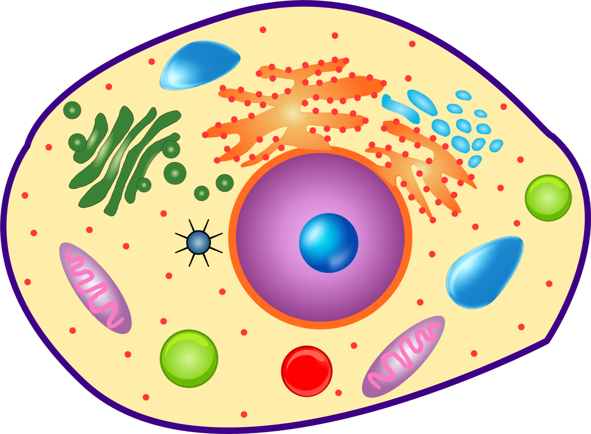
- The eukaryotic cell's organelles each have an orthopaedic role: the NUCLEUS holds DNA; ROUGH ER + RIBOSOMES synthesise secreted proteins (collagen-producing chondrocytes/osteoblasts are rich in them); the GOLGI modifies and packages; MITOCHONDRIA make ATP and trigger intrinsic apoptosis; LYSOSOMES degrade material (the osteoclast resorbs bone with lysosomal enzymes).
- The CELL CYCLE runs G1 -> S (DNA replication) -> G2 -> M (mitosis), regulated by CYCLINS/CDKs and policed at CHECKPOINTS (G1/S and G2/M) by tumour suppressors p53 and Rb; loss of this control underlies oncogenesis (relevant to bone/soft-tissue tumours).
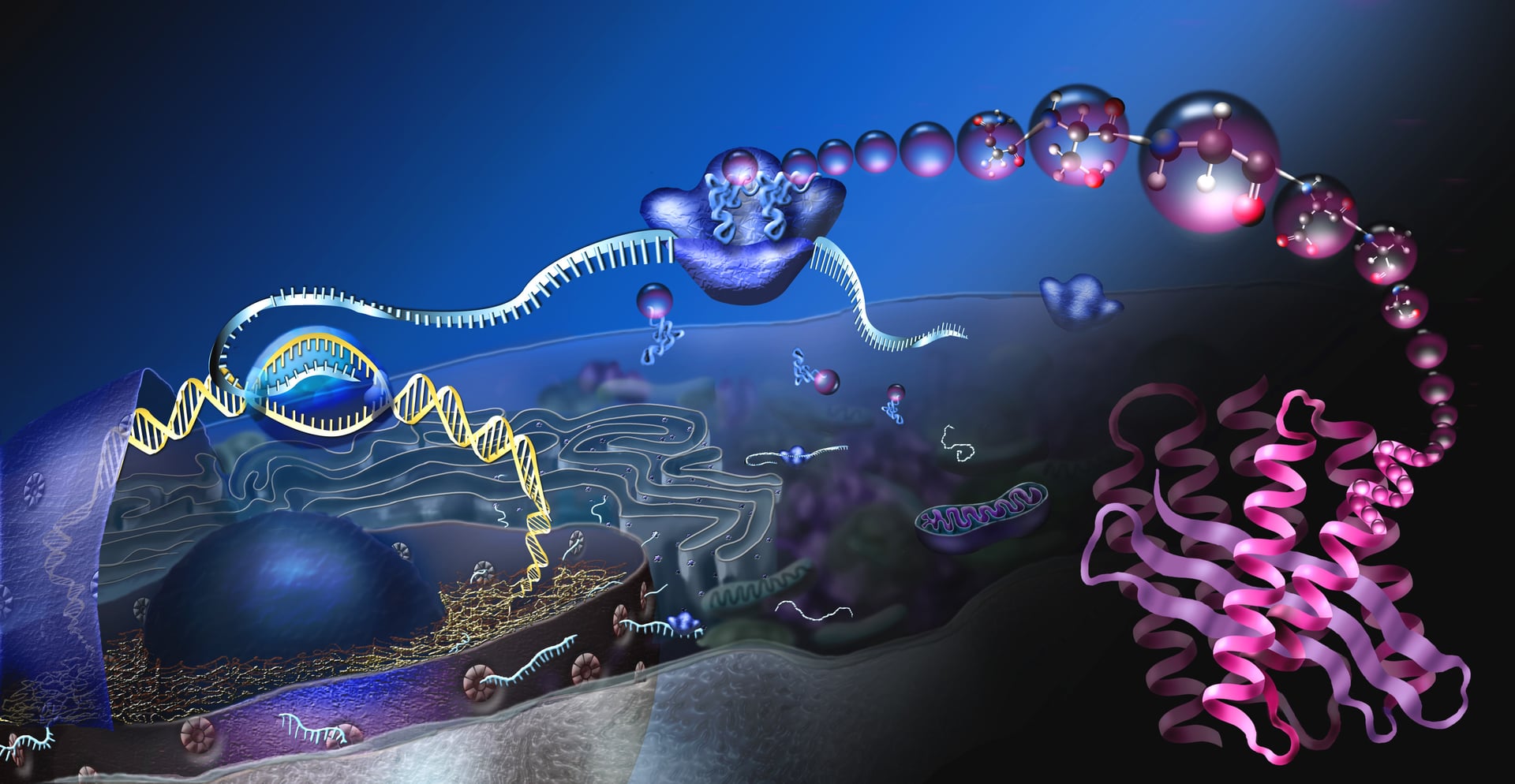
- The CENTRAL DOGMA is DNA -> (transcription) -> mRNA -> (translation by ribosomes) -> protein; a mutation at any step yields an abnormal protein - e.g. type I collagen gene (COL1A1/COL1A2) mutations cause osteogenesis imperfecta, FGFR3 causes achondroplasia, COMP causes pseudoachondroplasia.
- COLLAGEN is the orthopaedic exemplar of protein synthesis: a triple helix with a repeating GLYCINE-X-Y sequence, requiring POST-TRANSLATIONAL prolyl and lysyl HYDROXYLATION (vitamin C-dependent - hence scurvy), glycosylation, triple-helix assembly, secretion as procollagen, cleavage, and CROSS-LINKING; type I dominates bone/tendon, type II cartilage.
- Cell death comes in two principal forms: APOPTOSIS is programmed, energy-dependent and 'tidy' (cell shrinkage, no inflammation - via the intrinsic/mitochondrial and extrinsic/death-receptor pathways converging on caspases), whereas NECROSIS is passive cell swelling and lysis that provokes INFLAMMATION; apoptosis is essential physiologically (e.g. hypertrophic chondrocytes at the growth plate) and dysregulated in disease (e.g. chondrocyte apoptosis in osteoarthritis).
- Cells communicate by SIGNALLING - endocrine (bloodborne hormones), PARACRINE (local, e.g. growth factors in fracture healing), AUTOCRINE (self) and direct contact - acting through receptors and intracellular cascades (the substrate of the bone-signalling pathways).
- “Collagen needs vitamin C for prolyl/lysyl hydroxylation - this is why scurvy impairs collagen and causes bone/vascular fragility.
- “Apoptosis = programmed, ATP-dependent, NO inflammation (intrinsic mitochondrial + extrinsic death-receptor pathways, caspases); necrosis = passive, inflammatory.
- “Central dogma in one line: DNA -> mRNA (transcription) -> protein (translation); mutations explain the molecular dysplasias (COL1A1, FGFR3, COMP).
Active, ATP-dependent, regulated cell death: cell shrinkage, chromatin condensation, membrane blebbing, apoptotic bodies cleared by phagocytes - NO inflammation. Via the intrinsic (mitochondrial) and extrinsic (death-receptor) pathways converging on caspases. Physiological (e.g. growth-plate hypertrophic chondrocytes).
Passive, unregulated death from injury/ischaemia: cell swelling, membrane rupture, spillage of contents -> INFLAMMATION. Seen in infarction/AVN, severe trauma and infection.
The Cell & Its Organelles
The eukaryotic cell compartmentalises its work. The nucleus stores DNA and is the site of transcription; the rough endoplasmic reticulum, studded with ribosomes, synthesises secreted and membrane proteins; the smooth ER handles lipids and calcium; the Golgi apparatus modifies, sorts and packages proteins; mitochondria generate ATP and release factors that trigger intrinsic apoptosis; and lysosomes degrade material with acid hydrolases. In musculoskeletal tissue these are specialised: chondrocytes and osteoblasts have prominent rough ER and Golgi to manufacture and secrete the collagen-rich extracellular matrix, while the osteoclast is a multinucleated cell that resorbs bone using lysosomal enzymes (and acid) delivered at its ruffled border.
The Cell Cycle
Dividing cells progress through interphase - G1 (growth), S (DNA synthesis/replication), G2 (growth/preparation) - and then M (mitosis); non-dividing cells rest in G0. Progression is driven by cyclins and cyclin-dependent kinases (CDKs) and policed at checkpoints (principally G1/S and G2/M) that verify DNA integrity and replication before the cell commits. The tumour suppressors p53 (the 'guardian of the genome', which arrests the cycle or triggers apoptosis when DNA is damaged) and Rb are central. Loss of cell-cycle control - through oncogene activation or tumour-suppressor loss - is the basis of oncogenesis, relevant to bone and soft-tissue tumours.
The Central Dogma
Genetic information flows DNA -> RNA -> protein. In the nucleus, a gene's DNA is transcribed by RNA polymerase into messenger RNA (mRNA), which is processed (splicing, capping, poly-A tail) and exported to the cytoplasm. There, ribosomes translate the mRNA codons into a chain of amino acids (translation), using transfer RNA. The polypeptide then folds and undergoes post-translational modification into a functional protein. A mutation at any point yields an abnormal protein and disease - the molecular basis of many orthopaedic conditions: COL1A1/COL1A2 (osteogenesis imperfecta), FGFR3 (achondroplasia), COMP (pseudoachondroplasia/MED), EXT1/2 (multiple hereditary exostoses).
Collagen - The Orthopaedic Protein
Collagen is the dominant structural protein of the musculoskeletal system and the best orthopaedic example of protein synthesis and post-translational modification:
- Transcription/translation of pro-alpha chains on the rough ER, each with the repeating glycine-X-Y sequence (X often proline, Y often hydroxyproline).
- Hydroxylation of proline and lysine residues - which requires VITAMIN C as a cofactor (its lack in scurvy impairs collagen and causes bleeding and poor healing).
- Glycosylation and assembly of three chains into the triple helix (procollagen).
- Secretion, then cleavage of the propeptides to form tropocollagen.
- Fibril assembly and CROSS-LINKING (lysyl oxidase) for tensile strength. There are 28 collagen types; the fibrillar collagens (types I, II, III, V, XI...) build the load-bearing frameworks. Type I predominates in bone, tendon and skin; type II in hyaline cartilage.
| 0 | 1 | 2 |
|---|---|---|
| I | Bone, tendon, ligament, skin, annulus fibrosus | Most abundant; defective in osteogenesis imperfecta |
| II | Hyaline (articular) cartilage, nucleus pulposus | Cartilage; defective in some chondrodysplasias |
| III | Early wound/granulation tissue, vessels | Laid down early in healing, replaced by type I |
| V / XI | Regulate fibril diameter (with I / II) | Co-assemble with the major fibrillar collagens |
| X | Hypertrophic zone of the growth plate | Marker of hypertrophic chondrocytes (endochondral ossification) |
Cell Death & Cell Signalling
Apoptosis is programmed, ATP-dependent and tightly regulated: the cell shrinks, chromatin condenses, the membrane blebs into apoptotic bodies that are cleared by phagocytes without inflammation. It proceeds via the intrinsic (mitochondrial, Bcl-2 family/cytochrome c) and extrinsic (death-receptor, e.g. Fas) pathways, both converging on caspases. Necrosis is passive death from injury/ischaemia - the cell swells, the membrane ruptures and contents spill, provoking inflammation. Apoptosis is essential physiologically (hypertrophic chondrocytes apoptose at the growth plate) and is dysregulated in disease - e.g. chondrocyte apoptosis contributes to osteoarthritis, and necrosis dominates in osteonecrosis/infarction.
Evidence & Key Studies
Fibrillar collagens
- The fibrillar collagens (types I, II, III, V, XI, XXIV, XXVII) provide three-dimensional frameworks conferring mechanical strength; humans have 28 collagen types.
- The triple-helix structure depends on the amino-acid sequence (Gly-X-Y), hydrogen bonding and post-translational modifications such as prolyl 4-hydroxylation.
- Biosynthesis involves prolyl/lysyl hydroxylation, chaperones, trimerisation of procollagen, proteolytic maturation, fibril assembly and cross-linking.
The role of apoptosis in the pathogenesis of osteoarthritis
- Apoptosis is an important physiological process for development and tissue homeostasis; dysregulated chondrocyte apoptosis contributes to osteoarthritis (cartilage degeneration).
- Inflammatory mediators (ROS, NO, IL-1beta, TNF-alpha, Fas) and signalling pathways (NF-kappaB, Wnt, Notch) drive chondrocyte apoptosis and matrix degradation.
- Illustrates how core cell-death biology connects directly to a common orthopaedic disease.
According to PubMed, the collagen structure/biosynthesis facts come from the cited fibrillar-collagen review and the apoptosis-in-OA links from the cited International Orthopaedics review. The general cell-biology content (organelles, cell cycle, central dogma) is standard textbook science; the disease links (COL1A1, FGFR3, COMP) are covered in our Genetics, Bone Signaling and dysplasia topics.
Clinical Decision Scenarios
Practise clinical reasoning and management decisions out loud
“Walk me through the central dogma and use collagen to illustrate how a protein is made and modified. Why is vitamin C relevant?”
“Distinguish apoptosis from necrosis, and give orthopaedic examples of each. How do cells signal to one another?”
Mnemonics & Memory Aids
COLLAGEN
Hook:COLLAGEN: chains, Gly-X-Y, vitamin-C hydroxylation, triple helix, cross-linking - type I bone, type II cartilage.
APOPTOSIS
Hook:APOPTOSIS is active, orderly and caspase-driven with no inflammation - necrosis is the messy, inflammatory opposite.
Organelles (ortho roles)
- Nucleus = DNA/transcription; rough ER + ribosomes = protein synthesis (collagen)
- Golgi = modify/package; mitochondria = ATP + intrinsic apoptosis
- Lysosomes = degradation (osteoclast bone resorption)
Cell cycle & central dogma
- G1 -> S -> G2 -> M; checkpoints (G1/S, G2/M); cyclins/CDKs; p53 & Rb
- DNA -> mRNA (transcription) -> protein (translation)
- Mutations -> disease: COL1A1 (OI), FGFR3 (achondroplasia), COMP (pseudoachondroplasia)
Collagen
- Gly-X-Y triple helix; prolyl/lysyl hydroxylation needs VITAMIN C (scurvy)
- Procollagen -> secreted -> cleaved -> fibril -> cross-linked (lysyl oxidase)
- Type I bone/tendon, II cartilage, III early healing, X hypertrophic physis
Cell death & signalling
- Apoptosis: programmed, ATP-dependent, caspases, NO inflammation (intrinsic + extrinsic)
- Necrosis: passive, swelling/lysis, inflammation (osteonecrosis, infarction)
- Signalling: endocrine / paracrine (fracture healing) / autocrine / juxtacrine