A low-grade cartilage tumour with an abrupt transition to a juxtaposed high-grade non-cartilaginous sarcoma — the most lethal chondrosarcoma variant
- Defining histology is an ABRUPT junction between low-grade cartilage and a high-grade non-cartilaginous sarcoma — not a gradual grade transition.
- Both components share clonal genetic changes, including IDH1/IDH2 mutations, supporting a single precursor clone rather than collision of two tumours.
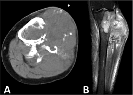
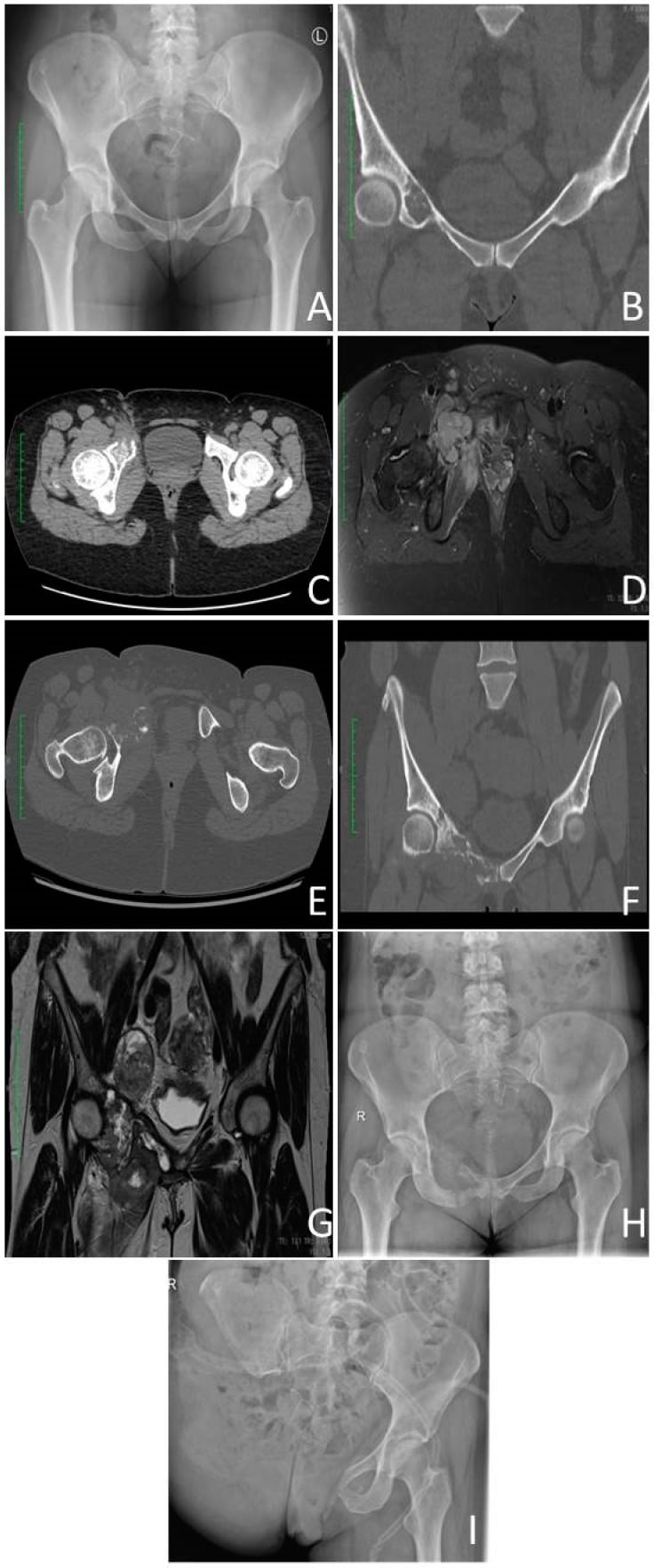
- Imaging is bimorphic: a ring-and-arc mineralised cartilage component adjacent to a purely lytic destructive area with cortical breach and soft-tissue mass.
- The biopsy must sample the LYTIC component. Sampling only the mineralised area yields low-grade cartilage and understages the tumour.
- Prognosis is dismal — early lung metastasis, median survival often under two years; local control alone rarely alters outcome.
- Pathological fracture is common at presentation and complicates limb salvage.
- “Longstanding dull ache for years that suddenly accelerates over weeks in an older patient equals dedifferentiation until proven otherwise.
- “A rising, expanding lytic area within a known atypical cartilaginous tumour on surveillance is the classic dedifferentiation signal.
- “Chemotherapy is often given to fit patients using osteosarcoma-type regimens, but registry and multicentre data show at best modest and inconsistent survival gain.
- “Radiotherapy is used for inoperable pelvic disease and palliation, not as a curative substitute for resection.
- “Amputation may be the only wide-margin option in a large pelvic or proximal femoral tumour with fracture haematoma contamination.
Image-guided biopsy targeted at the non-mineralised, destructive, contrast-enhancing component. A needle passed through the calcified cartilage returns bland low-grade cartilage and the patient is treated as a grade 1 lesion — a catastrophic understaging error. Plan the tract so it is excisable with the specimen.
Intralesional surgery for what is assumed to be an atypical cartilaginous tumour disseminates high-grade sarcoma. Any lytic, cortically destructive or soft-tissue-forming area within a cartilage lesion mandates staging and biopsy before any intralesional procedure.
Fracture haematoma contaminates compartments and neurovascular planes, converts a planned limb salvage to amputation in a substantial minority, and independently worsens survival. Never internally fix an undiagnosed destructive lesion in an older adult.
CT chest at presentation is mandatory; a significant proportion already have pulmonary metastases. Discovering metastatic disease after a morbid internal hemipelvectomy is an avoidable tragedy — staging precedes any resection planning.
Definition, Biology and Why It Matters
- Conventional chondrosarcoma grade 2 to 3
- Cartilaginous throughout, increasing cellularity, nuclear atypia and myxoid change
- Dedifferentiated chondrosarcoma
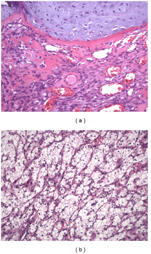
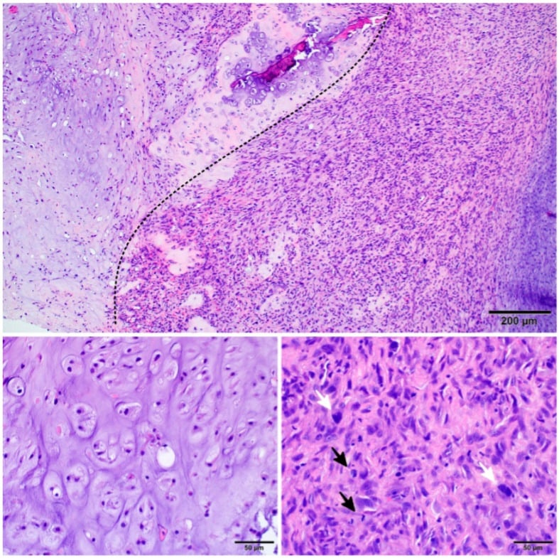
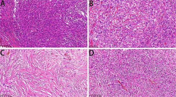
- Low-grade cartilage neoplasm abutting a high-grade non-cartilaginous sarcoma with an abrupt, sharply demarcated interface
- Mesenchymal chondrosarcoma
- Bimorphic small round blue cells with islands of well-differentiated hyaline cartilage
- Conventional chondrosarcoma grade 2 to 3
- Gradual grade progression, phenotype stays cartilaginous
- Dedifferentiated chondrosarcoma
- Knife-edge boundary with complete loss of cartilaginous phenotype, not simply grade progression
- Mesenchymal chondrosarcoma
- Intimate admixture of the two elements, not an abrupt front
- Conventional chondrosarcoma grade 2 to 3
- None, remains chondroid
- Dedifferentiated chondrosarcoma
- Most often undifferentiated pleomorphic sarcoma or fibrosarcoma-like spindle cell sarcoma; may be osteosarcomatous, rhabdomyosarcomatous or angiosarcomatous
- Mesenchymal chondrosarcoma
- Primitive mesenchymal round cell component
- Conventional chondrosarcoma grade 2 to 3
- IDH1 and IDH2 hotspot mutations in central tumours
- Dedifferentiated chondrosarcoma
- Clonally related components; shared IDH1 and IDH2 mutations in both elements in the majority, supporting dedifferentiation of one clone rather than a collision tumour, with TP53 alteration, CDKN2A loss and complex karyotype accumulating in the high-grade element
- Mesenchymal chondrosarcoma
- Distinct entity, not IDH driven
- Conventional chondrosarcoma grade 2 to 3
- Older adult, central skeleton and long bones
- Dedifferentiated chondrosarcoma
- De novo at presentation, within a pre-existing central chondrosarcoma or atypical cartilaginous tumour, in enchondromatosis (Ollier disease, Maffucci syndrome), or rarely in secondary peripheral chondrosarcoma from an osteochondroma
- Mesenchymal chondrosarcoma
- Younger patients, bone and extraskeletal sites
- Conventional chondrosarcoma grade 2 to 3
- Ring and arc chondroid matrix with endosteal scalloping
- Dedifferentiated chondrosarcoma
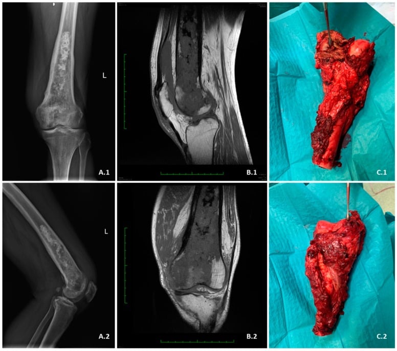
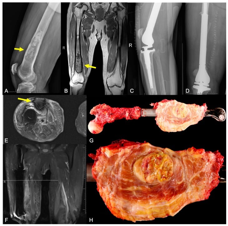
- Bimorphic lesion: calcified chondroid zone plus an adjacent lytic, destructive, soft tissue mass; the cartilage makes it look deceptively benign
- Mesenchymal chondrosarcoma
- Chondroid calcification within an aggressive round cell mass
- Conventional chondrosarcoma grade 2 to 3
- Wide surgical resection, chemoresistant and radioresistant
- Dedifferentiated chondrosarcoma
- Behaves as a high-grade pleomorphic sarcoma of bone in an older adult superimposed on a chemoresistant cartilage matrix; survival tracks the high-grade component and the cartilage component is prognostically irrelevant
- Mesenchymal chondrosarcoma
- Chemosensitive, treated as a round cell sarcoma with surgery
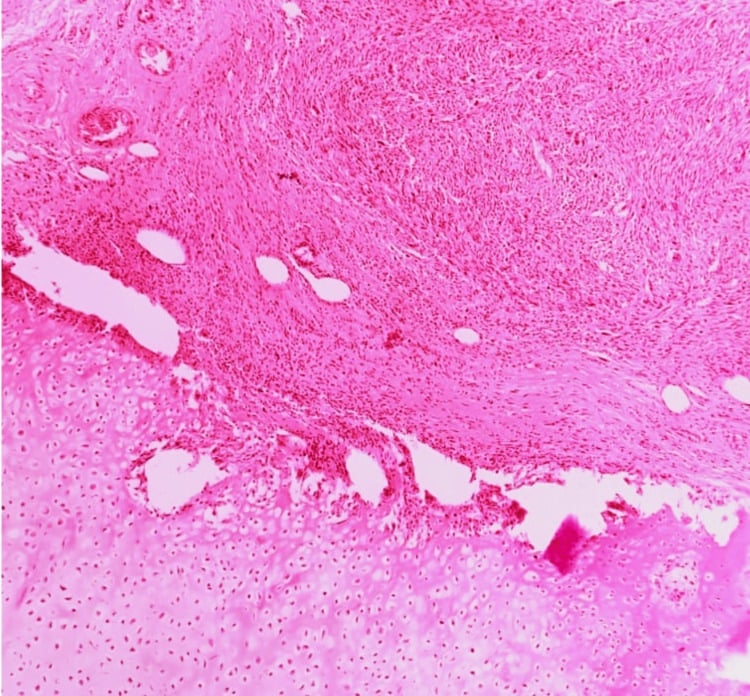
Dedifferentiated chondrosarcoma is defined by the juxtaposition of a low-grade cartilaginous neoplasm and a high-grade non-cartilaginous sarcoma with an abrupt, sharply demarcated interface. The high-grade component is most often an undifferentiated pleomorphic sarcoma or fibrosarcoma-like spindle cell sarcoma, but may be osteosarcomatous, rhabdomyosarcomatous or angiosarcomatous.
Key biological points
- The two components are clonally related. Shared mutations, including IDH1 and IDH2 hotspot mutations typical of conventional central cartilage tumours, are found in both the cartilage and dedifferentiated components in the majority of cases. This supports dedifferentiation of one clone, not a collision tumour.
- Additional events (TP53 alteration, CDKN2A loss, complex karyotype) accumulate in the high-grade element.
- Dedifferentiation may arise de novo at presentation, within a pre-existing central chondrosarcoma or atypical cartilaginous tumour, in enchondromatosis (Ollier disease, Maffucci syndrome), or in secondary peripheral chondrosarcoma from osteochondromas — the latter rarer.
- It is not simply "grade progression". A grade 1 lesion becoming grade 3 remains cartilaginous with a gradual transition; dedifferentiation means loss of cartilaginous phenotype altogether with a knife-edge boundary.
Clinical consequence: the biology of the tumour is that of a high-grade pleomorphic sarcoma of bone in an older adult, superimposed on a chemoresistant cartilage matrix. Survival tracks the high-grade component; the cartilage component is prognostically irrelevant except that it makes the tumour look deceptively benign.
Epidemiology and Presentation
- Accounts for roughly 10 to 15 percent of all chondrosarcomas.
- Peak in the sixth and seventh decades; rare before 40. Slight male predominance in most series.
- Sites in descending frequency: pelvis (especially periacetabular ilium), proximal femur, femoral diaphysis/distal femur, proximal humerus, scapula, ribs. Hands and feet almost never.
- Risk contexts: known central chondrosarcoma under surveillance, Ollier disease and Maffucci syndrome (lifetime malignant transformation risk substantial, and dedifferentiation reported), multiple hereditary exostoses (rarer route).
Staging and Prognostic Factors
- Enneking (MSTS) system: essentially all are stage IIB (high grade, extracompartmental) or III if metastatic.
- AJCC staging applies but adds little to decision-making beyond metastatic status.
Adverse prognostic factors
- Effect
- Dominant adverse factor; median survival months
- Effect
- Worse than appendicular — resection margins harder
- Effect
- Contamination, higher amputation rate, worse survival
- Effect
- Higher local recurrence and reduced survival
- Effect
- Associated with poorer outcome in several series
- Effect
- Limits ability to deliver chemotherapy or major resection
Achieving a wide margin without metastatic disease is the only variable consistently associated with prolonged survival. Local control by itself, in the presence of pulmonary metastases, does not rescue the patient — which is why the metastatic work-up must precede a decision about internal hemipelvectomy or amputation.
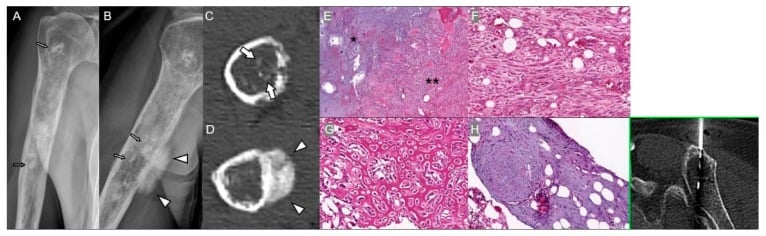
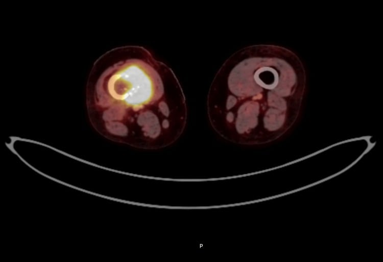
Imaging: The Bimorphic Hallmark
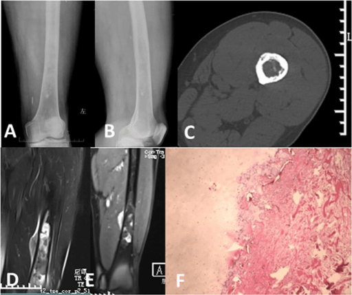
Sequenced imaging workup in dedifferentiated chondrosarcoma
Obtain orthogonal plain films of the whole involved bone, plus an AP pelvis for pelvic lesions.
Look for two adjacent but different territories in the same bone:
- Appearance
- Ring-and-arc / popcorn / stippled chondroid mineralisation, endosteal scalloping, fusiform expansion, relatively defined margins
- Appearance
- Purely lytic, geographic to permeative, no matrix, cortical destruction, periosteal reaction, extraosseous mass
The abrupt change from a mineralised, chronic-looking region to an aggressive lytic region in the same bone is the single most useful radiographic sign. Endosteal scalloping greater than two thirds of cortical thickness, cortical breach and any soft-tissue mass push a cartilage lesion out of the "atypical cartilaginous tumour" category.
If PET, dynamic MRI or plain films show a discrete lytic/solid area, biopsy must be steered there. A report of "low-grade cartilage tumour" from a needle placed in the calcified region does not exclude dedifferentiation. When histology and imaging disagree, the imaging wins — repeat the biopsy.
Biopsy: Technique and Rules
- 1Step 1 — Cartilage lesion identified
Suspicious cartilage-forming lesion on radiograph or MRI with rings-and-arcs mineralisation.
Biopsy haemorrhage and oedema degrade later MRI and obscure the true tumour boundary, so imaging must be banked first.
- 2Step 2 — Is there a lytic, non-mineralised area?
Scrutinise for a destructive lytic zone or soft-tissue mass abutting the mineralised cartilage — the bimorphic 'abrupt transition' sign.
The high-grade dedifferentiated component lives in the lytic zone, not in the calcified cartilage.
- 3Step 3 — Who performs the biopsy
Biopsy is done by, or in direct consultation with, the surgeon who will resect.
Reduces unplanned excisions and amputations.
- 4Step 4 — Target selection
Core needle biopsy under CT or ultrasound guidance.
Sampling only the cartilaginous portion yields a falsely low-grade diagnosis.
- 5Step 5 — Tract discipline
Longitudinal approach, single tract, shortest route through one compartment.
The tract is excised en bloc at definitive surgery.
- 6Step 6 — Does histology match the imaging?
Expect two populations with an abrupt border. Immunohistochemistry supports the dedifferentiated line: desmin and myogenin if rhabdomyosarcomatous; S100 absent in the high-grade area whereas the cartilage is S100 positive. IDH1/IDH2 mutation testing supports cartilage lineage and separates dedifferentiated chondrosarcoma from primary osteosarcoma or undifferentiated pleomorphic sarcoma of bone.
Concordant high-grade result confirms the diagnosis.
- 7Step 7 — Discordant low-grade report
A low-grade or benign cartilage result in the face of a destructive lytic zone means the lytic area was missed.
Imaging overrules a discordant low-grade histology report: a discordant benign result triggers re-biopsy, not reassurance.
Full imaging of the primary and chest CT first. Biopsy artefact (haemorrhage, oedema) degrades subsequent MRI interpretation and can obscure the true tumour boundary.
The tract must lie within the planned resection or flap sacrifice zone. Referral to a sarcoma unit before biopsy reduces unplanned excisions and amputations.
Core needle biopsy under CT or ultrasound guidance directed at the soft-tissue mass or lytic intraosseous area, avoiding necrotic centres. Take multiple cores; send fresh tissue for cytogenetics/molecular work and material for culture if infection is a differential.
Longitudinal approach, single tract, shortest route through one compartment, avoid neurovascular bundles and joints, mark the skin entry with a permanent marker/tattoo or note it precisely.
Discuss at sarcoma MDT with radiology and pathology together. A discordant benign result triggers re-biopsy, not reassurance.
Histology to expect: two populations with an abrupt border. Immunohistochemistry supports the dedifferentiated line (desmin/myogenin if rhabdomyosarcomatous, absent S100 in the high-grade area whereas the cartilage is S100 positive). IDH1/IDH2 mutation testing supports cartilage lineage and can help separate dedifferentiated chondrosarcoma from primary osteosarcoma or undifferentiated pleomorphic sarcoma of bone.
ABRUPTRecognising dedifferentiation
Hook:Everything about this tumour is ABRUPT — the histology, the imaging boundary, the clinical change, and the decline.
Differential Diagnosis
- Discriminating features
- Cartilaginous throughout with gradual increase in cellularity and atypia; no abrupt non-cartilaginous component; still lobular high T2 signal
- Consequence of getting it wrong
- Better survival than dedifferentiated; chemotherapy not offered
- Discriminating features
- Younger patient; malignant osteoid produced directly by tumour cells throughout; cartilage is malignant, not low grade; no abrupt interface
- Consequence of getting it wrong
- Chemosensitive — neoadjuvant chemotherapy is standard and outcome far better
- Discriminating features
- Young adults; small round blue cells with haemangiopericytoma-like vessels and islands of well-formed hyaline cartilage; HEY1-NCOA2 fusion
- Consequence of getting it wrong
- Chemosensitive; systemic therapy genuinely indicated
- Discriminating features
- Multiple lesions, known primary, monoclonal protein; no chondroid matrix
- Consequence of getting it wrong
- Radiotherapy and fixation appropriate — but never fix a solitary destructive lesion without a diagnosis
- Discriminating features
- No cartilage component anywhere on imaging or in generous sampling; IDH wild type
- Consequence of getting it wrong
- Similar high-grade management; distinction is prognostic and academic in practice
- Discriminating features
- Serpiginous infarct margins or coarse trabecular Paget bone with lytic destruction
- Consequence of getting it wrong
- Different setting, similarly poor prognosis
Management: Decision Thresholds
- Confirm diagnosis with an adequately targeted core biopsy reviewed at a sarcoma MDT.
- Stage — CT chest, cross-sectional imaging of the primary, PET-CT where available.
- Is the patient fit and the disease localised?
- Yes and a wide margin is achievable with acceptable function → wide en-bloc resection with reconstruction.
- Yes but a wide margin needs sacrifice of critical structures (sciatic nerve plus iliac vessels, extensive fracture contamination) → amputation / external hemipelvectomy may be the only oncologically sound option; discuss frankly against palliative alternatives given prognosis.
- Unresectable primary (extensive intrapelvic, sacral, or vascular encasement in a frail patient) → palliative approach: radiotherapy, analgesia, stabilisation of impending fracture, early palliative care involvement.
- Metastatic at presentation → the balance shifts sharply towards symptom control. Resection of the primary is justified mainly for pain, fungation or impending fracture, not for cure.
- Chemotherapy — consider in fit patients with localised, high-volume dedifferentiated disease as part of an MDT decision and ideally within a trial; counsel honestly that the evidence is weak.
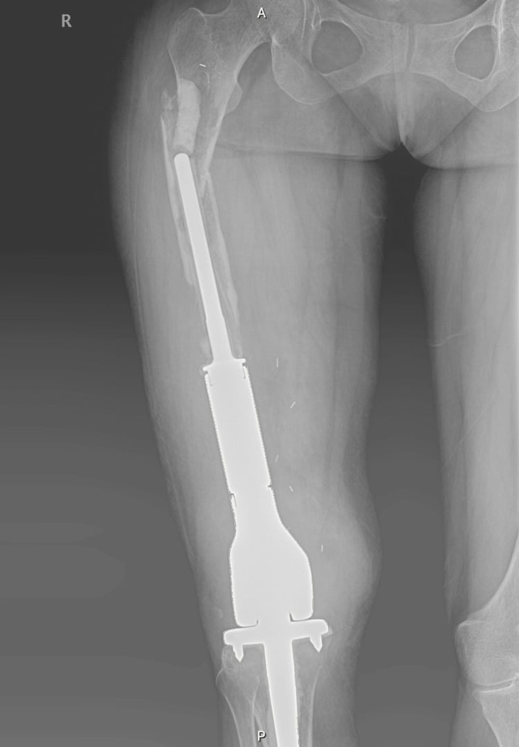
An older patient with a destructive proximal femoral lesion and a fracture is not automatically a metastasis. Intramedullary nailing of an undiagnosed dedifferentiated chondrosarcoma contaminates the entire femoral canal and converts a resectable tumour into a hindquarter or hip disarticulation problem. Investigate, biopsy, then decide.
STOPPre-treatment checklist before any bone lesion surgery in an older adult
Hook:STOP before the saw — the first operation determines whether cure is still possible.
Complications and Follow-Up
- Where it happens
- Needle biopsy of a large pelvic or femoral cartilage tumour
- Mechanism
- Sampling only the low-grade cartilage cap and missing the dedifferentiated high-grade component
- Avoid by
- Image-guided targeting of the lytic, non-cartilaginous, contrast-enhancing or PET-avid zone; multiple cores; correlate with imaging before accepting a low-grade result
- If it occurs
- Re-biopsy the aggressive area; treat as dedifferentiated and plan wide resection rather than intralesional surgery
- Where it happens
- Lesion mislabelled as enchondroma or atypical cartilaginous tumour
- Mechanism
- Intralesional surgery seeds the entire cavity and soft tissues with high-grade sarcoma
- Avoid by
- Never curette a cartilage lesion with cortical destruction, soft-tissue mass or a lytic component; pelvic and proximal femoral lesions merit staging first
- If it occurs
- Treat as an unplanned intralesional excision: restage with MRI and CT chest, then wide re-excision of the whole contaminated field or amputation
- Where it happens
- Proximal femur, periacetabular bone
- Mechanism
- Haematoma disseminates tumour along fascial planes; strongly associated with local recurrence and systemic disease
- Avoid by
- Protected weight-bearing and prompt definitive resection once high-grade disease is suspected; do not internally fix a suspicious lesion
- If it occurs
- Do not nail — resect en bloc with the fracture haematoma and any prior fixation tracks; endoprosthetic reconstruction
- Where it happens
- Lung, most within 12 to 24 months
- Mechanism
- The dominant cause of death; occult nodules present at diagnosis make radical local surgery futile
- Avoid by
- Staging CT chest before any resection; CT chest every 3 months for 2 years, 6-monthly to 5 years, then annually
- If it occurs
- Reassess intent of treatment; metastasectomy only for few, resectable, late nodules with a controlled primary, otherwise palliative and symptom-directed care
- Where it happens
- Internal hemipelvectomy and pelvic reconstruction
- Mechanism
- Nerve encased by tumour or divided to achieve a wide margin
- Avoid by
- Pre-operative MRI mapping of the neurovascular relationship; counsel and consent for deficit before surgery
- If it occurs
- Pre-warn the patient, provide ankle-foot orthosis or knee-extension orthosis and gait retraining; do not compromise the margin to spare the nerve
- Where it happens
- Non-specialist centre; previously irradiated or gluteal flap territory
- Mechanism
- Marginal or intralesional margins contaminate the field; superior gluteal artery sacrifice and prior radiotherapy cause flap necrosis and deep infection after custom pelvic implants
- Avoid by
- Refer all suspicious cartilage lesions to a sarcoma unit before biopsy; plastic surgical input at the planning stage; abduction bracing, constrained or dual-mobility bearings and mesh soft-tissue reconstruction to prevent instability
- If it occurs
- Wide re-excision of the whole surgical field; for deep infection, debridement with targeted antibiotics and implant removal with conversion to flail hip if uncontrolled; re-resection or amputation for isolated local recurrence only in the absence of metastases
- Notes and management
- Commonest after marginal/intralesional margins and after fracture; usually heralds systemic disease. Re-resection or amputation only if isolated and the patient has no metastases
- Notes and management
- The dominant cause of death; typically within 12 to 24 months. Metastasectomy considered only for few, resectable, late nodules with controlled primary
- Notes and management
- High rates reported after custom pelvic implants; manage with debridement, targeted antibiotics, and implant removal with conversion to flail hip if uncontrolled
- Notes and management
- Abduction bracing, constrained or dual-mobility bearings, soft-tissue reconstruction with mesh
- Notes and management
- Superior gluteal artery sacrifice, prior radiotherapy; plastic surgical involvement at the planning stage
- Notes and management
- Sciatic or femoral nerve sacrifice — pre-warn the patient, provide orthoses and gait retraining
- Notes and management
- Anthracycline cardiotoxicity, nephrotoxicity, myelosuppression — significant in the seventh decade; weigh against unproven benefit
Surveillance: clinical review with local imaging and CT chest every 3 months for 2 years, then 6-monthly to 5 years, then annually — reflecting the front-loaded risk. In practice surveillance often shifts to symptom-directed and supportive care once metastases appear.
Guidelines, Registries & Global Practice
Global epidemiology. Chondrosarcoma is the second or third commonest primary bone sarcoma worldwide and is proportionally more important in ageing populations; the dedifferentiated variant, at roughly 10 to 15 percent of chondrosarcomas, is therefore encountered in almost every regional sarcoma unit but rarely in general orthopaedic practice. Incidence appears broadly similar across regions; differences in reported outcome largely reflect stage at presentation and access to specialist surgery.
Society guidance — where it genuinely applies
- What it says relevant to this topic
- Management of bone sarcoma in reference centres; biopsy by the treating team; wide surgical excision is the mainstay for chondrosarcoma; chemotherapy of unproven benefit in conventional and dedifferentiated chondrosarcoma outside trials
- What it says relevant to this topic
- Dedifferentiated chondrosarcoma may be treated along osteosarcoma lines in selected fit patients; wide excision remains primary; radiotherapy for unresectable or close-margin disease
- What it says relevant to this topic
- Any suspicious bone lesion referred to a sarcoma diagnostic service before biopsy; imaging pathway with plain radiograph then MRI; strict prohibition on local biopsy or excision of suspected bone sarcoma
- What it says relevant to this topic
- Defines dedifferentiated chondrosarcoma by the abrupt bimorphic pattern; separates it from grade 3 conventional and mesenchymal chondrosarcoma
- What it says relevant to this topic
- Emphasise centralisation of bone sarcoma surgery and multidisciplinary decision-making
Registry evidence. Bone sarcoma registries and national cancer datasets (for example the Netherlands and Scandinavian bone tumour registries, and large population datasets) consistently show that dedifferentiated chondrosarcoma has the worst survival of all chondrosarcoma subtypes, that pelvic location and metastatic presentation dominate outcome, and that treatment at high-volume centres is associated with better margin rates. Arthroplasty registries (NJR, AJRR, AOANJRR, SHAR, NZJR) contribute mainly by documenting the high revision, infection and dislocation burden of tumour endoprostheses and custom pelvic reconstructions relative to primary arthroplasty — data worth quoting when counselling about reconstruction.
Practice variation by resource setting.
- High-resource centres: navigation and 3D-printed patient-specific guides and implants, particle therapy for unresectable axial disease, molecular profiling and trial access.
- Middle-resource settings: wide resection with off-the-shelf endoprostheses, allografts or hip transposition; radiotherapy for unresectable disease; chemotherapy availability variable.
- Low-resource settings: late presentation with fungating or fractured tumours; amputation or hindquarter amputation is often the only feasible oncological operation, and palliative care provision becomes the central issue. Prognostic honesty and pain control are universal obligations regardless of setting.
Controversies & Areas of Uncertainty
- Does chemotherapy help? The central controversy. Retrospective data conflict; there is no randomised evidence. Some centres treat all fit patients as high-grade osteosarcoma, others reserve chemotherapy for trials only. Selection bias plausibly explains most reported benefit.
- Radical surgery versus palliation in metastatic disease. With a median survival often under a year in metastatic cases, an internal or external hemipelvectomy may consume the patient's remaining functional life. Some units offer it for uncontrolled local symptoms only.
- Reconstruct or not after pelvic resection. Custom implants restore better limb length and function but carry high infection and mechanical failure rates; hip transposition and flail hip are lower-morbidity options that some argue better suit a short prognosis.
- Margin width. No agreed millimetre threshold. Contaminated fields after fracture or prior surgery make "wide" a judgement rather than a measurement.
- Surveillance of atypical cartilaginous tumours. How intensively and for how long to image indolent cartilage lesions to catch dedifferentiation early is unsettled; over-surveillance is costly and anxiety-provoking, under-surveillance misses the treatable window — and it is unproven that earlier detection of dedifferentiation improves survival.
- Role of particle therapy and IDH-directed agents. Promising but with limited access and immature outcome data.
MCQ Practice Points
Q: What single histological feature defines dedifferentiated chondrosarcoma?
A: An abrupt transition between a low-grade cartilaginous tumour and a high-grade non-cartilaginous sarcoma. The criterion is about the interface, not about either component: a gradual increase in cellularity and atypia within cartilage is high-grade conventional chondrosarcoma, not dedifferentiation.
Q: Which area must be biopsied and why?
A: The lytic, non-mineralised, avidly enhancing area. Sampling the calcified cartilage yields low-grade histology and understages the tumour, risking inappropriate intralesional treatment. Mercuri's point sharpens this: the non-cartilaginous component may be a very small fraction of the tumour, so a core through cartilage is a negative test that carries no information.
Q: What is the approximate five-year survival?
A: Grim, and quoted across series as roughly 10 to 25 per cent. The one figure you can attach to a named series is Mercuri's 13 per cent in 74 cases at the Rizzoli Institute. Early pulmonary metastasis is the dominant cause of death. What improved survival in that series was prompt diagnosis and achievement of a wide or radical margin, which is also why pelvic tumours do worse — the margin is harder to get, not the biology different.
Q: What molecular finding supports a common clonal origin of both components?
A: Identical IDH1 or IDH2 hotspot mutations in both the cartilaginous and the dedifferentiated component. Yang's paired-component study adds the more direct evidence: the same X-chromosome inactivation pattern in both halves of the same tumour, which is evidence against a collision tumour rather than merely consistent with a shared origin.
Q: A young adult has a bone tumour with small round cells and islands of hyaline cartilage. What is the diagnosis?
A: Mesenchymal chondrosarcoma, characterised by the HEY1-NCOA2 fusion. It is treated with multi-agent chemotherapy and carries a better prognosis than dedifferentiated disease — but do not call it "chemosensitive" as though a survival benefit were established: Xu's systematic review of 107 patients (PMID 25849226) found anthracycline-based chemotherapy did not improve overall survival, and it was the negative margin that predicted local control and event-free survival. Its biphasic pattern is small round cells beside cartilage — not the abrupt low-grade-cartilage-to-high-grade-sarcoma interface of dedifferentiation. Age separates them too: mesenchymal is a young adult tumour, dedifferentiated is a tumour of the fifth decade onwards.
Q: What is the role of radiotherapy?
A: Non-curative. It is used for unresectable axial or pelvic disease, close or positive margins, and palliation of pain or impending fracture; particle therapy is used for unresectable axial disease in selected centres. Mavrogenis states the same limit plainly — high-dose radiotherapy is confined to inaccessible sites and to palliation.
Exam Viva Scenarios
Practise clinical reasoning and management decisions out loud
“A 68-year-old man has had a dull right hip ache for four years, previously labelled osteoarthritis. Over ten weeks the pain has become severe and wakes him at night. Radiographs show a proximal femoral lesion with ring-and-arc mineralisation in the diaphysis and an adjacent purely lytic area with cortical destruction in the subtrochanteric region. How do you proceed?”
“A CT-guided core biopsy of a large periacetabular lesion in a 62-year-old woman reports 'low-grade cartilage neoplasm, features of atypical cartilaginous tumour'. The MRI, however, shows a 9 cm avidly enhancing solid extraosseous mass beside a lobulated calcified intraosseous component. The referring team proposes curettage and cement. What is your response?”
“A 70-year-old woman presents to the emergency department with a displaced femoral neck fracture after a trivial stumble. Radiographs of the pelvis show chondroid calcification in the femoral neck and a lytic destructive area extending into the intertrochanteric region. She is already scheduled for hemiarthroplasty. What do you do?”
“A fit 58-year-old man with a localised dedifferentiated chondrosarcoma of the ilium asks whether he should have chemotherapy, having read that osteosarcoma patients do well with it. How do you counsel him?”
Definition and biology
- Low-grade cartilage tumour with ABRUPT junction to high-grade non-cartilaginous sarcoma
- High-grade element: undifferentiated pleomorphic sarcoma, fibrosarcoma-like, osteosarcomatous or rhabdomyosarcomatous
- Single clone — shared IDH1/IDH2 mutations in both components
- 10 to 15 percent of all chondrosarcomas; sixth to seventh decade; pelvis and femur
Presentation
- Years of indolent ache then abrupt escalation over weeks
- Rest and night pain, rapidly enlarging deep mass
- Pathological fracture in up to a third at presentation
- Arises de novo, in existing chondrosarcoma, or in Ollier/Maffucci disease
Imaging
- Radiograph: ring-and-arc mineralisation beside a purely lytic destructive area with cortical breach
- MRI: lobulated high T2 cartilage with septal enhancement, adjacent solid avidly enhancing mass
- CT primary for matrix and pelvic geometry; CT chest mandatory
- PET-CT identifies the hottest area as the biopsy target
Biopsy rules
- Stage before biopsy; biopsy by or with the resecting surgeon
- Target the lytic/solid enhancing component
- Single longitudinal tract, excisable en bloc, avoid neurovascular bundles and joints
- Discordant benign result equals re-biopsy, not reassurance
Management
- Wide en-bloc resection is the only curative-intent treatment
- Amputation or hindquarter amputation where wide margin otherwise unachievable
- Chemotherapy: osteosarcoma-type regimens in fit patients, weak and conflicting evidence, prefer trials
- Radiotherapy for unresectable disease, close margins and palliation
- No curettage, no cementing, no nailing of an undiagnosed lesion
Prognosis and pitfalls
- Five-year survival roughly 10 to 25 percent; early lung metastasis
- Adverse: metastasis at diagnosis, pelvic site, fracture, positive margin, large dedifferentiated volume
- Surveillance: clinical review and CT chest 3-monthly for 2 years then tapering
- Key error: treating it as a grade 1 cartilage lesion because the biopsy missed the lytic zone
Evidence Base
Dedifferentiation of Low-Grade Chondrosarcomas
- Named the entity: a low-grade cartilage tumour abutting an anaplastic non-cartilaginous sarcoma, with an abrupt interface rather than a gradual change in grade.
- The indexed sites span the skeleton the page describes - femur, humerus, tibia, scapula, ribs and lumbar spine - and amputation is indexed as a treatment, which is what the outlook was in 1971.
- Framed the lesion as a change in behaviour within a known cartilage tumour, not as a new tumour arising beside one.
Dedifferentiated Chondrosarcoma - 74 Cases with a Radiographic Classification
- 74 cases of central and peripheral dedifferentiated chondrosarcoma reviewed at the Rizzoli Institute.
- Overall five-year survival 13 per cent - not a range, a single figure from the largest early series.
- Three radiographic patterns among the central cases: TYPE 1 (36 cases) looks like a central chondrosarcoma PLUS a region of very aggressive change; TYPE 2 (20) looks like a benign enchondroma PLUS destruction or a large soft-tissue mass; TYPE 3 (8) is not distinctive at all and presents as a high-grade destructive lesion, diagnosed only after biopsy or resection.
- The non-cartilaginous component may make up a very small OR a very large proportion of the tumour, so diagnosis often requires histological evaluation of the entire specimen.
- Survival improved where diagnosis was prompt and a wide or radical margin was achieved. No benefit was found from adjuvant chemotherapy or radiotherapy.
Clonality Analysis and IDH1/IDH2 Mutation Detection in Both Components
- DNA was extracted SEPARATELY from the cartilaginous and the dedifferentiated component of the same tumour in 18 patients.
- X-chromosome inactivation analysis in the 9 female patients showed the same inactivation pattern in both components.
- IDH1 and IDH2 mutation status was consistent between the two components across all 18.
- Concluded that the two components arise from the same primitive cell - dedifferentiated chondrosarcoma is monoclonal in origin, not a collision of two tumours.
Dedifferentiated Chondrosarcoma Revisited
- Wide or radical surgical resection is stated as mandatory - the review offers no alternative curative route.
- High-dose radiotherapy is confined to inaccessible sites and to palliation.
- Chemotherapy is given 'whenever the dedifferentiated component is chemosensitive and the patient is in good general condition' - the decision follows the histology of the high-grade half, not the diagnosis of chondrosarcoma.
- Despite this approach, metastases result in poor survival.
The Hazards of the Biopsy, Revisited
- Errors in diagnosis, technique and complications from biopsy were several times more frequent when the biopsy was performed outside the treating tumour centre.
- In a significant proportion of patients the biopsy adversely altered the treatment plan or outcome, including unnecessary amputation.
- A re-audit of the same society's 1982 study, finding the problem essentially unchanged over more than a decade.