Diffusely dense bones — a pattern, not a diagnosis
- Confirm the sclerosis is real: check exposure, compare cortex-to-medulla differentiation, and look for loss of the normal trabecular pattern rather than simply a 'white' film.
- Age splits the list brutally. Under 20 years think sclerosing bone dysplasia (osteopetrosis, pyknodysostosis, Camurati–Engelmann). Over 50 think metastasis, myelofibrosis, renal osteodystrophy, Paget disease.
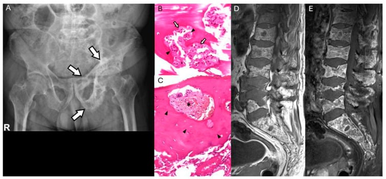
- Axial-predominant sclerosis (spine, pelvis, ribs, skull base) favours marrow-replacement processes — metastases, myelofibrosis, mastocytosis.
- Renal osteodystrophy gives the rugger-jersey spine: sclerotic vertebral endplate bands with a relatively lucent central body, plus subperiosteal resorption of the radial side of the middle phalanges.
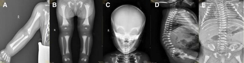
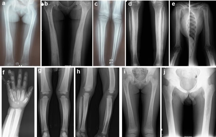
- Osteopetrosis gives bone-within-bone appearance, Erlenmeyer flask femora and, paradoxically, transverse fractures through dense bone - the high bone mass does NOT mean strong bone, and repair is often delayed or fails.
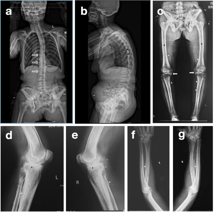
- Fluorosis is misnamed as a sclerosing disease: in 127 patients with endemic fluorosis, osteosclerosis appeared in only 43% and OSTEOPENIA in 40%, while ossification of ligament, tendon and interosseous membrane attachments was present in 89% of adults. Look for the entheseal ossification, not for dense bone.
- Paget disease is rarely truly generalised: look for bone expansion and cortical thickening, which no metastasis or dysplasia produces to the same degree.
- Diffuse sclerosis with hepatosplenomegaly and a leucoerythroblastic film is myelofibrosis until proven otherwise.
- “Sclerosis that respects the enthesis and thickens the cortex is Paget; sclerosis that ignores anatomy and blurs the corticomedullary junction is marrow disease.
- “Bone expansion is the single most useful discriminator: present in Paget and thalassaemia, absent in metastases and renal osteodystrophy.
- “A normal serum alkaline phosphatase makes widespread Paget disease and osteoblastic metastases both unlikely.
- “In a child, always look at the metaphyses: banding equals lead, healed rickets or bisphosphonate; uniform density equals osteopetrosis.
An underpenetrated film makes every bone look dense. Check that soft-tissue planes are visible and that the disc spaces and endplates are distinguishable. True sclerosis loses the trabecular pattern; a dark film keeps it.
Diffuse osteoblastic disease in a man over 60 is usually prostate — but myelofibrosis, mastocytosis and fluorosis all mimic it. Do the film and the PSA before committing to a bone biopsy.
Endplate banding is easily read as degenerative endplate sclerosis. The band in renal osteodystrophy is broad, symmetrical at every level, and paired above and below a lucent centre. Degenerative sclerosis is focal and asymmetric.
Osteopetrosis, fluorosis and bisphosphonate-associated bone all fracture. Osteopetrotic bone fractures transversely with poor healing, and reaming for a nail may be impossible — plan for burrs, sharp reamers and long operative times.
Recognising the Pattern
Definition. Generalised osteosclerosis is a diffuse increase in bone radiodensity affecting multiple bones or the whole skeleton, with loss of the normal distinction between cortex and medulla, or coarsening and thickening of trabeculae to the point where individual trabeculae are no longer resolved.
Confirming it is genuinely present. Three checks before you commit:
- Exposure check. Are the soft-tissue shadows, gas pattern and disc spaces visible? If the whole film is white, the technique is at fault.
- Corticomedullary junction. In normal bone the medulla is clearly less dense than the cortex. In true sclerosis this junction is lost or blurred.
- Multiplicity. Look at more than one region. A dense pelvis alone may be Paget or a single blastic deposit; dense pelvis, ribs and vertebrae together is a generalised process.
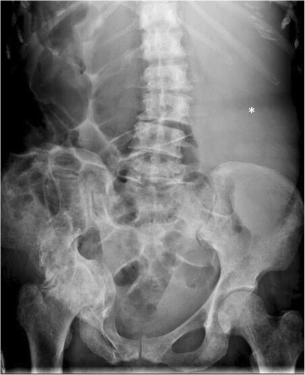
Words to use out loud. "This is a plain radiograph of the pelvis and lumbar spine in a skeletally mature patient. There is diffuse increased density of the axial skeleton with loss of the normal corticomedullary differentiation and obliteration of the trabecular pattern. There is no bone expansion and no cortical thickening. The endplates show broad symmetrical sclerotic bands. The appearances are those of generalised osteosclerosis; my leading diagnoses are renal osteodystrophy and osteoblastic metastatic disease."
Then always add the completing sentence: "I would want the patient's age, renal function, calcium, phosphate, alkaline phosphatase, PTH, a full blood count with film, and a PSA in a man."
Mimics — the false positives.
- Technical underexposure — the commonest mimic by far.
- Physiological neonatal density and the dense metaphyseal bands of normal infancy.
- Overlying contrast or barium in the bowel projecting over the pelvis.
- Diffuse degenerative change with vacuum phenomena and osteophytes, giving a busy dense-looking spine but preserved trabeculae.
- Osteopenia elsewhere making a normal bone look relatively dense — a relative, not absolute, increase.
Next Investigation
- What it shows
- Leucoerythroblastic picture, tear-drop cells
- What it changes
- Triggers urgent haematology referral and marrow biopsy
- What it shows
- Separates renal, hypoparathyroid and Paget patterns
- What it changes
- Directs medical rather than oncological workup
- What it shows
- Very high in Paget and widespread blastic metastases; normal in dysplasias
- What it changes
- A normal ALP largely excludes both aggressive causes
- What it shows
- Focal versus diffuse marrow replacement, cord compression
- What it changes
- Emergency decompression or radiotherapy if cord threatened
- What it shows
- Reticulin fibrosis, mast cells, plasma cells, tumour
- What it changes
- Definitive diagnosis when imaging is equivocal
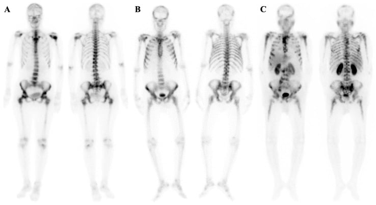
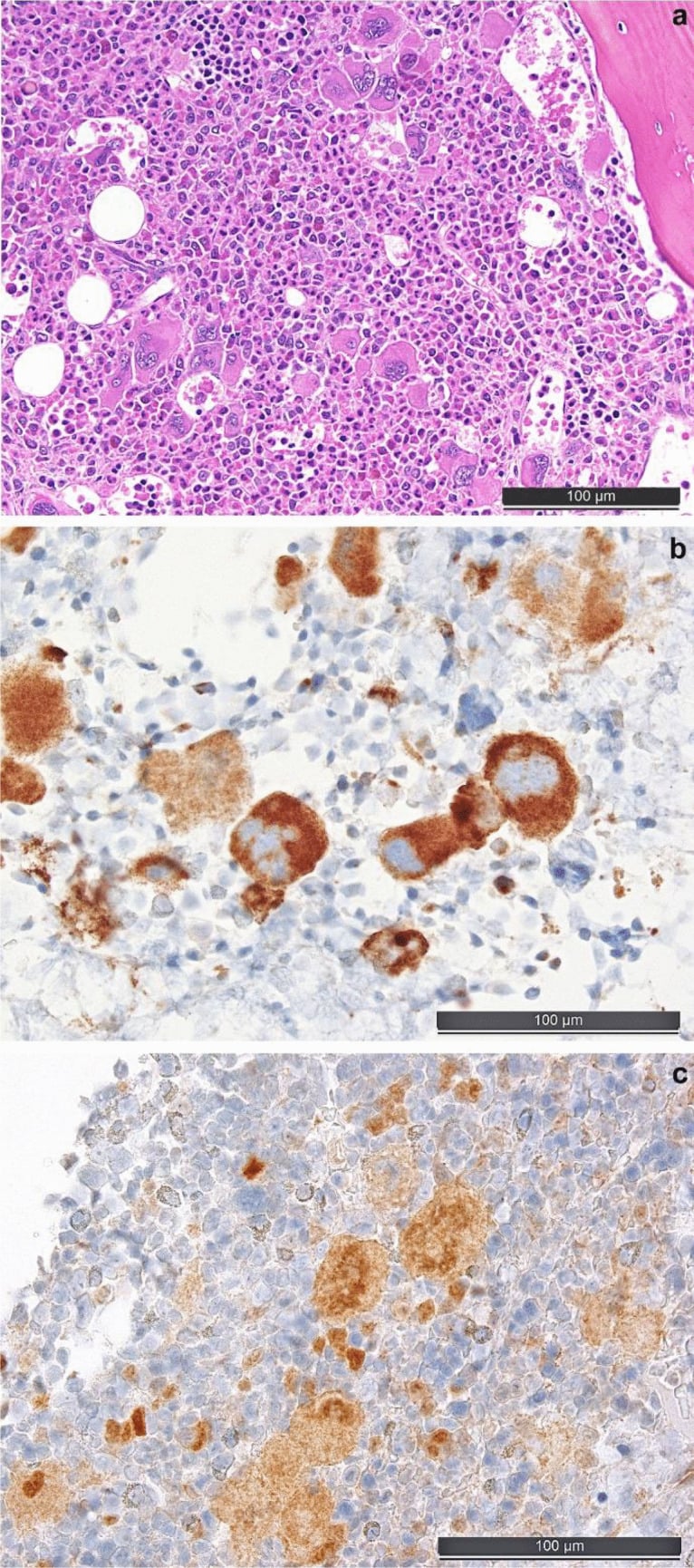
Mets, Myelofibrosis, Mastocytosis; Paget, Pyknodysostosis (and osteoPetrosis); Fluorosis — plus Renal osteodystrophyGeneralised osteosclerosis — 3 M's, 2 P's, 1 F
Hook:A widely taught grouping for the dense-bone differential. The three M's are all marrow-based and axial-predominant. The P group are the expanded or constitutional causes. Fluorosis is remembered separately because of its soft-tissue ossification. Renal osteodystrophy is added as the metabolic cause you must never miss in a dialysis patient.
The Differential
- Typical age / setting
- Over 50, known or occult primary
- Discriminating feature
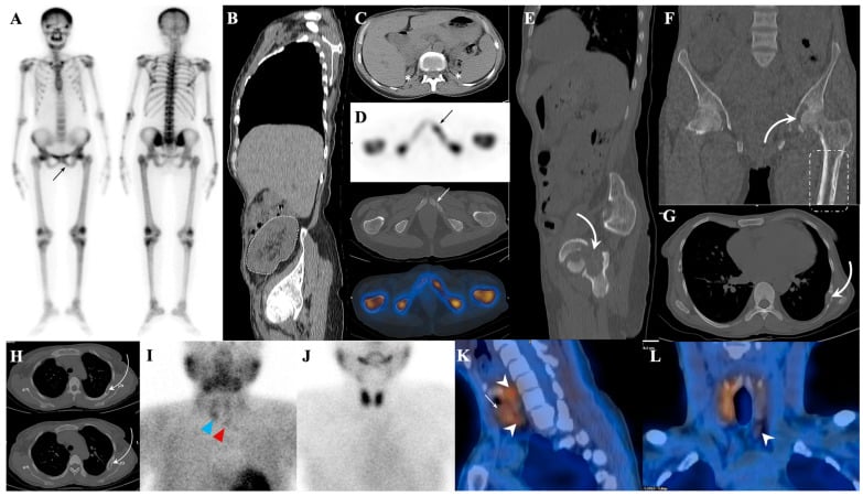
- Patchy, asymmetric, spares no anatomic rule; vertebral pedicle involvement and an ivory vertebra amid normal neighbours
- What confirms it
- Bone scan or NaF PET showing multifocal uptake; PSA; biopsy of an accessible lesion
- Typical age / setting
- Age 60 plus, fatigue, massive splenomegaly
- Discriminating feature
- Uniform axial sclerosis WITH hepatosplenomegaly and a leucoerythroblastic blood film with tear-drop poikilocytes
- What confirms it
- Bone marrow trephine showing reticulin fibrosis; JAK2 mutation
- Typical age / setting
- Age 50 plus, POEMS syndrome in sclerotic myeloma
- Discriminating feature
- Sclerotic myeloma is usually solitary or few lesions with peripheral neuropathy and organomegaly, not lytic punched-out disease
- What confirms it
- Serum paraprotein, VEGF level, marrow biopsy
- Typical age / setting
- Adult, flushing, diarrhoea, urticaria pigmentosa
- Discriminating feature
- Mixed sclerotic and lucent axial lesions with skin lesions and raised serum tryptase
- What confirms it
- Serum tryptase; marrow biopsy with mast cell aggregates; KIT D816V
- Typical age / setting
- Dialysis or chronic kidney disease, any adult age
- Discriminating feature
- Rugger-jersey spine — broad paired endplate bands with lucent central body — plus subperiosteal resorption of radial middle phalanges and a lamina dura loss
- What confirms it
- Creatinine, phosphate high, calcium low, PTH markedly raised
- Typical age / setting
- Over 55, Northern European descent
- Discriminating feature
- Bone EXPANSION with cortical thickening and coarse trabeculae; skull cotton-wool lesions; rarely truly generalised
- What confirms it
- Markedly raised alkaline phosphatase with normal calcium; bone scan showing intense uptake in expanded bones
- Typical age / setting
- Afro-Caribbean, childhood onward
- Discriminating feature
- H-shaped (Lincoln log) vertebrae from central endplate infarction, plus avascular necrosis of femoral heads
- What confirms it
- Haemoglobin electrophoresis
- Typical age / setting
- Endemic areas, industrial exposure, chronic high fluoride water
- Discriminating feature
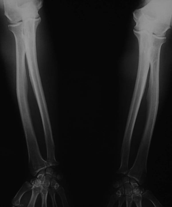
- Dense bone PLUS ossification of the interosseous membrane of the forearm and leg and paraspinal ligaments
- What confirms it
- Urinary and serum fluoride; occupational and geographical history
- Typical age / setting
- Any age; short fourth metacarpal in pseudo form
- Discriminating feature
- Sclerosis with low calcium, HIGH phosphate and basal ganglia calcification
- What confirms it
- Calcium, phosphate, PTH (low in true, high in pseudo)
- Typical age / setting
- Adolescent to adult, incidental or fracture
- Discriminating feature
- Bone-within-bone vertebrae, sandwich vertebra, Erlenmeyer flask distal femur, transverse fractures through dense bone
- What confirms it
- Family history, radiographic gestalt; carbonic anhydrase II testing in select forms
- Typical age / setting
- Child; short stature, dense bone
- Discriminating feature
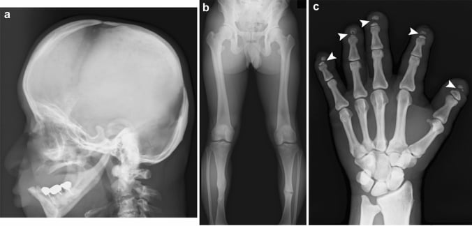
- Acro-osteolysis of the distal phalanges and an obtuse mandibular angle with persistent open fontanelles
- What confirms it
- Cathepsin K gene testing
- Typical age / setting
- Child aged 4 to 10, waddling gait, leg pain
- Discriminating feature
- Symmetrical DIAPHYSEAL cortical thickening sparing the epiphyses and metaphyses entirely
- What confirms it
- TGFB1 mutation; symmetrical diaphyseal pattern on skeletal survey
- Typical age / setting
- Any age, usually incidental
- Discriminating feature
- Osteopoikilosis gives symmetric round periarticular foci; osteopathia striata gives linear metaphyseal streaks; melorheostosis is a dripping candle wax cortex in one limb
- What confirms it
- Radiographic appearance alone — no further imaging needed
- Typical age / setting
- Child with pica or chronic ingestion
- Discriminating feature
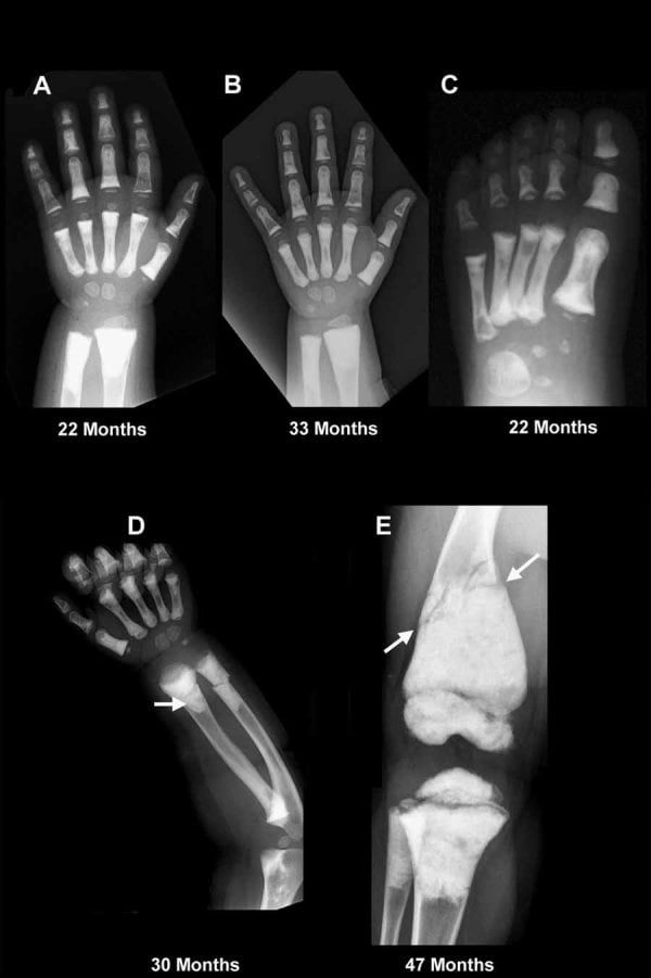
- Dense transverse METAPHYSEAL bands at fast-growing ends (distal femur, proximal tibia) with normal diaphyses
- What confirms it
- Blood lead level; vitamin D and calcium levels
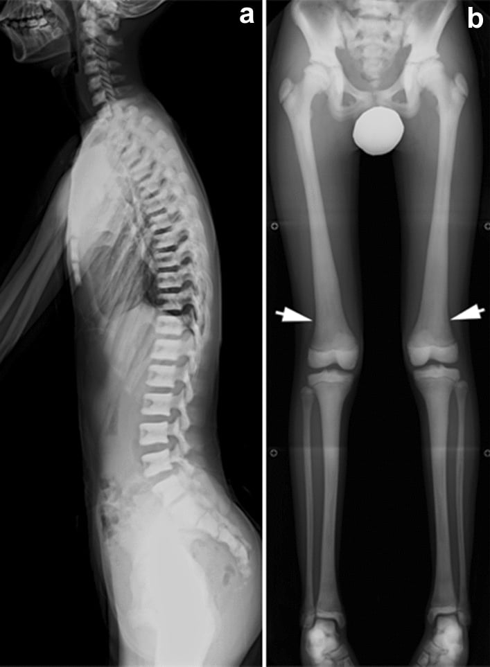
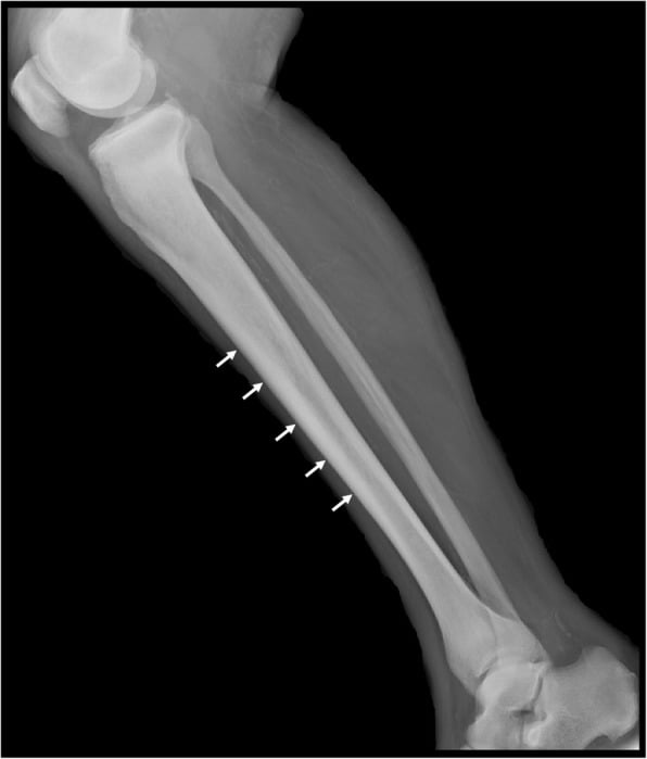
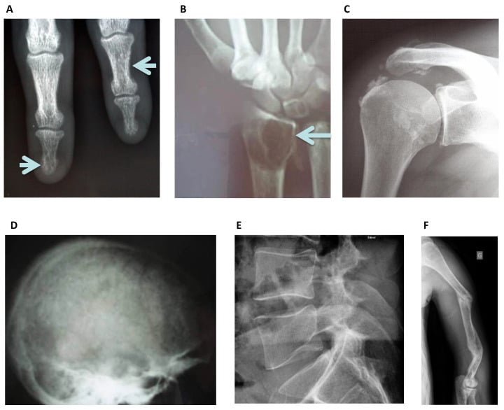

Narrowing It Down
- 1Step 1 - Is it real, and how old is the patient?
Exclude underexposure first, then split the list at skeletal maturity.
Under 20 years: sclerosing dysplasia, healed rickets, lead, chronic anaemia. Over 50 years: metastasis, myelofibrosis, renal osteodystrophy, Paget, fluorosis. Age alone removes more than half the differential.
- 2Step 2 - Axial only, or whole skeleton?
Establish the distribution before naming a cause - you need films beyond the one that prompted the question.
Sclerosis confined to spine, pelvis, ribs, skull base and proximal femora points to RED MARROW disease: metastases, myelofibrosis, mastocytosis. Uniform density reaching the peripheral skeleton, including hands and feet, points to a constitutional dysplasia or fluorosis.
- 3Step 3 - Where within the bone?
Localise the sclerosis to a zone rather than calling it diffuse.
Endplate bands are renal osteodystrophy. Metaphyseal bands are lead, healed rickets, bisphosphonate or treated leukaemia. Diaphyseal cortical thickening is Camurati-Engelmann. Bone-within-bone is osteopetrosis. Random and patchy is metastases.
- 4Step 4 - Is the bone expanded?
Compare the cortical outline against the expected width of that bone.
Expansion with cortical thickening and coarse trabeculae is Paget disease or, in a young patient with an anaemia history, thalassaemic marrow hyperplasia. Metastases, renal disease and osteopetrosis do NOT expand bone. This single question is the highest-yield in the algorithm.
- 5Step 5 - Are the soft tissues abnormal?
Look deliberately outside the bone: entheses, interosseous membranes, vessels, spleen and skin.
Ligamentous and interosseous membrane ossification is FLUOROSIS - and it is the most reliable sign of it, present in 89 per cent of adults in a 127-patient series, whereas osteosclerosis itself appeared in only 43 per cent. Vascular and basal ganglia calcification is renal osteodystrophy or hypoparathyroidism. Splenomegaly is myelofibrosis, sickle disease or mastocytosis. Skin lesions are mastocytosis.
- 6Step 6 - What do the bloods say?
Calcium, phosphate, ALP, PTH, PSA and a full blood count with film.
Raised ALP with normal calcium is Paget. Low calcium, high phosphate and high PTH is renal osteodystrophy. Low calcium, high phosphate and LOW PTH is hypoparathyroidism. A leucoerythroblastic film is marrow infiltration or fibrosis. Raised PSA is prostatic metastasis.
- 7Step 7 - Still unresolved?
Proceed to marrow sampling rather than repeating imaging.
A bone marrow trephine answers myelofibrosis, mastocytosis, sclerotic myeloma and marrow-infiltrating metastasis in a single procedure. Note that in osteopetrosis the diagnosis has moved the other way: radiographic appearance alone is no longer sufficient, and molecular classification now determines prognosis and treatment.

MCQ Practice Points
Q: Which single radiographic feature best separates polyostotic Paget disease from diffuse osteoblastic metastases?
A: Bone EXPANSION with cortical thickening. Paget disease enlarges the bone and thickens the cortex with coarse disorganised trabeculae; metastases increase density without altering bone size or contour.
Q: A 61-year-old has diffuse axial sclerosis, fatigue and a spleen palpable 8 cm below the costal margin. Diagnosis and confirmatory test?
A: Primary myelofibrosis. The blood film shows a leucoerythroblastic picture with tear-drop poikilocytes; bone marrow trephine demonstrates reticulin fibrosis and there is often a dry tap. JAK2 V617F is positive in around half.
Q: Which cause of generalised osteosclerosis is associated with ossification of the forearm interosseous membrane, and how reliable is that sign?
A: Skeletal fluorosis - and the soft-tissue ossification is not merely the discriminator, it is the most COMMON finding. In 127 patients with endemic fluorosis, calcification or ossification of ligament, tendon, muscle and interosseous membrane attachments was present in 89 per cent of adults, while osteosclerosis itself appeared in only 43 per cent and osteopenia in 40 per cent. Fluorosis is therefore not reliably a sclerosing disease at all: look for the entheseal ossification, not for dense bone.
Q: In a 3-year-old with dense transverse metaphyseal bands at the distal femur and proximal tibia but normal diaphyses, what is the differential?
A: Lead or heavy metal poisoning, hypervitaminosis D, healed rickets, treated leukaemia, and bisphosphonate therapy. Blood lead level and dietary history are the first steps. Uniform whole-bone sclerosis rather than banding would point instead to osteopetrosis.
Q: Which biochemical triad identifies renal osteodystrophy as the cause of a rugger-jersey spine?
A: Raised phosphate, low or low-normal calcium, and markedly raised parathyroid hormone, with impaired renal function. Alkaline phosphatase may also be raised, reflecting high bone turnover.
Exam Viva Scenarios
Practise clinical reasoning and management decisions out loud
“You are shown this lateral lumbar spine radiograph of a 64-year-old man with 3 months of back pain. The vertebrae are diffusely sclerotic. Describe and give a differential.”
“You are shown this pelvic radiograph of a 14-year-old with a transverse subtrochanteric femoral fracture after a minor fall. The bones are uniformly dense with obliteration of the medullary canal.”
“You are shown this lateral thoracolumbar radiograph of a 48-year-old on haemodialysis. There are dense bands adjacent to each endplate with a relatively lucent central vertebral body.”
Confirm the pattern
- Check exposure — soft tissues and disc spaces must be visible
- Loss of corticomedullary differentiation equals true sclerosis
- Confirm in at least two separate regions before calling it generalised
- Describe: density, distribution, expansion, trabecular pattern, soft tissues
Split by age
- Under 20: osteopetrosis, pyknodysostosis, Camurati–Engelmann, lead, healed rickets
- 20 to 50: renal osteodystrophy, sickle disease, mastocytosis, fluorosis
- Over 50: metastases, myelofibrosis, Paget, sclerotic myeloma
Site-specific signs
- Rugger-jersey spine — renal osteodystrophy
- Sandwich vertebra and bone-within-bone — osteopetrosis
- Cotton-wool skull and expanded pelvis — Paget disease
- H-shaped vertebrae — sickle cell disease
- Diaphyseal cortical thickening sparing epiphyses — Camurati–Engelmann
- Acro-osteolysis with obtuse mandibular angle — pyknodysostosis
Biochemistry map
- High ALP, normal calcium — Paget
- High phosphate, low calcium, high PTH — renal osteodystrophy
- High phosphate, low calcium, low PTH — hypoparathyroidism
- Leucoerythroblastic film — myelofibrosis or marrow infiltration
- Raised PSA — prostatic metastases
- Raised tryptase — systemic mastocytosis
Do not miss
- Cord compression from blastic metastatic deposits — MRI urgently
- Marrow failure in severe osteopetrosis and myelofibrosis
- Osteopetrotic fractures: transverse, poor healing, obliterated canal, high infection risk
- Cranial nerve palsies from foraminal encroachment in osteopetrosis
No further imaging required
- Osteopoikilosis — symmetric periarticular round foci, incidental
- Osteopathia striata — linear metaphyseal streaks
- Melorheostosis — dripping candle wax cortex in one limb, if asymptomatic
- Established dialysis patient with classic rugger-jersey spine and known PTH
Evidence Base
Endemic Fluorosis of the Skeleton - Radiographic Features in 127 Patients
- 127 patients with clinically proven endemic fluorosis had radiographs of chest, spine, pelvis, elbow, forearm and knee, classified for osteosclerosis, osteopenia, growth lines, diaphyseal widening and soft-tissue ossification
- THE COMMONEST FINDING IS NOT SCLEROSIS: calcification or ossification of the attachments of ligaments, tendons, muscles and interosseous membranes was present in 98 patients - 89 per cent of the adults
- OSTEOSCLEROSIS WAS SEEN IN ONLY 54 PATIENTS (43%), while OSTEOPENIA was seen in 51 (40%) - of those, an osteoporotic pattern in 28 and an osteomalacic pattern in 23
- Growth lines were found in 89 patients (70%) and diaphyseal widening in 35 (28%); metaphyseal osteomalacic zones occurred in children
- The authors conclude that endemic skeletal fluorosis has a WIDE VARIETY of radiographic appearances, of which osteosclerosis is only one
Osteopetrosis and Related Osteoclast Disorders in Adults - a Review and Knowledge Gaps
- A review on behalf of the European Calcified Tissue Society and ERN BOND, addressing the adult presentation specifically
- THE CENTRAL PARADOX, STATED PLAINLY: despite the high bone mass, SKELETAL STRENGTH IS COMPROMISED and fracture risk is high, particularly in the long bones
- The autosomal dominant form, typically diagnosed in adolescence or young adulthood, is caused by mutations in CLCN7 encoding the ClC-7 chloride channel; the recessive forms are severe and present in the first years of life
- Diagnosis was TRADITIONALLY made on radiographic appearance alone, but molecular and genetic advances now allow far greater fidelity in subtype classification
- The major complications of the adult dominant form are ATYPICAL FRACTURES with delay or failure of repair, and difficulty in orthopaedic management; marrow failure, dental abscess, deafness and visual loss are described as often underestimated and neglected
- Apart from haematopoietic stem cell transplantation in certain infantile forms, treatment is NOT standardised and remains supportive
Genetics of Osteopetrosis
- Osteopetrosis is a GROUP of rare skeletal diseases sharing one hallmark - a generalised increase in bone density from defective bone resorption - and is clinically and genetically heterogeneous
- A precise MOLECULAR classification is relevant for prognosis and treatment, not merely for academic completeness
- Next-generation sequencing has expanded the spectrum: novel mutations in known genes, defects in entirely new genes, and molecular classification of complex phenotypes in which osteopetrosis is accompanied by additional clinical features
- Novel mutation types have been recognised which, by their nature or genomic location, are at high risk of being MISSED by standard testing
- In some patients the causative mutation remains unknown, so the genetics of osteopetrosis is still an open field