The Posterior Arm of the Force Couple
- Origin: medial two-thirds of the infraspinous fossa plus the tough overlying infraspinous fascia; bipennate architecture.
- Insertion: middle facet of the greater tuberosity and, per Mochizuki, much of the superior facet as well.
- Innervation: suprascapular nerve (C5, C6) after it passes the spinoglenoid notch beneath the inferior transverse scapular ligament.
- Provides roughly 60 per cent of external rotation torque with the arm adducted.
- Fatty infiltration of the infraspinatus is the single strongest muscle-quality predictor of failure after cuff repair.
- “A spinoglenoid notch lesion wastes infraspinatus ONLY; a suprascapular notch lesion wastes both fossae.
- “ER lag sign at 0 degrees abduction implicates supraspinatus and upper infraspinatus; the drop sign at 90 degrees isolates infraspinatus; hornblower's sign isolates teres minor.
- “The interval between infraspinatus (suprascapular nerve) and teres minor (axillary nerve) is a true internervous plane.
- “Teres minor hypertrophy on MRI is a compensatory good sign in massive posterosuperior tears.
Overview
The infraspinatus occupies the infraspinous fossa below the scapular spine and is the largest of the posterior cuff muscles. It is the workhorse external rotator and, with the teres minor, forms the posterior arm of the transverse force couple that centres the humeral head. Where the supraspinatus dominates surgical conversation because of impingement and repair, the infraspinatus dominates prognosis: its fatty infiltration is the muscle-quality variable that most consistently predicts failure of a cuff repair and poor external rotation after reverse arthroplasty.
Its clinical signature is distinctive. Because the suprascapular nerve reaches it only after negotiating a second bony corner at the spinoglenoid notch, the infraspinatus can be denervated in isolation while the supraspinatus is untouched — a pattern seen with paralabral cysts and with repetitive traction in overhead athletes, and one that no other cuff muscle reproduces.
The infraspinatus appears in both of the shoulder's force couples, and examiners will ask you to place it in each.
- Transverse (axial) force couple. The subscapularis anteriorly is balanced by the infraspinatus and teres minor posteriorly. Their equal and opposite pull compresses and centres the head on the glenoid, generating the fulcrum the deltoid needs. An intact transverse couple can keep a shoulder functional even with a large supraspinatus defect.
- Coronal force couple. The deltoid pulls superiorly; the inferior cuff (subscapularis, infraspinatus and teres minor) pulls inferiorly and medially. Their resultant is a rotational moment rather than superior translation.
- What failure looks like. Loss of the posterior arm unbalances the transverse couple: the unopposed subscapularis pulls the head anteriorly and the deltoid shear drives it superiorly. Clinically the patient loses external rotation first, then loses the ability to hold the arm in space, then develops static superior migration.
- Why the couple must be restored, not just the tendon. In a massive posterosuperior tear, a partial repair that re-establishes the posterior arm often restores function even when the supraspinatus defect is left open. Balance beats completeness.
Lag, Drop, HornLocalising Posterior Cuff Weakness
Hook:Test progressively higher in abduction and the lesion localises progressively lower in the posterior cuff.
Attachments, Innervation and Relations
Origin
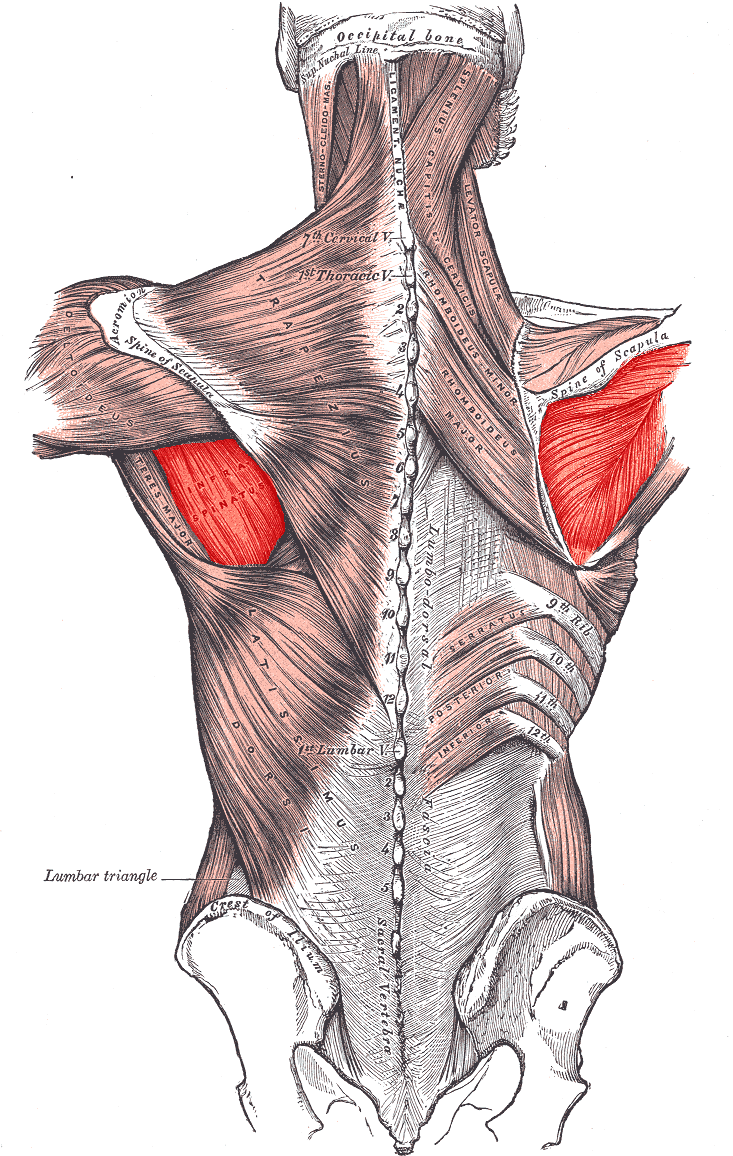
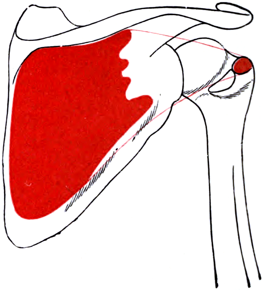
- Medial two-thirds of the infraspinous fossa on the dorsal surface of the scapula, below the scapular spine.
- Also from the deep surface of the dense infraspinous fascia, which roofs the muscle and separates it from teres minor; a genuine osteofascial compartment, and the reason a haematoma here can produce a compartment-like presentation.
- The muscle is bipennate, with an oblique central tendinous raphe that is often mistaken arthroscopically for the plane between infraspinatus and teres minor.
Insertion — the footprint
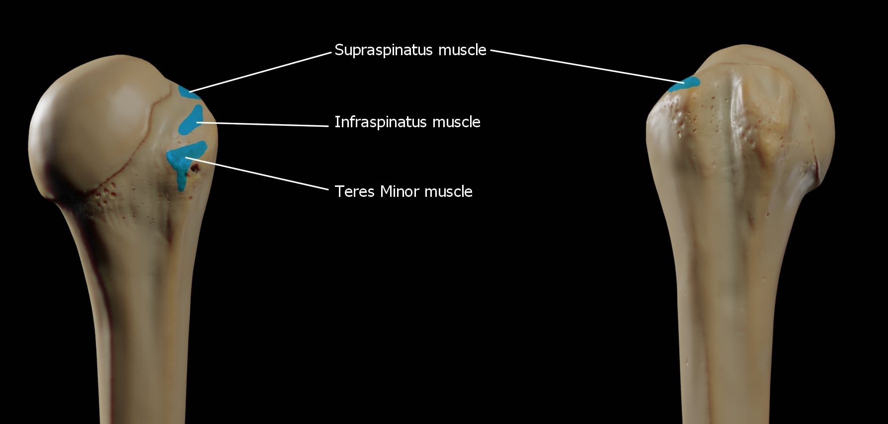
- Middle facet of the greater tuberosity, and — per Mochizuki and colleagues (JBJS Am 2008) — a substantial part of the superior facet as well, extending anteriorly to overlie the supraspinatus insertion.
- Mochizuki's measured footprints, from 26 shoulders: the infraspinatus is a trapezoid, average maximum 32.7 mm anteroposterior by 10.2 mm medial-to-lateral. The supraspinatus is a triangle of just 12.6 mm by 6.9 mm — and in 21 per cent of specimens its tendon also reached the superiormost lesser tuberosity.
- Put those two side by side and the conclusion is unavoidable: the supraspinatus footprint is much smaller than the textbooks had it, and the greater tuberosity area attributed to it is substantially occupied by infraspinatus.
- Surgical translation: a defect on the posterior part of the superior facet, which surgeons habitually call a supraspinatus tear, is anatomically often an infraspinatus tear. This reframes the significance of posterior extension of a cuff tear — and it is the anatomical explanation Mochizuki set out to find for a clinical observation, that infraspinatus atrophy is common even with small and medium tears.
- The tendon interdigitates with the supraspinatus anteriorly and with the joint capsule deep to it; the two cannot be separated by blunt dissection at the insertion.
- The rotator cable inserts posteriorly at the inferior border of the infraspinatus — the posterior anchor of the suspension bridge.
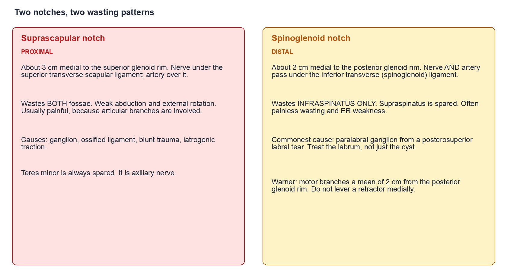
Proximal lesion.
- Lies about 3 cm medial to the superior glenoid rim.
- Nerve under the superior transverse scapular ligament, artery over it.
- Lesion here wastes both supraspinous and infraspinous fossae.
- Causes: ganglion, ossified ligament with a complete bony foramen, blunt trauma, iatrogenic traction.
- Typically painful, because articular branches are involved.
Distal lesion.
- Lies about 2 cm medial to the posterior glenoid rim.
- Nerve and artery both pass under the inferior transverse (spinoglenoid) ligament.
- Lesion here wastes the infraspinatus only; supraspinatus is spared.
- Commonest cause: paralabral ganglion from a posterosuperior labral tear.
- Often painless, presenting purely as wasting and external rotation weakness.
Action and Biomechanics
Actions by plane
- External rotation: the principal action. With the arm at the side, the infraspinatus supplies roughly 60 per cent of external rotation torque; teres minor contributes proportionally more as the arm abducts to 90 degrees.
- Concavity compression: its horizontally directed vector compresses the head into the glenoid and, together with subscapularis, forms the transverse couple.
- Posterior stabilisation: the upper fibres resist anterior translation in the abducted, externally rotated position; the lower fibres resist posterior translation in flexion and internal rotation.
- Head depression: the inferiorly directed component of the lower fibres opposes deltoid shear.
Moment arm and length-tension
- The external rotation moment arm is greatest with the arm adducted and decreases with abduction, which is precisely why testing at 0 degrees loads the infraspinatus most efficiently and testing at 90 degrees shifts the demand toward teres minor.
- Chronic retraction shortens the muscle-tendon unit, moves it down the ascending limb of the length-tension curve, and reduces the force it can generate even if the tendon is subsequently repaired. This is one mechanism, alongside fatty infiltration, by which delayed repair yields poor strength.
Synergists and antagonists
- Synergists: teres minor (external rotation), posterior deltoid (external rotation in abduction), supraspinatus (compression).
- Antagonists: subscapularis, pectoralis major, latissimus dorsi, teres major (all internal rotators).
Describe the progression rather than a single sign.
- External rotation weakness at the side — an ER lag sign appears, and the patient reports difficulty reaching behind the head or holding a phone to the ear.
- Loss of the abducted, externally rotated position — a positive drop sign at 90 degrees, and functionally the inability to hold a plate or steer a car.
- Loss of the transverse couple — the head is drawn anteriorly and superiorly by the unopposed anterior cuff and the deltoid.
- Combined loss of elevation and external rotation — when the tear extends to include teres minor, hornblower's sign becomes positive, and the patient is a candidate for a reverse arthroplasty with a concomitant external rotation transfer rather than a reverse alone.
Surface Anatomy and Examination
Palpation and positioning
- The infraspinous fossa is inspected and palpated below the scapular spine; hollowing here is the most visible sign of chronic posterior cuff pathology and should be compared with the opposite side with the patient's shirt off and arms relaxed.
- The spinoglenoid notch is deep and not directly palpable, but deep pressure just lateral to the base of the scapular spine, medial to the posterior glenohumeral joint line, reproduces the pain of a ganglion.
- The tendon insertion at the middle facet is palpated just posterior to the posterolateral acromial corner with the arm in flexion and internal rotation.
Named tests
- How to perform
- Elbow 90 degrees, arm at the side, passively place near-maximal external rotation and release
- Positive finding
- The arm springs back into internal rotation
- What it means
- Supraspinatus and upper infraspinatus tear; the size of the lag grades severity
- False positives
- Stiffness prevents the passive position being achieved, masking the lag
- How to perform
- Arm 90 degrees abduction in the scapular plane, elbow 90 degrees, passive full external rotation, then release
- Positive finding
- The arm drops into internal rotation
- What it means
- Infraspinatus deficiency, usually severe with fatty infiltration
- False positives
- Pain inhibition; deltoid substitution if the arm is unsupported
- How to perform
- Ask the patient to bring the hand to the mouth
- Positive finding
- The patient must abduct the shoulder to 90 degrees to do so
- What it means
- Teres minor deficiency; severe posterior cuff loss
- False positives
- Elbow flexion contracture forcing the same posture
- How to perform
- Elbows tucked in, resist ER
- Positive finding
- Weakness or pain
- What it means
- Infraspinatus load, the most efficient isolation position
- False positives
- Pain inhibition from posterior capsular pathology
- How to perform
- Arm 90 degrees abduction in the scapular plane, resist external rotation
- Positive finding
- Weakness
- What it means
- Posterior cuff at the abducted position, biased to teres minor and lower infraspinatus
- False positives
- Fatigue on repeated testing
- How to perform
- Inspect the infraspinous fossa from behind, arms relaxed
- Positive finding
- Visible hollowing
- What it means
- Chronic tear or denervation; distinguish by whether the supraspinous fossa is also wasted
- False positives
- Constitutional thinness; always compare sides
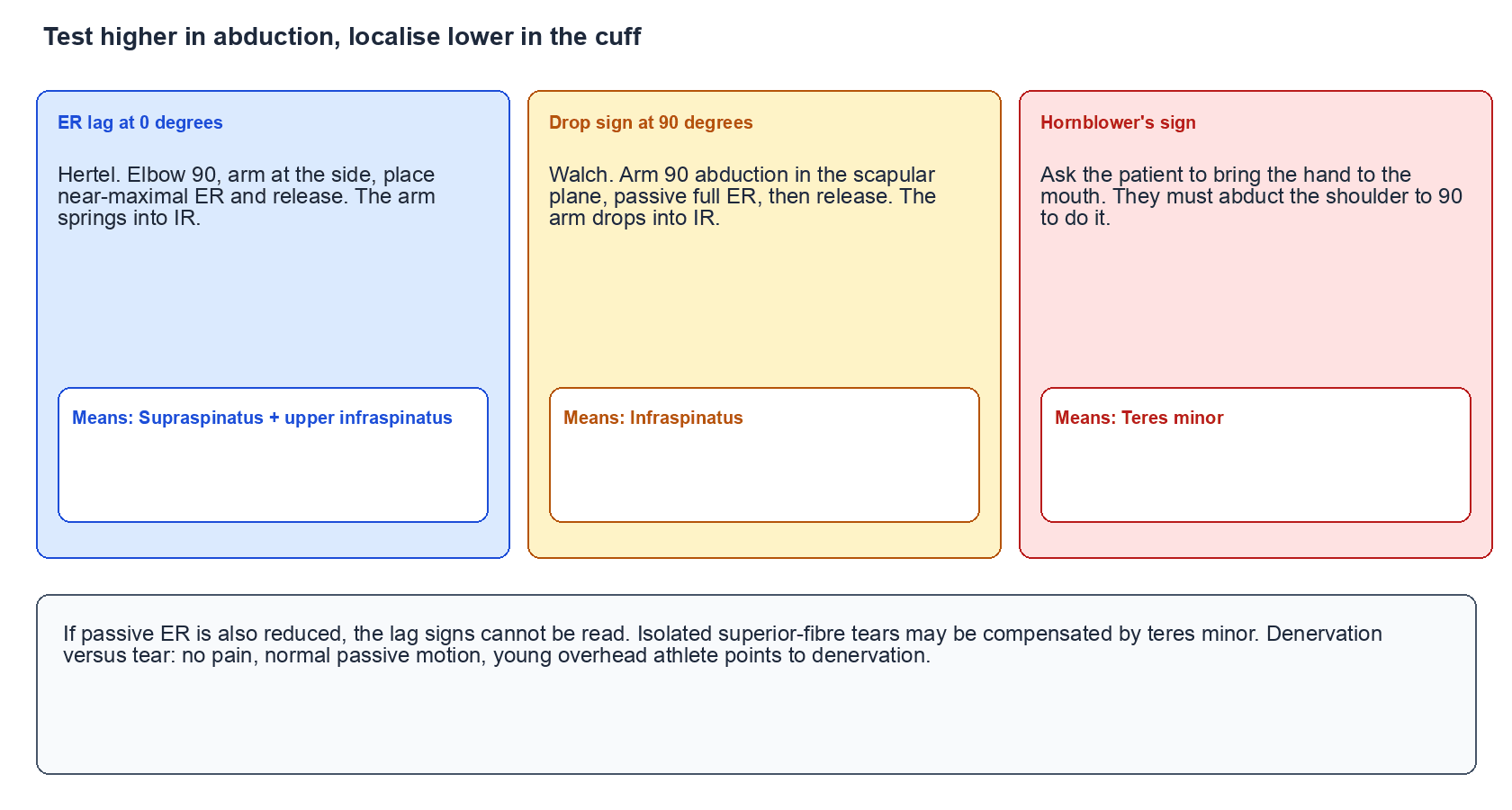
The single most useful examination principle on the posterior cuff: test progressively higher in abduction and the lesion localises progressively lower in the muscle group. External rotation lag at the side implicates supraspinatus and upper infraspinatus; the drop sign at 90 degrees isolates infraspinatus; hornblower's sign, where the patient must lift the elbow to 90 degrees to bring the hand to the mouth, isolates teres minor. Walch reported very high sensitivity and specificity for the dropping sign in identifying irreparable degeneration of the infraspinatus and for hornblower's sign in identifying teres minor loss.
Grading and pitfalls
- Compare passive and active external rotation. If passive external rotation is also reduced, the problem includes stiffness and the lag signs cannot be interpreted.
- False negatives: an isolated tear of the superior part of the infraspinatus may be compensated by teres minor, giving normal strength at 90 degrees of abduction while the lag at the side is subtle.
- Denervation versus tear: both waste the fossa. Absence of pain, normal passive motion, and a young athletic patient point toward denervation; imaging then differentiates.
Complications
Suprascapular neuropathy — iatrogenic
- Mechanism: medial retractor pressure at the posterior glenoid, dissection medial to the 2 cm limit, or traction when a chronically retracted infraspinatus is pulled laterally to the footprint.
- Presentation: external rotation weakness with an intact repair, progressive infraspinous wasting, no sensory deficit.
- Avoidance: respect the 2 cm rule, place retractors intracapsularly, release retraction intermittently, and accept a partial repair rather than force excursion.
- Management: EMG at six weeks; if there is a structural cause such as an unrecognised ganglion, decompress; otherwise observe with therapy, since traction neurapraxia usually recovers.
Denervation from splitting the wrong plane
- Splitting the intramuscular raphe of the bipennate infraspinatus instead of the infraspinatus-teres minor interval denervates the inferior half of the muscle. This is a silent complication that presents months later as unexplained external rotation weakness.
- Avoidance: identify the true interval by following the inferior border of infraspinatus laterally and by locating the circumflex scapular vessels emerging from the triangular space.
Axillary nerve injury
- Occurs when a posterior interval is carried inferiorly past teres minor into the quadrangular space, or when a posterior deltoid split extends more than about 5 cm below the scapular spine.
- Produces deltoid weakness and teres minor loss, compounding the very deficit the operation was intended to treat.
Failure of repair and progression
- Posterior cuff tears with Goutallier 3 to 4 change fail predictably. Fatty infiltration does not reverse after repair, so the decision to repair is really a decision about muscle quality.
- Untreated, tears extend posteriorly, the transverse couple is lost, and the shoulder progresses to static superior migration and cuff tear arthropathy.
Post-operative external rotation deficit after arthroplasty
- In reverse arthroplasty, a degenerate teres minor with an absent infraspinatus predicts persistent external rotation loss despite a technically perfect implant. Patients report inability to reach the back of the head and difficulty with hand-to-mouth activity when the elbow must be lifted.
- Avoidance: identify the deficit pre-operatively with hornblower's sign and the sagittal MRI, and plan a combined latissimus dorsi and teres major transfer at the time of arthroplasty.
Clinical Relevance
Differential of infraspinatus wasting and external rotation weakness
- Supraspinous Fossa
- Normal
- Infraspinous Fossa
- Wasted
- Sensory
- Normal
- Confirmatory Finding
- Paralabral cyst with posterosuperior labral tear on MRI
- Supraspinous Fossa
- Wasted
- Infraspinous Fossa
- Wasted
- Sensory
- Normal
- Confirmatory Finding
- Ganglion or ossified transverse ligament; both fossae denervated on EMG
- Supraspinous Fossa
- Normal or mildly reduced
- Infraspinous Fossa
- Wasted, often painless
- Sensory
- Normal
- Confirmatory Finding
- EMG denervation of infraspinatus only, no structural lesion
- Supraspinous Fossa
- Wasted
- Infraspinous Fossa
- Wasted
- Sensory
- Normal
- Confirmatory Finding
- Tendon discontinuity, Goutallier 3 to 4, positive tangent sign
- Supraspinous Fossa
- Variable, patchy
- Infraspinous Fossa
- Variable, patchy
- Sensory
- May be abnormal
- Confirmatory Finding
- Severe prodromal pain then patchy multi-nerve weakness, often including serratus anterior
- Supraspinous Fossa
- Wasted
- Infraspinous Fossa
- Wasted
- Sensory
- Dermatomal loss present
- Confirmatory Finding
- Neck pain, reduced biceps reflex, deltoid also weak
- Supraspinous Fossa
- Normal
- Infraspinous Fossa
- Normal
- Sensory
- Lateral deltoid patch may be abnormal
- Confirmatory Finding
- Isolated teres minor fatty change; infraspinatus normal
A paralabral ganglion at the spinoglenoid notch is the classic cause of isolated infraspinatus wasting. It matters because the cyst is a symptom, not the disease.
- Source: a posterosuperior labral tear acting as a one-way valve, decompressing joint fluid into the notch.
- Presentation: painless or dull posterior shoulder ache, visible infraspinous hollowing, weak external rotation, no sensory deficit anywhere, normal abduction.
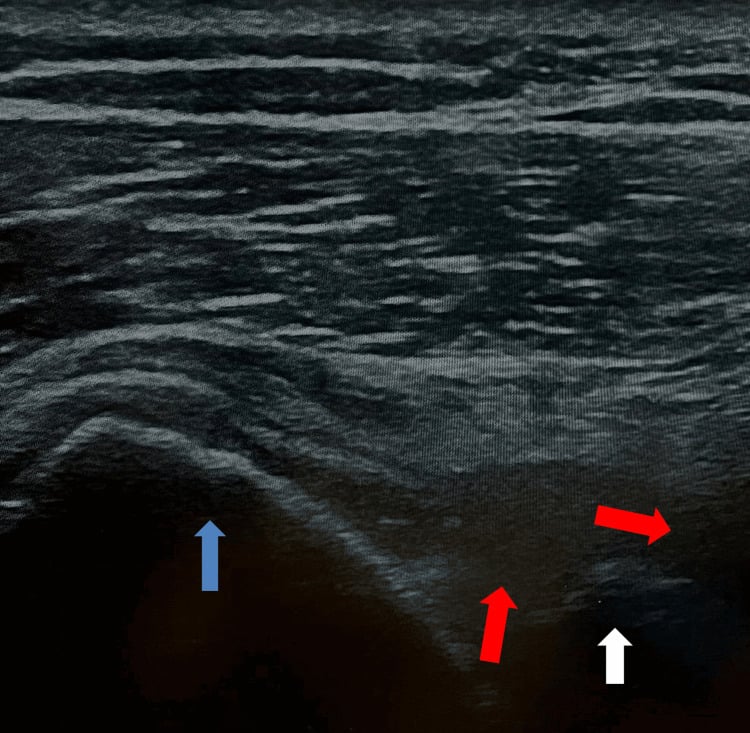
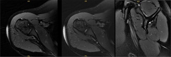
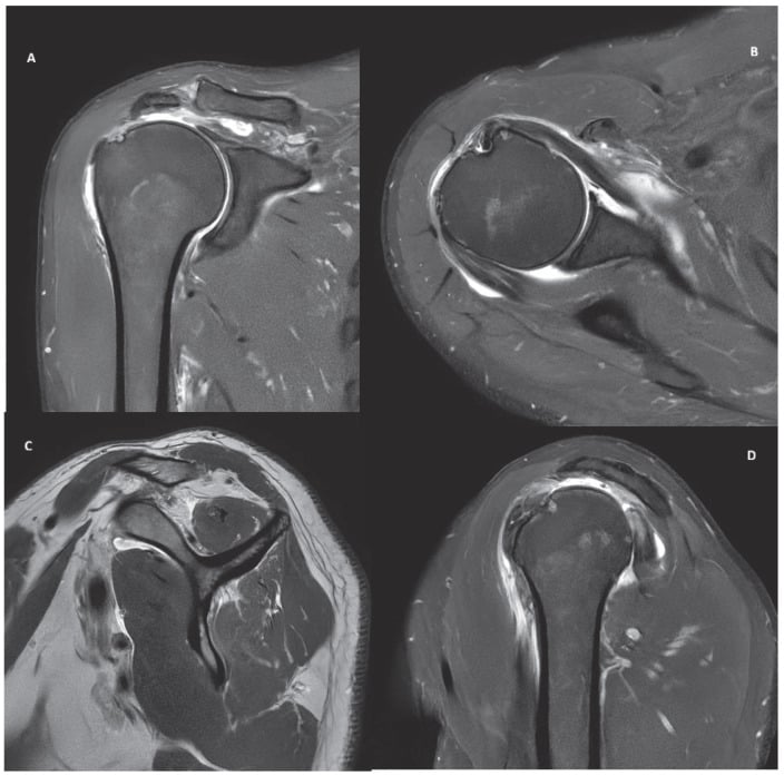
- Imaging: MRI shows a well-defined T2-bright lobulated cyst at the notch, denervation oedema in infraspinatus early, fatty change late. Look explicitly for the labral tear.
- Management: address the labral tear. Arthroscopic labral repair with cyst decompression treats the source. Isolated aspiration under ultrasound or CT has a high recurrence rate because the valve remains.
- Contrast: a suprascapular notch ganglion is usually more painful and wastes both fossae; open or arthroscopic release of the superior transverse scapular ligament is the operation there.
Staging systems
Imaging
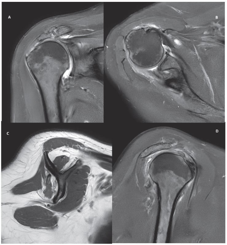
- MRI sagittal oblique at the level of the scapular spine is the working slice: it displays all four cuff bellies in cross-section for Goutallier grading, the tangent sign for supraspinatus, and the occupation ratio. It also reveals teres minor hypertrophy or atrophy, which is prognostically important.
- Coronal oblique and axial sequences show the infraspinatus tendon and any posterior extension of a cuff tear.
- Denervation appears as diffuse T2 hyperintensity of the muscle belly (oedema-like signal) in the subacute phase, followed by fatty replacement. Denervation change is uniform across the whole belly, whereas disuse atrophy from a tear preferentially thins the muscle with a preserved fascicular pattern.
- Always look at the spinoglenoid notch on the axial and sagittal images when the infraspinatus is selectively affected.
Surgical Relevance
The infraspinatus-teres minor interval
The interval between infraspinatus (suprascapular nerve) and teres minor (axillary nerve) is a genuine internervous plane — one of the few in the posterior shoulder. It is the working interval of the posterior approach to the shoulder (Judet) and of posterior glenoid exposure for posterior Bankart repair, posterior bone block procedures and glenoid osteotomy.
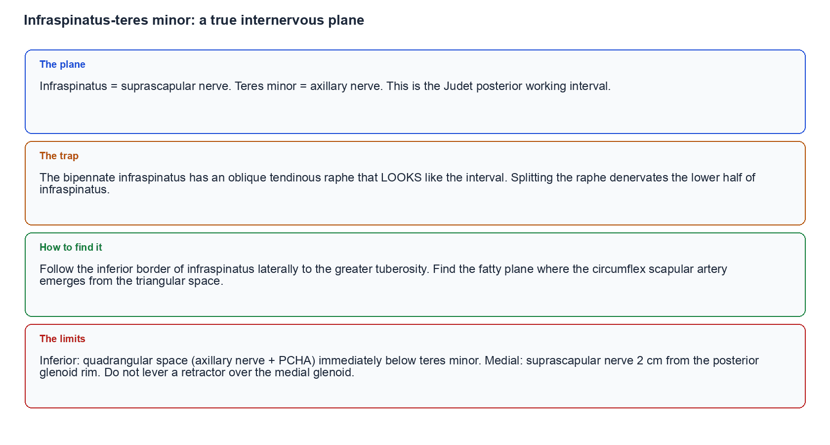
- The trap: the oblique tendinous raphe within the bipennate infraspinatus looks exactly like the interval. Splitting the raphe rather than the true interval denervates the lower half of the infraspinatus, because the branches enter from medial to lateral.
- How to find the true plane: identify the inferior border of infraspinatus by following it laterally to the greater tuberosity and by finding the fatty plane in which the circumflex scapular artery emerges from the triangular space below.
- The inferior limit: the quadrangular space lies immediately below teres minor and contains the axillary nerve and posterior circumflex humeral vessels. Do not carry the interval inferiorly past teres minor.
- The medial limit: the suprascapular nerve at the spinoglenoid notch, roughly 2 cm medial to the posterior glenoid rim. Retractors placed medially over the posterior glenoid rim can compress it.
Approaches and portals
- Relationship to infraspinatus
- Enters through the soft spot between infraspinatus and teres minor
- Structure at risk
- Axillary nerve inferiorly, suprascapular nerve medially
- Distance rule
- 2 cm inferior and 1 cm medial to the posterolateral acromial corner
- Relationship to infraspinatus
- Passes below the infraspinatus through teres minor territory
- Structure at risk
- Axillary nerve
- Distance rule
- Reported to lie roughly 2 to 4 cm from the axillary nerve; keep close to the glenoid rim
- Relationship to infraspinatus
- Reflects deltoid, then works through the infraspinatus-teres minor interval
- Structure at risk
- Suprascapular nerve medially, axillary nerve inferiorly
- Distance rule
- Do not dissect more than 2 cm medial to the posterior glenoid rim
- Relationship to infraspinatus
- Split deltoid, retract, enter the same interval
- Structure at risk
- Axillary nerve as it exits the quadrangular space
- Distance rule
- Split no more than 5 cm distal to the scapular spine
- Relationship to infraspinatus
- Works medial to the infraspinatus muscle belly
- Structure at risk
- The nerve itself and the suprascapular artery
- Distance rule
- Nerve lies about 2 cm medial to the posterior glenoid rim
The commonest iatrogenic suprascapular nerve injury in posterior shoulder surgery is not from the knife but from a retractor placed over the medial edge of the posterior glenoid, compressing the nerve against the base of the scapular spine. Place retractors intracapsularly on the glenoid rim, release them intermittently, and do not lever medially. A post-operative external rotation deficit with an intact repair should prompt EMG rather than reassurance.
Repair of infraspinatus tears
- Posterior extension of a cuff tear into the infraspinatus converts a manageable crescent tear into a massive posterosuperior tear. The priority is to restore the posterior anchor of the rotator cable at the inferior border of infraspinatus.
- Partial repair that reconstitutes the posterior arm of the transverse couple, leaving a central defect, reliably restores active elevation and external rotation and is preferable to a complete repair under excessive tension.
- Margin convergence is used where the tear geometry is U-shaped; the posterior leaf of an L-shaped tear is usually the infraspinatus.
- Releases must respect the measured anatomy: the infraspinatus motor branches lie a mean of only 2 cm from the posterior glenoid rim, and safe lateral advancement is of the order of 1 cm through a standard approach and up to 3 cm with a formal muscle advancement.
The infraspinatus as a donor
- The infraspinatus is not used as a free transfer donor, but it is advanced and rotated in some reconstructive strategies for irreparable posterosuperior tears, and its inferior border is the target to which a latissimus dorsi or lower trapezius transfer is sutured.
- The infraspinatus fossa periosteum and fascia have been used as local augmentation patches.
- More important is the reverse relationship: the infraspinatus is the muscle a lower trapezius transfer is designed to replace, because the trapezius line of pull most closely reproduces the infraspinatus vector.
- 1Establish which muscles are goneER lag at the side, drop sign at 90 degrees, and hornblower's sign each localise a different level of the posterior cuff. Hornblower positive means teres minor is involved.
- 2Grade the muscle on the sagittal MRIGoutallier grade of infraspinatus and teres minor. Teres minor hypertrophy is a favourable compensatory finding; teres minor fatty infiltration is a strong negative predictor.
- 3Decide whether a fulcrum can be preservedYoung patient, intact subscapularis, preserved passive motion and no arthritis favour tendon transfer. Established arthropathy and pseudoparalysis favour reverse arthroplasty.
- 4If a reverse is chosen, decide whether it needs augmentingA reverse alone restores elevation but does not restore external rotation when teres minor is degenerate. Combine with a latissimus dorsi and teres major transfer (modified L'Episcopo) for combined loss.
Guidelines, Registries & Global Practice
Anatomical variation across populations
- The spinoglenoid (inferior transverse scapular) ligament is not universally present; cadaveric series report it in a majority but not all scapulae, and it is more consistently identified in males. Its presence converts the notch into a fibro-osseous tunnel and is relevant to traction neuropathy in overhead athletes.
- The circumflex scapular artery and the size of the triangular space vary, which matters for scapular free flap planning as much as for shoulder exposure.
- Teres minor hypertrophy in the presence of a massive posterosuperior tear is a well-described compensatory phenomenon whose reported prevalence differs between imaging cohorts; it is consistently associated with better retained external rotation.
Side-by-side guidance
- Position Relevant to the Posterior Cuff
- Cuff guidance stratifies by tear acuity and patient activity; muscle quality and retraction are recognised determinants of reparability rather than tear size alone.
- Position Relevant to the Posterior Cuff
- Recommend structured non-operative care for atraumatic degenerative tears and early referral for acute traumatic tears; support reverse arthroplasty for cuff tear arthropathy with pseudoparalysis.
- Position Relevant to the Posterior Cuff
- Emphasises the internervous infraspinatus-teres minor plane in posterior approaches and the 2 cm suprascapular nerve limit at the spinoglenoid notch.
- Position Relevant to the Posterior Cuff
- Support combining an external rotation tendon transfer with reverse arthroplasty when hornblower's sign is positive and teres minor is degenerate.
Registry and outcome signals
- National joint registries record reverse total shoulder arthroplasty as the dominant growth area in shoulder arthroplasty, with cuff tear arthropathy the leading indication and good medium-term survivorship. Registries capture revision but not external rotation, so the functional cost of a degenerate teres minor is visible only in institutional cohorts.
- Across imaging cohorts internationally, the consistent structural finding is that posterior cuff involvement and infraspinatus fatty infiltration predict re-tear more reliably than tear width, fixation construct, or surgeon volume.
- Suprascapular nerve decompression, whether open or arthroscopic, is performed in low volumes; published series are small, and there is no registry-level evidence.
High-resource versus limited-resource practice
- Well-resourced settings: routine sagittal MRI for Goutallier grading, arthroscopic labral repair with cyst decompression for spinoglenoid ganglia, and reverse arthroplasty with concomitant transfer for combined deficits.
- Limited-resource settings: clinical localisation using the ER lag, drop and hornblower's signs achieves most of what the MRI would achieve for decision-making; ultrasound identifies large paralabral cysts; open posterior approaches through the internervous plane remain safe and reproducible without arthroscopic equipment.
- Where reverse arthroplasty is unavailable, partial repair to restore the posterior arm of the transverse couple is a genuinely effective, low-cost operation with a strong evidence base.
MCQ Practice Points
Q: Onto which facet of the greater tuberosity does the infraspinatus insert? A: The middle facet, and per Mochizuki a large part of the superior facet as well. The superior facet is not the exclusive territory of supraspinatus.
Q: What is the relationship of the suprascapular nerve and artery at the spinoglenoid notch? A: Both pass beneath the inferior transverse (spinoglenoid) ligament. This differs from the suprascapular notch, where the nerve passes under and the artery over the superior transverse ligament.
Q: What proportion of external rotation torque does the infraspinatus provide with the arm at the side? A: Roughly 60 per cent. Teres minor contributes proportionally more as the arm abducts to 90 degrees, which is why testing position changes the muscle you are loading.
Q: A patient has a positive drop sign but a negative hornblower's sign. Which muscle is deficient? A: Infraspinatus. A positive hornblower's sign would additionally implicate teres minor.
Q: Which two nerves define the internervous plane used in the posterior approach to the shoulder? A: The suprascapular nerve (infraspinatus) and the axillary nerve (teres minor).
Q: How far medial to the posterior glenoid rim does the suprascapular nerve lie? A: Approximately 2 cm, at the spinoglenoid notch. Dissection and retractor placement must respect that limit.
Q: What passes through the triangular space and what are its boundaries? A: The circumflex scapular artery. Boundaries are teres minor above, teres major below, and the long head of triceps laterally.
Q: Which cuff muscle's fatty infiltration best predicts failure of a rotator cuff repair? A: The infraspinatus. Goutallier grade 3 or 4 change here is a strong independent predictor of re-tear and poor function, and does not reverse after repair.
Q: Where does the rotator cable insert posteriorly? A: At the inferior border of the infraspinatus. Loss of that anchor converts a balanced tear into a functionally massive one.
Q: How does denervation change differ from disuse atrophy on MRI? A: Denervation produces diffuse, uniform T2 hyperintensity across the whole muscle belly in the subacute phase before fatty replacement. Disuse atrophy from a tear thins the muscle with a preserved fascicular pattern and without early oedema-like signal.
Exam Viva Scenarios
Practise clinical reasoning and management decisions out loud
“A 32-year-old right-handed tennis coach has a visibly hollow right infraspinous fossa, normal supraspinous bulk, weak resisted external rotation and entirely normal sensation. Take me through your reasoning.”
“Three months after a posterior bone block procedure for posterior instability, a patient has a well-reduced shoulder on CT but marked external rotation weakness and new infraspinous hollowing. What has happened and how would you have prevented it?”
“A 74-year-old woman with cuff tear arthropathy cannot elevate past 50 degrees and cannot bring her hand to her mouth without lifting the elbow to shoulder height. You plan a reverse total shoulder arthroplasty. What extra assessment does that examination finding force you to make?”
Attachments
- Origin: medial two-thirds infraspinous fossa plus fascia
- Insertion: middle facet plus much of the superior facet
- Footprint about 32 mm AP, larger than supraspinatus
- Bipennate with a decoy central raphe
- Posterior anchor of the rotator cable at its inferior border
Nerve and Vessels
- Suprascapular nerve C5, C6
- Spinoglenoid notch: nerve AND artery under the ligament
- Notch 2 cm medial to the posterior glenoid rim
- Three to four motor branches enter the deep surface
- Suprascapular, circumflex scapular and dorsal scapular arteries
- No cutaneous sensory territory
Biomechanics
- Roughly 60 per cent of external rotation torque at the side
- Posterior arm of the transverse force couple
- Inferior arm of the coronal couple against deltoid shear
- ER moment arm greatest in adduction
- Partial repair restoring the couple beats tensioned closure
Localising Signs
- ER lag at 0 degrees: supraspinatus plus upper infraspinatus
- Drop sign at 90 degrees: infraspinatus
- Hornblower's sign: teres minor
- Both fossae wasted: suprascapular notch lesion
- Infraspinatus only: spinoglenoid notch lesion
Surgical Rules
- Internervous plane: infraspinatus (suprascapular) and teres minor (axillary)
- Do not dissect beyond 2 cm medial to the posterior glenoid rim
- Do not carry the interval below teres minor into the quadrangular space
- Split the interval, never the intramuscular raphe
- Assess teres minor before promising ER after a reverse
Evidence Base
Humeral Insertion of the Supraspinatus and Infraspinatus: New Anatomical Findings Regarding the Footprint of the Rotator Cuff
- Cadaveric dissection separating the two tendons at their insertions
- The infraspinatus footprint is a trapezoid occupying the middle facet and much of the superior facet
- Its anterior-posterior dimension is substantially greater than that of the supraspinatus
- Many insertional defects previously attributed to supraspinatus are anatomically infraspinatus
The Dropping and Hornblower's Signs in Evaluation of Rotator Cuff Tears
- 54 patients operated on for combined supraspinatus and infraspinatus tears; the signs were correlated with Goutallier stage 3 or 4 fatty degeneration
- The dropping sign had 100 per cent sensitivity and 100 per cent specificity for irreparable degeneration of the infraspinatus
- Hornblower's sign had 100 per cent sensitivity and 93 per cent specificity for irreparable degeneration of the teres minor
- Seven patients showed hypertrophy of the teres minor, which can give useful function for activities of daily living
Anatomy and Relationships of the Suprascapular Nerve: Anatomical Constraints to Mobilisation of the Supraspinatus and Infraspinatus Muscles
- 31 shoulders in 18 cadavera dissected to define the anatomical limits of cuff mobilisation
- The infraspinatus had three or four motor branches of the same size in 48 per cent of shoulders; its branches were more numerous, larger and significantly longer than those to supraspinatus
- Mean distance from the posterior rim of the glenoid to the infraspinatus motor branches was 2 cm
- A standard anterosuperior approach permitted only 1 cm of safe lateral advancement of either tendon; a Debeyre-type muscle advancement permitted up to 3 cm, limited by tension in the motor branches
Suprascapular Neuropathy in Volleyball Players
- 96 top-level volleyball players from eight teams examined at the 1985 European Championships
- Twelve players had asymptomatic isolated paralysis of the infraspinatus on the dominant side
- Electromyography and isokinetic dynamometry in three players confirmed infraspinatus denervation with approximately 22 per cent loss of external rotation strength
- Attributed to repeated stretching of the nerve during cocking and follow-through when serving
Impact of Fatty Infiltration of the Teres Minor Muscle on the Outcome of Reverse Total Shoulder Arthroplasty
- 42 shoulders treated with reverse arthroplasty for cuff tear arthropathy or irreparable cuff deficiency with pseudoparesis, followed for a minimum of 24 months
- Teres minor fatty infiltration graded pre-operatively on MRI using the Goutallier system
- The 30 shoulders with stage 0 to 2 infiltration had significantly better final Constant scores, subjective shoulder values and improvement than the 12 shoulders with stage 3 or 4
- Relative Constant score rose by 41 per cent in the low-grade group against 32 per cent in the high-grade group, and the low-grade group gained 9 degrees of external rotation
Fatty Muscle Degeneration in Cuff Ruptures: Pre- and Postoperative Evaluation by CT Scan
- Pre-operative five-stage CT grading in 63 patients scheduled for cuff repair, with post-operative comparison at a mean of 17.7 months in 57
- Infraspinatus fatty degeneration occurred even when its own tendon was intact, in the presence of large anterosuperior tears, and worsened with time
- The subscapularis rarely degenerated and did so only moderately
- Moderate supraspinatus degeneration regressed in six of 14 patients after effective repair, so moderate change is not always fixed while advanced change behaves as though it is
Partial Repair of Irreparable Rotator Cuff Tears
- Series of massive cuff tears treated by arthroscopic debridement and partial repair
- Restoration of the anterior and posterior force-couple attachments was the operative aim
- Most patients regained satisfactory active elevation and strength despite a residual defect
- Function correlated with a balanced force couple rather than complete closure of the defect