The Medial Anchor of the Scapula
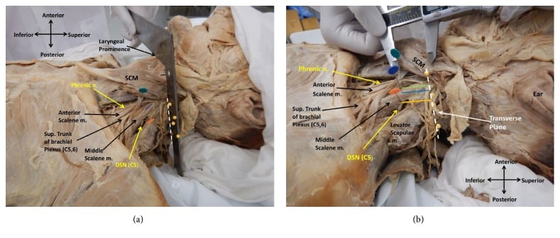
- Both rhomboids are supplied by the dorsal scapular nerve, which arises from the C5 root proximal to the upper trunk and pierces the middle scalene - but say 'usually': in Nguyen's 23 dissected nerves about 70 per cent arose from C5, 74 per cent pierced the middle scalene, and 48 per cent supplied levator scapulae alone.
- The dorsal scapular nerve and artery descend together 1 to 2 cm medial to the medial scapular border, on the deep surface of the rhomboids - the structure divided or avulsed in any careless interscapular dissection.
- Rhomboids are DOWNWARD rotators of the glenoid, antagonists of the trapezius-serratus upward-rotation couple.
- Rhomboid palsy produces a subtle winging - the scapula sits slightly laterally and inferiorly displaced, and the winging is best seen on the eccentric phase of lowering the arm from forward flexion.
- The medial border is the interval of the Judet posterior approach to the scapula; the rhomboids are detached flush with bone so the pedicle is left undisturbed.
- “A C5 root avulsion abolishes rhomboid function; an upper trunk lesion distal to the dorsal scapular branch spares it, so preserved rhomboids on EMG mean the lesion is postganglionic and distal to the C5 root. The rule assumes the nerve reliably reaches the rhomboids off C5, which held in only about half of Nguyen's specimens, so corroborate an isolated rhomboid finding with serratus anterior and paraspinal EMG.
- “The dorsal scapular nerve and the long thoracic nerve both pierce the middle scalene, which is why an interscalene block can produce transient scapular winging.
- “Dorsal scapular nerve entrapment gives a deep, dull interscapular ache radiating to the lateral arm with no dermatomal sensory loss, because the nerve is purely motor.
- “Divide the rhomboids within a few millimetres of the medial scapular border in the Judet approach - the artery lies 1 to 2 cm medial and retracts irretrievably if cut.
Overview
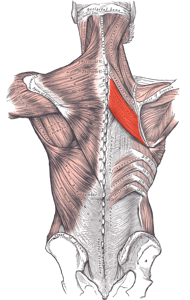
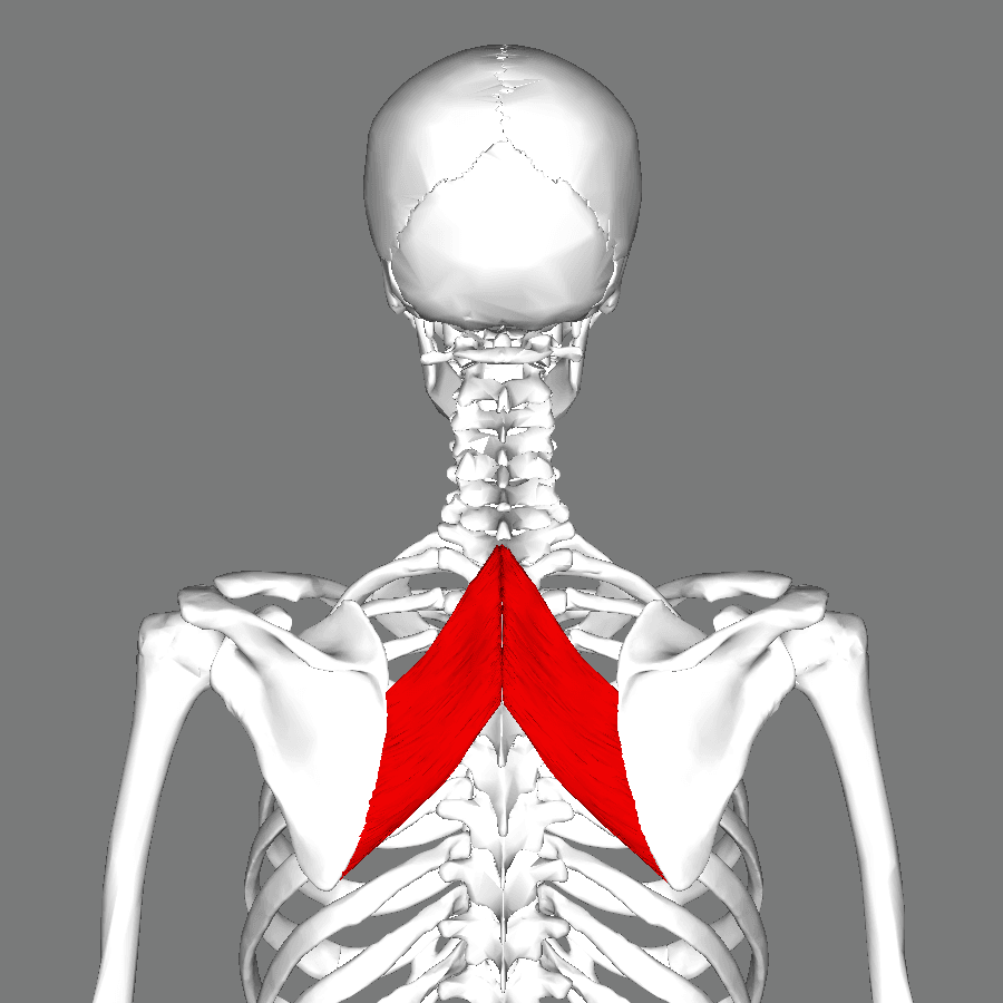
The rhomboids are two flat, quadrilateral sheets running obliquely downward and laterally from the vertebral spinous processes to the medial border of the scapula. They lie deep to the trapezius, with serratus posterior superior and the erector spinae deep to them. They are the scapula's medial tether: everything that pulls the scapula forward around the chest wall, whether serratus anterior, pectoralis minor or the weight of the arm, is resisted by these two muscles.
Rhomboid minor is the narrow upper band. Rhomboid major is the broader lower sheet, roughly twice the size of the minor, and reaches the inferior angle.
Why they matter. Three surgical settings recur in examinations:
- They define the interval and the danger zone of the posterior (Judet) approach to the scapula.
- Their nerve, the dorsal scapular nerve, is the single most useful electrodiagnostic marker for the level of a brachial plexus lesion.
- They are the donor muscles of the Eden-Lange transfer, so their integrity dictates whether an irreparable trapezius palsy is reconstructable at all.
Attachments, Innervation and Relations
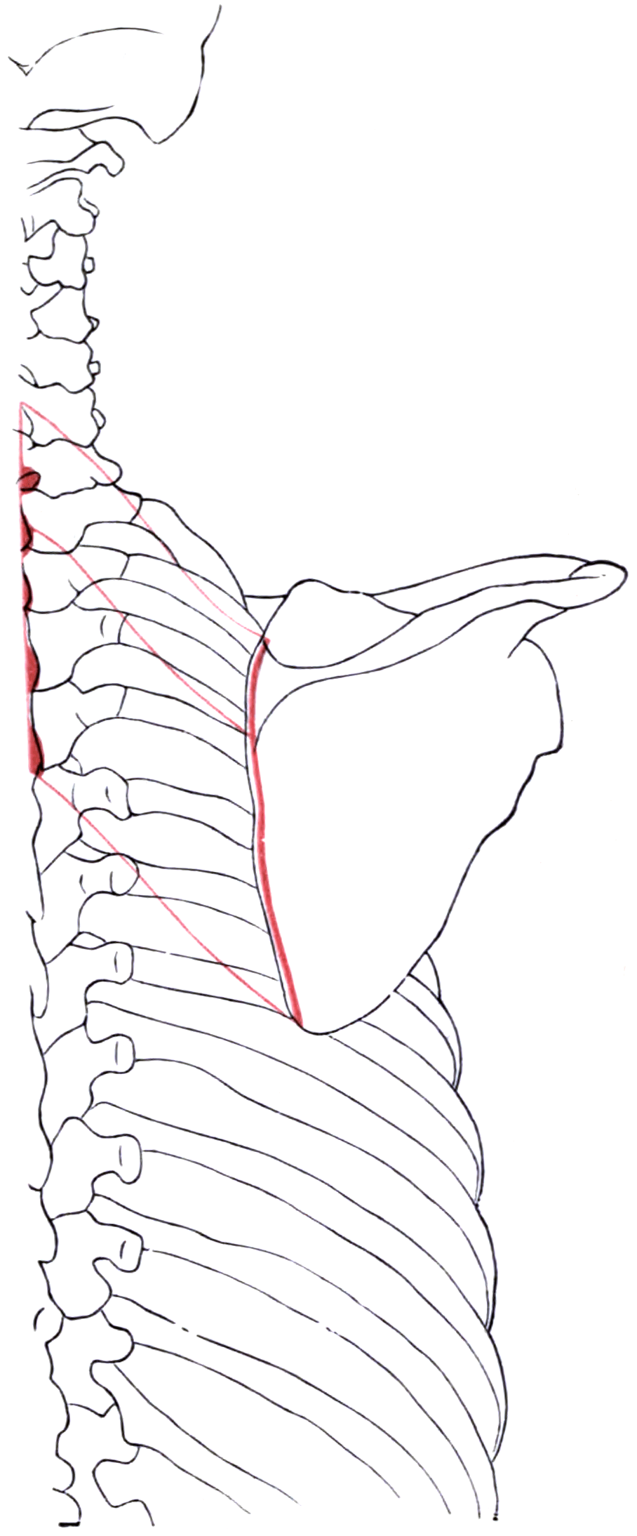
Rhomboid minor. It arises from the lower part of the ligamentum nuchae and the spinous processes of C7 and T1, with their supraspinous ligaments. It inserts as a narrow band, roughly 1 to 2 cm wide, onto the medial border of the scapula at the root (base) of the scapular spine, the smooth triangular area where the spine meets the medial border.
Rhomboid major. It arises from the spinous processes of T2 to T5 and the intervening supraspinous ligaments, and inserts along the medial border from the root of the scapular spine down to the inferior angle. Much of the insertion is through a tendinous arch that attaches firmly at its upper and lower ends with a thinner membranous middle, a detail that matters when you are looking for suture purchase.
Fused more often than not. A thin fascial cleft may separate the two, but rhomboid minor is frequently fused with rhomboid major. In a minority of people the two are entirely separate with a clear interval, and in others rhomboid minor is doubled.
Fibre direction. Both run inferolaterally from spine to scapula at roughly 45 degrees. That oblique orientation means their pull retracts and elevates the medial border, producing downward rotation of the glenoid.
Landmarks for the incision. With the arm at the side, the root of the scapular spine lies at the level of the T3 spinous process and the inferior angle at T7, which lets you site an incision without imaging. The medial border is subcutaneous in a thin patient and is the direct route to both muscles.
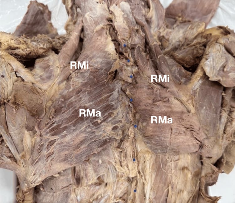
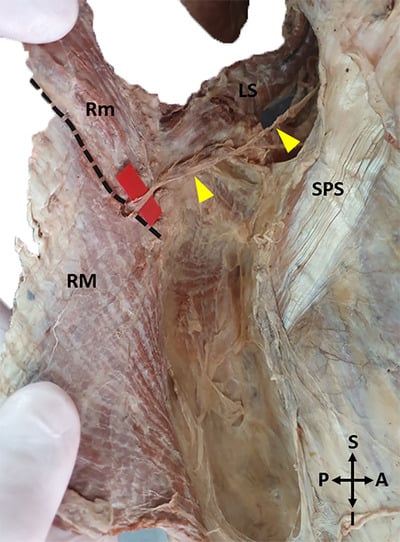
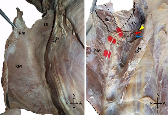
Every interscapular operation, whether Judet approach, scapulothoracic fusion, Eden-Lange harvest or rhomboid repair, shares one hazard: the dorsal scapular neurovascular bundle running vertically 1 to 2 cm medial to the medial border of the scapula, deep to the rhomboids.
Three rules follow.
- Detach the rhomboids flush with bone, subperiosteally, leaving a cuff of tendon on the muscle for later repair. Never divide them in their belly.
- Work from lateral to medial, so the bundle always lies beyond your dissection rather than within it.
- If bleeding occurs, do not blindly diathermy medially, because the artery retracts into the paraspinal mass. Pack, extend the exposure, and find the vessel.
Action and Biomechanics
Two opposing couples. Scapular position at rest and during elevation is a tug of war between two couples:
- Upward rotators - upper trapezius, lower trapezius and serratus anterior - swing the glenoid up and the inferior angle laterally.
- Downward rotators - rhomboid minor and major, levator scapulae, pectoralis minor and latissimus dorsi - swing the glenoid down and the inferior angle medially.
Middle trapezius joins the rhomboids in retraction, and levator scapulae shares their nerve. Serratus anterior opposes them in protraction as well as rotation.
The rhomboid-serratus couple. These two are a true antagonistic pair across the scapula: serratus pulls the medial border forward around the ribs, the rhomboids pull it back to the spine. Either failing produces winging, but in opposite directions.
- Mechanism
- Direct medial pull on the medial border
- Functional Significance
- Fixes the scapula for pulling and rowing activity
- Loss Produces
- Weak rowing; protracted, forward-drifted scapula
- Mechanism
- Oblique inferolateral fibre direction
- Functional Significance
- Balances the depressing pull of the lower trapezius
- Loss Produces
- Loss of the resting scapular set point
- Mechanism
- Superomedial pull above the rotation axis
- Functional Significance
- Controls the return from overhead elevation
- Loss Produces
- Uncontrolled eccentric lowering; scapular dyskinesis
- Mechanism
- Anteriorly directed component toward the vertebrae
- Functional Significance
- Maintains the scapulothoracic articulation
- Loss Produces
- Subtle medial border lift - rhomboid winging
- Mechanism
- Antagonist co-contraction
- Functional Significance
- Provides a fixed base for deltoid and cuff
- Loss Produces
- Loss of a stable proximal base; effort intolerance overhead
Why rhomboid failure wings the scapula. The rhomboids are the only downward rotators that also press the medial border against the chest wall. Their vector is directed superomedially and anteriorly, toward the vertebral column, which lies anterior to the plane of the scapular blade. That anterior component is why rhomboid failure produces winging at all.
Stabilisers more than movers. They have a short moment arm for rotation but a large physiological cross-sectional area relative to their excursion, which marks them as postural stabilisers rather than prime movers. Total excursion is only a few centimetres, which limits how far a rhomboid can be lateralised in an Eden-Lange transfer.
Why the winging is subtle. Because the muscle is a stabiliser, a patient loses scapular control long before they lose scapular power. The trapezius overlies the rhomboids and can retract the scapula in their place, so an isolated rhomboid palsy is partially compensated from the outset; what is not compensated is control.
The eccentric phase. The rhomboids are maximally loaded eccentrically, controlling the scapula as the arm is lowered and as the trunk is pulled forward in a rowing stroke. During the lowering phase of forward flexion they control the scapula's return from upward rotation, and that is the phase in which a weak rhomboid lets the medial border lift.
Surface Anatomy and Examination
Palpation. Locate the root of the scapular spine and the inferior angle, the two vertebral-level landmarks given with the attachments. Rhomboid major occupies the medial border between them; rhomboid minor sits at and just above the spine root.
The palpation manoeuvre. Ask the patient to place the hand behind the back and then lift the hand away from the back. This retracts and downwardly rotates the scapula, lifting the medial border away from the chest wall and making both rhomboids palpable as firm oblique bands.
Where it is tender. Tenderness at the medial border insertion is common and non-specific. Tenderness in the muscle belly, midway between spine and scapula, is more suggestive of a genuine rhomboid problem.
Hand-on-hip resisted retraction is the standard isolation manoeuvre. The patient stands with the hand of the affected side on the hip and the elbow pointing backwards, then pushes the elbow backwards against your resistance. If the medial border lifts off the chest wall and the scapula translates laterally, the rhomboids are insufficient: the trapezius alone cannot hold the medial border down.
Eccentric lowering is where rhomboid winging is actually seen. The patient forward-flexes both arms to full elevation, then lowers them slowly over about five seconds while you watch both medial borders from behind through the whole descent. Subtle lifting of the medial border and lateral drift of the scapula on the descent only is loss of eccentric control by the downward rotators, the most sensitive sign of rhomboid palsy, and the phase most examiners forget to watch.
Two control tests. They exclude the commoner wings:
- Wall push-up, with the elbows extended. Frank medial winging of the whole medial border and inferior angle is serratus anterior palsy, the far commoner diagnosis; an obvious, gross wing is serratus, and rhomboid winging is never gross.
- Resisted abduction in the coronal plane, with the shrug tested as well. The inferior angle rotating laterally with a drooped shoulder and a weak shrug is trapezius palsy, which is much commoner than rhomboid palsy and shares its lateral direction, so it must be excluded first.
The commonest error is calling a rhomboid palsy when the patient has a trapezius palsy, since both produce a lateral pattern. The discriminators are the shrug, weak in trapezius palsy and normal in rhomboid palsy, and the shoulder height, drooped in trapezius palsy and level in rhomboid palsy.
False positives and false negatives. Interscapular myofascial pain, thoracic facet or costotransverse joint pain and C5 cervical radiculopathy can all produce interscapular ache with normal muscles. The false negative is the well-compensated patient examined only at rest or only concentrically: always watch the descent.
EMG is the arbiter. Needle examination of the rhomboids is technically demanding because the needle must traverse the trapezius. It must be performed by an operator who will document the depth and confirm that the units recorded are rhomboid rather than trapezius.
Complications
Iatrogenic dorsal scapular nerve injury. It happens at two sites: the middle scalene, in scalenectomy, first rib resection, supraclavicular plexus exploration and interscalene block, and the medial scapular border, in the Judet approach, scapulothoracic surgery and rhomboid harvest. The result is rhomboid and often levator scapulae denervation, giving a subtle lateral wing and, more troublesome to the patient, a chronic interscapular ache. In the neck, split the middle scalene under direct vision and identify both the dorsal scapular and long thoracic nerves before dividing anything; at the scapula, stay flush with bone.
Dorsal scapular artery haemorrhage. Division during medial detachment causes brisk bleeding into the paraspinal muscle with retraction of the vessel. It may present post-operatively as an expanding interscapular haematoma with disproportionate pain. Control it at the time: extend the exposure and ligate rather than diathermy blindly.
Pleural injury. Pneumothorax after scapulothoracic fusion, deep interscapular injection or rhomboid dry needling is a genuine and reported complication. Suspect it with new pleuritic pain or desaturation and image immediately.
Failure and attenuation of transfers. Recurrent winging after Eden-Lange usually reflects an unrecognised second palsy or a dorsal scapular nerve lesion missed pre-operatively, rather than failure of the transfer itself. Loss of retraction strength after a rhomboid transfer is expected and should be part of consent, particularly for a rower or a climber.
Scapular dysfunction after the Judet approach. Detaching and repairing the rhomboids and levator, and raising the infraspinatus flap, both produce measurable periscapular weakness. The modified Judet was developed specifically to avoid this and is preferred wherever the fracture pattern allows.
Misdiagnosis. The commonest "complication" in this territory is failing to make the diagnosis: an interscapular ache in an athlete labelled myofascial for two years when it is a dorsal scapular nerve entrapment, or a subtle rhomboid wing labelled postural when the patient has an evolving C5 lesion.
Clinical Relevance
Causes. The nerve or its root can be injured in five settings:
- Dorsal scapular nerve injury at the middle scalene, from traction, direct trauma or a penetrating injury to the posterior triangle
- Surgery: a posterior approach to the cervicothoracic spine, first rib resection or scalenectomy for thoracic outlet syndrome, or the medial dissection of a scapulothoracic procedure
- An interscalene brachial plexus block, because the dorsal scapular and long thoracic nerves both pierce the middle scalene and can be injured by needle trauma or by local anaesthetic neurotoxicity
- Neuralgic amyotrophy (Parsonage-Turner syndrome), typically as part of a patchy multi-nerve pattern with severe prodromal pain
- C5 root pathology: foraminal stenosis, disc herniation, or root avulsion in a traction plexus injury
Presentation. The patient rarely presents complaining of weakness; they present with pain and a shoulder that feels wrong. The complaint is a vague, dull, deep interscapular ache, worse with sustained postural loading, with fatigue and a sense of instability on pulling and overhead activity and the sense that "the shoulder blade feels loose".
What you see. On careful observation the scapula sits slightly lateral and slightly inferior, and the medial border lifts subtly, most visibly during slow lowering from forward flexion and on resisted retraction with the hand on the hip.
Differential of interscapular pain with a wing. The alternatives each have a discriminating feature:
- Trapezius (spinal accessory) palsy - drooped shoulder, weak shrug
- Serratus (long thoracic) palsy - gross medial winging on the wall push
- C5 cervical radiculopathy - dermatomal sensory change, weak deltoid
- Scapulothoracic bursitis or snapping scapula - audible or palpable crepitus
- Osteochondroma of the ventral scapular surface - a fixed, non-dynamic pseudo-wing
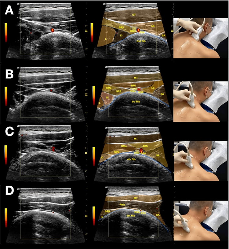
The dorsal scapular nerve leaves C5 before the upper trunk forms, so the state of the rhomboids on EMG places a brachial plexus lesion relative to that take-off.
Rhomboids denervated. The lesion is at or proximal to the C5 root, which raises the possibility of a preganglionic root avulsion. That is not amenable to grafting, so plan nerve transfers. Corroborate the proximal level with denervation of the paraspinal muscles (posterior primary ramus) and the serratus anterior (long thoracic, also very proximal, C5-C7), a raised hemidiaphragm (phrenic) and a Horner sign (T1 sympathetic).
Rhomboids preserved. The take-off is spared, so the lesion is postganglionic, at or beyond the upper trunk; a proximal stump exists and the lesion is potentially graftable. The same finding is also consistent with an isolated suprascapular or axillary nerve lesion rather than a plexus injury.
The caveat. A C4-dominant dorsal scapular nerve can preserve the rhomboids despite a C5 lesion, so never rest the whole decision on rhomboid EMG alone.
Surgical Relevance
The posterior approach to the scapula (Judet and modified Judet)
Indication. Open reduction and internal fixation of displaced scapular body, neck and glenoid fossa fractures, and access to the posterior glenoid.
Position and incision. Position the patient lateral decubitus or prone, with the arm draped free so it can be placed in flexion and internal rotation to relax the deltoid. The boomerang (Judet) incision runs along the scapular spine laterally to the acromion and turns distally along the medial border to the inferior angle.
The classical Judet exposure. It is developed in two layers:
- Superficially, the deltoid is elevated off the scapular spine and reflected laterally, protecting the axillary nerve as it emerges from the quadrangular space, and the trapezius is elevated medially.
- In the medial parascapular interval, the rhomboids and levator scapulae are detached flush with the medial border of the scapula, subperiosteally, leaving a repairable tendinous cuff. This releases the medial tether and allows infraspinatus and teres minor to be raised as a laterally based flap on their pedicle, exposing the whole dorsal scapula.
The modified (minimally invasive) Judet. The muscle flap is not raised. The work is done through two windows: the interval between infraspinatus and teres minor for the lateral border and glenoid neck, and the plane along the medial border deep to the rhomboids for the medial border. This spares the rhomboid detachment entirely and has largely replaced the full Judet in most units.
The rule for the rhomboid detachment. Detach with a knife on bone, hugging the medial border: the dorsal scapular nerve and artery lie 1 to 2 cm medial to your blade. Retract the released muscle medially with a broad retractor placed against the muscle, never with a self-retainer biting into the paraspinal mass.
The rhomboids as transfer donors
The role of the rhomboids. In the Eden-Lange transfer for irreparable trapezius palsy the rhomboids provide two of the three limbs:
- Rhomboid minor is detached from the medial border and transferred laterally onto the infraspinatus fossa, substituting for the middle trapezius.
- Rhomboid major is detached and transferred laterally onto the infraspinatus fossa below the minor, substituting for the lower trapezius.
- Levator scapulae, the third limb, goes laterally onto the scapular spine to substitute for the upper trapezius. In the modified transfer it is carried further laterally, toward the lateral spine or acromion, to lengthen its upward-rotation moment arm.
Technical requirements. The technique has four requirements:
- Confirm the dorsal scapular nerve is intact on EMG. All three donors depend on it, and transferring denervated muscle is a wasted operation.
- Detach each muscle with a wafer of bone or a tendinous cuff for suture purchase; the membranous middle of rhomboid major is poor suture tissue.
- Protect the dorsal scapular pedicle, which enters the deep surface medially and must not be tethered or divided as the muscle is swung laterally. The pedicle, too, limits excursion to a few centimetres.
- Fix through bone tunnels or troughs in the infraspinatus fossa with transosseous sutures, tensioned with the arm at about 30 degrees of abduction.
Outcome. Expect reliable pain relief with improvement in scapular position and cosmesis; abduction improves but is rarely normalised. Long-term follow-up shows the correction is durable where the trapezius palsy is truly isolated, and the reported failures had a coexisting long thoracic and dorsal scapular nerve lesion.
Guidelines, Registries & Global Practice
Anatomical variation that matters
- Fusion of rhomboid minor and major is common; a clean fascial plane between them is present in only a proportion of specimens, which complicates raising them as two independent limbs in an Eden-Lange transfer.
- Dorsal scapular nerve origin: approximately 70 per cent arise from C5, and 74 per cent pierce the middle scalene in the largest cadaveric series; a contribution from C4 is described and a C4-dominant nerve would preserve rhomboid function despite a C5 root lesion - the caveat to the plexus-localisation rule.
- Middle scalene penetration is the usual pattern, but the nerve may pass anterior or posterior to the muscle, altering the entrapment risk and the decompression plan.
- Dorsal scapular artery origin is from the thyrocervical trunk, as the deep branch of the transverse cervical artery, in the majority, and directly from the subclavian artery in a substantial minority; the direct pattern passes between the plexus trunks and can be encountered during supraclavicular exploration.
- Rhomboideus occipitalis and other accessory slips are described and are of no clinical consequence except as a source of confusion at operation.
Practice framing across bodies
- Position Relevant to the Rhomboids and the Dorsal Scapular Nerve
- Favours interval-based posterior approaches where the fracture allows, minimising periscapular muscle detachment and denervation.
- Position Relevant to the Rhomboids and the Dorsal Scapular Nerve
- Recognise three distinct winging patterns - serratus, trapezius and rhomboid - and recommend testing all three, including an eccentric lowering manoeuvre.
- Position Relevant to the Rhomboids and the Dorsal Scapular Nerve
- Rhomboid needle EMG is a standard part of localising a brachial plexus injury; denervation raises suspicion of preganglionic avulsion.
- Position Relevant to the Rhomboids and the Dorsal Scapular Nerve
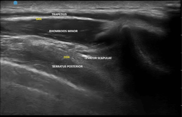
- Interscalene needle placement should avoid the substance of the middle scalene, where the dorsal scapular and long thoracic nerves lie; ultrasound guidance reduces but does not abolish the risk.
Resource-dependent practice
- Well-resourced settings: ultrasound-guided diagnostic scalene blocks, high-resolution MR neurography of the dorsal scapular nerve, and CT-based planning of the posterior scapular approach.
- Limited-resource settings: the diagnosis remains clinical and is entirely reliable if all three winging patterns are tested; the modified Judet approach requires only standard retractors; Eden-Lange requires nothing beyond heavy suture and a drill.
Epidemiological framing
- Isolated rhomboid palsy is rare relative to serratus and trapezius palsy, and published experience consists of case series rather than trials.
- Interscapular pain is extremely common and overwhelmingly musculoskeletal; dorsal scapular nerve entrapment is a diagnosis of careful exclusion, supported by electrophysiology and a diagnostic block, not by history alone.
- The modified, muscle-sparing posterior scapular approach has become the default in most trauma units internationally, with the classical Judet flap reserved for complex articular patterns.
MCQ Practice Points
Q: What is the root value of the dorsal scapular nerve, and where does it arise? A: C5 in about 70 per cent of dissected nerves, with a C4 contribution described in some, arising from the ventral ramus proximal to the formation of the upper trunk.
Q: Which muscle does the dorsal scapular nerve pierce? A: The middle scalene - together with, or just above, the long thoracic nerve. This is the site of entrapment and of block-related injury.
Q: How far medial to the medial scapular border does the dorsal scapular neurovascular bundle run? A: 1 to 2 cm, on the deep surface of the rhomboids.
Q: Where exactly do the two rhomboids insert? A: Rhomboid minor at the medial border at the root of the scapular spine; rhomboid major along the medial border from the spine root to the inferior angle.
Q: Do the rhomboids upwardly or downwardly rotate the glenoid? A: Downwardly. They are antagonists of the trapezius-serratus upward-rotation couple.
Q: Which spinous processes correspond to the root of the scapular spine and the inferior angle? A: T3 for the root of the spine and T7 for the inferior angle, with the arm at the side.
Q: What is the cutaneous territory of the dorsal scapular nerve? A: None. It is purely motor, which is why entrapment causes a deep ache without sensory loss.
Q: Which muscle forms the floor of the triangle of auscultation? A: Rhomboid major, with trapezius medially, latissimus dorsi inferiorly and the medial scapular border laterally.
Q: What does denervation of the rhomboids tell you in a brachial plexus injury? A: The lesion is at or proximal to the C5 root - suspect a preganglionic avulsion, which is not graftable.
Q: Which parts of the trapezius do the two rhomboids substitute for in Eden-Lange? A: Rhomboid minor for the middle trapezius and rhomboid major for the lower trapezius, both transferred laterally onto the infraspinatus fossa.
Exam Viva Scenarios
Practise clinical reasoning and management decisions out loud
“A 25-year-old competitive rower complains of a deep ache between the shoulder blades and a feeling that the right shoulder blade is loose. The wall push-up is normal and the shrug is strong. What is your assessment?”
“You are fixing a displaced scapular body and glenoid neck fracture through a posterior approach. Your assistant is retracting hard medially and there is sudden brisk bleeding from deep to the rhomboids. What has happened and what do you do?”
“A 22-year-old motorcyclist has a flail arm after a high-energy fall. You need to know whether his C5 and C6 lesions are graftable. How do the rhomboids help you, and what else do you look for?”
Anatomy
- Minor: ligamentum nuchae, C7-T1 to the medial border at the spine root
- Major: T2-T5 to the medial border from spine root to inferior angle
- Nerve: dorsal scapular (C5, sometimes C4)
- Artery: dorsal scapular, 1-2 cm medial to the medial border
Nerve Course
- Leaves C5 proximal to the upper trunk
- Pierces middle scalene with the long thoracic nerve
- Supplies levator scapulae; in about half it supplies levator alone and never reaches the rhomboids
- Purely motor - no sensory territory
Clinical
- Action: retraction, elevation, downward rotation, medial border compression
- Palsy: subtle lateral wing, seen on eccentric lowering
- Entrapment at middle scalene: deep interscapular ache, normal sensation
- Denervated rhomboids on EMG suggest a preganglionic C5 lesion
Surgical
- Judet approach: detach flush with the medial border, lateral to medial
- Modified Judet uses intervals and avoids rhomboid detachment
- Triangle of auscultation: rhomboid major in the floor
- Eden-Lange: minor replaces middle trapezius, major replaces lower trapezius
Evidence Base
A Cadaveric Investigation of the Dorsal Scapular Nerve
- 23 dorsal scapular nerves documented in 20 embalmed adult cadavers
- Approximately 70 per cent of the nerves originated from C5, and 74 per cent pierced the middle scalene muscle
- About 48 per cent supplied levator scapulae alone, while 52 per cent supplied both levator scapulae and the rhomboids
- Measured from a transverse plane at the laryngeal prominence, the nerve entered, crossed and exited the middle scalene at mean distances of 1.50 cm, 1.79 cm and 2.08 cm
Role of Dorsal Scapular Nerve Entrapment in Unilateral Interscapular Pain
- 55 consecutive patients with unilateral interscapular pain compared with 30 healthy controls, assessed clinically and electrophysiologically
- Electrophysiological abnormalities consistent with a dorsal scapular nerve lesion were detected in 29 patients (52.7 per cent)
- 25 patients had electromyographic abnormalities in rhomboid major and levator scapulae; prolonged compound muscle action potential latency was found in only 4
- Scapular winging was observed in 9 patients (16.4 per cent)
- The authors concluded entrapment at scalenus medius, or nerve stretch during overhead activity, is a frequent contributor to interscapular pain
A Modified Judet Approach to the Scapula
- Description of a modified Judet approach for operative exposure of displaced and intra-articular scapular fractures
- The approach aims to expose all bony elements of the scapula with adequate bone stock for internal fixation
- It is designed to cause minimal trauma to the rotator cuff musculature
- It explicitly protects the suprascapular nerve superiorly and the axillary nerve laterally
- The stated advantage is limiting muscular dissection, potentially improving rehabilitation and reducing operative morbidity
Transfer of the Levator Scapulae, Rhomboid Major and Rhomboid Minor for Paralysis of the Trapezius
- 22 patients with trapezius paralysis from spinal accessory nerve injury underwent transfer of levator scapulae and both rhomboids
- Aetiology was cervical node biopsy in 13, trauma in 7 and radical neck dissection in 2
- All had pain, neckline asymmetry, a drooping girdle with lateral displacement of the scapula and weak active elevation
- 14 patients had been given an incorrect clinical diagnosis and 12 had had an inaccurate or incomplete electromyographic examination
- At a mean of 7.5 years the result was excellent in 13, satisfactory in 6 and unsatisfactory in 3, with adequate pain relief and functional improvement in all but 3
- A LONG THORACIC NERVE PALSY developed in THREE patients - a complication of the transfer itself, and one that removes the serratus half of the upward-rotation couple the operation is trying to restore
Brachial Plexopathies - Classification, Causes and Consequences
- Review of the brachial plexus as the most complex structure of the peripheral nervous system
- The high incidence of plexopathy reflects its vulnerability to trauma and to secondary involvement by adjacent disease
- Combining anatomic, pathophysiological and neuromuscular knowledge with detailed clinical and ancillary studies yields diagnostic and prognostic information
- Because most plexus disorders show a regional predilection rather than involving the whole plexus, a regional approach to assessment is required
Scapulothoracic Fusion for Facioscapulohumeral Muscular Dystrophy
- Eleven scapulothoracic fusions in eight patients with infantile and adolescent facioscapulohumeral muscular dystrophy, mean age 17 years
- The scapula was fused in 25 degrees of abduction using 16-gauge wires with a plate or washers to prevent wire pull-out, plus iliac crest autograft
- Winging, shoulder fatigue and pain were eliminated in every case; abduction improved from a mean of 75 to 145 degrees and flexion to 144 degrees
- At a mean of 6.3 years motion was maintained in seven shoulders but had fallen to a mean of 48 degrees in four because of progressive deltoid weakness
- The only complication was prominent subcutaneous wires requiring trimming in two cases