A lump in the foot is usually a ganglion — but the foot is the commonest site in the body for synovial sarcoma
- A mass that is small, deep to fascia, firm, or has grown is malignant until proven otherwise — size less than 5 cm does NOT exclude sarcoma in the foot, and now there is a number for it: 65 per cent of acral sarcomas measured under 5 cm at presentation, median 3 cm. Dean's authors state it outright — size 'should not be used as a diagnostic threshold for referral'.
- The threshold that DOES carry meaning in the foot is 3 cm. In 84 hand and foot synovial sarcomas, tumours under 3.0 cm had 95 per cent five-year progression-free and 100 per cent overall survival; at 5.0 cm or more, progression-free survival was 53 per cent (hazard ratio 7.99).
- Examine the skin and the nail, not just the lump: in a 195-tumour foot series the commonest MALIGNANT soft tissue lesion was malignant melanoma, not a sarcoma. Acral and subungual melanoma are frequently amelanotic and are mistaken for a wart, an ulcer or a fungal nail.
- Ultrasound answers one question well: cystic or solid. A solid mass in the foot needs MRI with gadolinium.
- Never shell out (marginal excise) an undiagnosed solid soft-tissue mass — unplanned excision compromises limb salvage and worsens local control.
- Plantar fibromatosis (Ledderhose) arises from the plantar aponeurosis, is often multinodular and bilateral, and is the classic benign mimic of a plantar sarcoma.
- Morton's neuroma is a mechanical, symptomatic diagnosis in the 3rd (then 2nd) web space; imaging is confirmatory, not diagnostic.
- Refer to a sarcoma unit BEFORE biopsy: the biopsy tract must lie in the line of the definitive resection.
- “Foot and ankle is the single commonest site for synovial sarcoma; the classic story is a young adult with a painful lump present for years.
- “Calcification within a soft-tissue mass in a young adult narrows to synovial sarcoma, haemangioma with phleboliths, myositis ossificans or tumoural calcinosis.
- “Fluid-fluid levels on MRI suggest a haemorrhagic or vascular lesion — but also occur in sarcoma; they are not a reassurance sign.
- “Blooming on gradient-echo sequences equals haemosiderin: think pigmented villonodular synovitis / tenosynovial giant cell tumour.
- “A lesion that is bright on T2 and shows NO enhancement after gadolinium is a true cyst; any internal enhancement means it is solid.
Foot sarcomas are small at presentation because the compartments are tight and the lump is noticed early in a shoe. Synovial sarcoma is frequently under 3 cm. Size is a staging variable, not a diagnostic one.
Most benign soft-tissue tumours are painless; synovial sarcoma is the notorious exception and is painful in roughly a third. "It hurts, so it cannot be a tumour" is a wrong answer.
A ganglion must be anechoic, non-vascular on Doppler, and transmit sound. Internal echoes, vascularity or incompressibility mean solid — proceed to MRI with contrast.
Marginal excision of an unrecognised sarcoma seeds the whole compartment, mandates re-excision and often converts a limb-salvage case into an amputation. Image first, refer, then biopsy.
Recognising the Pattern
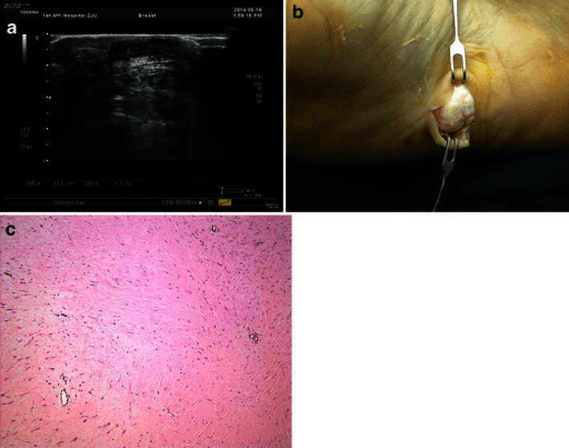
The pattern: a discrete soft-tissue swelling of the foot, either palpable clinically or seen radiographically as a focal soft-tissue density that displaces fat planes, with or without matrix mineralisation, pressure erosion of adjacent cortex, or underlying bony change.
Confirming it is genuinely a mass
- On plain film, a true mass displaces the normal fat planes and has convex margins. Diffuse thickening without a convex border is oedema or cellulitis, not a mass.
- Confirm it is not simply a bony prominence or exostosis — the plain film should show whether the density is continuous with cortex.
- On ultrasound, confirm three things: is it cystic or solid, is it compressible, and is there internal Doppler flow.
- On MRI, confirm the mass has a discrete margin on T1 (fat-suppressed sequences exaggerate oedema and overcall size).
Words to use out loud
"These are weight-bearing radiographs of the foot. There is a well-defined soft-tissue mass in the plantar soft tissues measuring approximately X cm, deep to the plantar fascia. There is no matrix mineralisation and no underlying cortical destruction or periosteal reaction. The joint spaces are preserved. My differential is a benign lesion such as a ganglion or plantar fibroma, but a soft-tissue sarcoma — particularly synovial sarcoma, for which the foot is the commonest site — must be excluded. I would take a history for duration, growth and pain, examine for depth, fixity and size, and arrange MRI with gadolinium before any intervention."
Mimics — what is NOT a tumour
- Giveaway
- Follows skeletal muscle signal on every sequence; no enhancement beyond muscle
- Giveaway
- Symmetrical on the contralateral side if imaged
- Giveaway
- Rim enhancement with non-enhancing centre, systemic features, raised inflammatory markers
- Giveaway
- Intermediate to low T2 signal, dual-energy CT shows urate, periarticular erosions with overhanging edges
- Giveaway
- Known seropositive disease, subluxed MTP joints
- Giveaway
- History of injury; must resolve — a "haematoma" that persists beyond six weeks is a sarcoma until proven otherwise
- Giveaway
- Pulsatile, Doppler shows yin–yang flow with a neck
Next Investigation
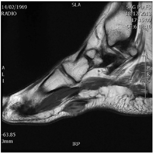
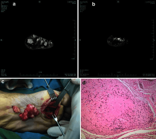
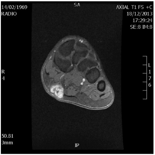
The single most useful MRI sequence for a foot mass is post-gadolinium fat-suppressed T1. It answers the one question that changes everything: does the lesion enhance internally. No enhancement equals cyst. Enhancement equals solid tissue and a possible sarcoma.
The Differential
- Typical age / setting
- 15–40 years; foot and ankle is the commonest site
- Discriminating feature
- Small (often less than 3 cm), slow-growing over years, PAINFUL, juxta-articular but not intra-articular; calcifies in about a third; triple signal on T2
- What confirms it
- MRI with contrast then core biopsy at a sarcoma unit; SS18 (SYT) translocation t(X;18)
- Typical age / setting
- 20–40 years; tendons and aponeuroses of foot and ankle
- Discriminating feature
- Attached to tendon or aponeurosis; T1 signal HIGHER than muscle due to melanin — unusual for a sarcoma
- What confirms it
- Biopsy: t(12;22) EWSR1–ATF1; S100 and HMB-45 positive
- Typical age / setting
- Over 50 years; deep or subfascial
- Discriminating feature
- Larger, heterogeneous, necrotic centre; myxofibrosarcoma shows an infiltrative enhancing 'tail' along fascia
- What confirms it
- MRI with contrast then core biopsy; tail must be included in resection
- Typical age / setting
- Over 50 years; sole, heel or nail bed
- Discriminating feature
- It is a SKIN lesion — pigmentation, ulceration, Hutchinson's sign at the nail fold; amelanotic variants mimic granuloma
- What confirms it
- Excisional or incisional skin biopsy; not an imaging diagnosis
- Typical age / setting
- Over 55 years; known lung, renal or breast primary
- Discriminating feature
- Rare distal to the knee — but lung is the classic primary; usually painful with bone destruction rather than a pure soft-tissue lump
- What confirms it
- Staging CT chest–abdomen–pelvis, bone scan, biopsy
- Typical age / setting
- Any age; dorsum of foot, sinus tarsi, tendon sheath
- Discriminating feature
- Anechoic and compressible on ultrasound, no Doppler flow, NO internal enhancement after gadolinium; transilluminates clinically
- What confirms it
- Ultrasound alone is sufficient if unequivocally anechoic and avascular
- Typical age / setting
- 30–60 years; associated with Dupuytren, epilepsy, diabetes, alcohol
- Discriminating feature
- Arises FROM the plantar aponeurosis, medial cord, often multinodular and bilateral; low T1 and low-to-intermediate T2 (collagen)
- What confirms it
- MRI showing continuity with the plantar fascia; observation, not excision, is first-line
- Typical age / setting
- 40–60 years, female predominance; 3rd then 2nd web space
- Discriminating feature
- Plantar to the intermetatarsal ligament, LOW signal on T2, positive Mulder click, radiating burning into the toes
- What confirms it
- Ultrasound or MRI in prone plantar-loaded position; diagnostic local anaesthetic block
- Typical age / setting
- 30–50 years; along flexor or extensor tendon sheaths, digits
- Discriminating feature
- Blooming artefact on gradient-echo from haemosiderin; low signal on T1 and T2 with avid enhancement; may cause smooth pressure erosion of cortex
- What confirms it
- MRI including gradient-echo; excision biopsy — CSF1 rearrangement
- Typical age / setting
- Childhood to young adult; enlarges with dependency or Valsalva
- Discriminating feature
- Phleboliths on plain film are close to pathognomonic; serpiginous T2-bright channels with fat interspersed; fluid–fluid levels
- What confirms it
- Plain film for phleboliths plus MRI; Doppler distinguishes high-flow from low-flow
- Typical age / setting
- 40–60 years; dorsum or plantar
- Discriminating feature
- Follows subcutaneous fat on EVERY sequence and suppresses completely with fat saturation; no thick septa, no nodules
- What confirms it
- MRI alone; septa thicker than 2 mm or nodular enhancement means atypical lipomatous tumour — biopsy
- Typical age / setting
- Any age; barefoot injury, glass or thorn
- Discriminating feature
- Central hyperechoic focus with posterior acoustic shadowing and a hypoechoic halo on ultrasound
- What confirms it
- Ultrasound (detects radiolucent wood and glass that plain film misses)
- Typical age / setting
- 20–50 years; along the course of a named nerve
- Discriminating feature
- Fusiform, entering and exiting nerve ('tail sign'), split-fat sign, target sign on T2; Tinel on percussion
- What confirms it
- MRI; schwannoma is eccentric and shellable, neurofibroma is central and is not
- Typical age / setting
- Renal failure, hyperparathyroidism (calcinosis); long-standing gout (tophus)
- Discriminating feature
- Amorphous periarticular calcification with fluid–calcium levels on horizontal-beam film (calcinosis); low T2 with urate on dual-energy CT (tophus)
- What confirms it
- Plain film plus serum calcium, phosphate, PTH, urate; dual-energy CT for gout
Narrowing It Down
- 11. Is it truly a mass — and have you looked at the skin and the nail?
Examine the whole foot including the nail beds and the interdigital web skin before palpating the lump, and check temperature, erythema and inflammatory markers.
Fever, erythema, raised CRP and rim enhancement point to abscess. Continuity with cortex on plain film points to osteochondroma or an accessory ossicle. A convex fat-plane-displacing density with a discrete margin is a genuine mass. And do not skip the skin: in a 195-tumour foot series the commonest malignant soft tissue lesion was MELANOMA, frequently amelanotic here and routinely mistaken for a wart, an ulcer or a fungal nail.
- 22. Cystic or solid?
Ultrasound first — it answers this in five minutes and needs no contrast.
Anechoic, compressible, avascular and posteriorly enhancing equals ganglion or bursa, and you can stop. Anything solid, vascular or with internal echoes goes to MRI with gadolinium. On MRI the definitive test is contrast: a true cyst shows a thin enhancing rim only, with no internal enhancement.
- 33. Which anatomical structure does it arise from?
Name the structure of origin before naming the lesion.
Plantar aponeurosis equals fibromatosis. Web space plantar to the intermetatarsal ligament equals Morton neuroma — for which ultrasound matches MRI (pooled sensitivity 0.91 versus 0.90) and is cheaper, dynamic and injectable in the same visit. Along a tendon sheath equals tenosynovial giant cell tumour or ganglion. Fusiform along a named nerve equals schwannoma. Juxta-articular but arising from NONE of these is the pattern that should raise sarcoma.
- 44. What is the signal or matrix?
Read T1, T2 and post-contrast fat-suppressed T1 together, and look for gradient-echo blooming.
Fat signal that suppresses completely equals lipoma. Low on T1 AND T2 equals collagen, haemosiderin or urate — fibroma, tenosynovial giant cell tumour or tophus. Very bright T2 with serpiginous channels and phleboliths equals vascular malformation. Heterogeneous intermediate signal with necrosis and avid patchy enhancement equals sarcoma until proven otherwise.
- 55. How has it behaved — and resist using size to reassure yourself
Ask duration and growth explicitly, measure the lesion, and record the measurement as a STAGING variable rather than a diagnostic one.
Anything growing, anything deep to fascia, anything recurrent after previous excision, and any 'haematoma' persisting beyond six weeks is treated as sarcoma. Size will not help you decide: 65 per cent of acral sarcomas were under 5 cm with a median of 3 cm, and Dean's authors state that size should not be used as a referral threshold. It does predict outcome once the diagnosis is made — under 3 cm gave 100 per cent five-year survival in foot synovial sarcoma against 53 per cent progression-free survival at 5 cm or more.
- 66. Solitary or multiple, and are there systemic features?
Examine the other foot, the hands and the skin, and take a renal and oncological history.
Multiple nodules along the medial plantar cord with palmar Dupuytren equals Ledderhose — and if surgery is later considered, the recurrence gradient is 67 per cent after local resection, 42 per cent wide, 27 per cent fasciectomy. Multiple lesions in neurofibromatosis equals neurofibromas. Multiple periarticular calcified masses with renal failure equals tumoural calcinosis. Multiple lesions with a known primary equals metastases.
- 77. If sarcoma remains on the list — stop and refer, before anything else
Do not biopsy and do not excise. Refer to the regional sarcoma multidisciplinary team and let the team that will resect plan the tract in the line of the definitive incision, staged with CT chest.
This is the step with the measured cost of failure: 43 per cent of acral sarcomas in Dean's series arrived having already been inadvertently excised, and although survival and local recurrence were not significantly different, those patients were significantly MORE LIKELY TO UNDERGO AMPUTATION (p equals 0.008). The whoops procedure does not usually kill the patient; it costs them part of the foot.
MCQ Practice Points
Q: Standard guidance is to refer any soft-tissue mass over 5 cm. Why is that rule unsafe in the foot?
A: Because it would miss most foot sarcomas. In Dean's 63 acral soft tissue sarcomas, 65 per cent measured under 5 cm at presentation, with a median largest dimension of 3 cm — the compartments are tight and a lump is noticed early inside a shoe, so these tumours present small. The authors state it plainly: size "should not be used as a diagnostic threshold for referral". Size remains a prognostic variable — over 5 cm predicted lower survival (p = 0.04) and higher local recurrence (p = 0.009) — but it is not a diagnostic one. Single tertiary centre, 63 patients over 16 years, so the proportions carry referral bias; the principle does not.
Q: How often does an acral sarcoma reach the sarcoma unit already excised, and what is the consequence?
A: 43 per cent — 27 of Dean's 63 patients had undergone inadvertent excision before referral. The consequence is specific and is not what most people assume: survival and local recurrence were not significantly different, but those patients were significantly more likely to undergo amputation (p = 0.008). Overall, 24 per cent came to partial amputation and 19 per cent to a more proximal one. The unplanned excision does not usually cost the patient their life; it costs them part of the foot, because the contaminated field can no longer be resected with a limb-preserving margin.
Q: In foot synovial sarcoma, at what size does outcome start to diverge?
A: At 3 cm, not 5. In 84 hand and foot synovial sarcomas, tumours under 3.0 cm had 95 per cent five-year progression-free survival and 100 per cent overall survival; 3.0 to 4.9 cm gave 84 per cent PFS; 5.0 cm or more gave 53 per cent (p = 0.007), with a hazard ratio for progression of 7.99. Local control was excellent throughout — 100 per cent local recurrence-free survival — so the problem is metastasis, not the operation. The authors recommend considering systemic therapy from 3.0 cm. Caveat: retrospective over 40 years at one cancer centre, and the hazard ratio's interval runs from 1.68 to 37.91.
Q: What is the commonest malignant soft-tissue lesion of the foot?
A: In a 195-tumour foot series drawn from 4,997 orthopaedic oncology records, it was malignant melanoma — not a sarcoma. Acral and subungual melanoma present late, are frequently amelanotic in this site, and are routinely mistaken for a wart, a chronic ulcer or a fungal nail. Examine the skin and the nail beds on every foot lump. Caveat on the denominator: that series comes from an oncology unit's referrals, so roughly one in four of its foot tumours being malignant vastly overstates the risk of a lump presenting in primary care.
Q: Which imaging modality should be used first for a suspected Morton neuroma?
A: Ultrasound. A meta-analysis of 14 studies found pooled sensitivity of 0.91 for ultrasound against 0.90 for MRI, with no significant difference (p = 0.88) — and ultrasound is cheaper, quicker, allows dynamic assessment and permits guided injection at the same visit. The one asymmetry: MRI's pooled specificity was 1.00 against 0.854 for ultrasound, so reserve MRI for the atypical picture. Important limitation: every included study used surgery as the reference standard, so only operated patients were verified and both sensitivities are inflated.
Q: A patient with symptomatic plantar fibromatosis asks about recurrence after surgery. What do you tell them?
A: That recurrence is likely and depends on how much is taken. Pooled across six studies and 109 feet: 67 per cent after local resection, 42 per cent after wide resection, 27 per cent after fasciectomy. Even the most extensive procedure recurs in more than a quarter, which is why non-operative management comes first and surgery is reserved for genuine pain or functional limitation. In the authors' own cohort those recurrences were only captured over a mean 7.8 years of follow-up, so short-follow-up series understate the problem — and the size gradient is an across-study comparison, confounded by surgeons choosing wider excisions for worse disease.
Q: Which soft-tissue sarcoma has the foot and ankle as its commonest site of origin?
A: Synovial sarcoma. It typically affects patients aged 15 to 40, is juxta-articular rather than intra-articular, is frequently painful, may be present for years before diagnosis, calcifies in a minority, and carries the t(X;18) SS18 translocation. It was the commonest foot subtype in Dean's acral series.
Q: What MRI feature reliably distinguishes a ganglion from a solid mass?
A: Absence of internal enhancement on post-gadolinium fat-suppressed T1. A ganglion shows at most a thin peripheral rim of enhancement. Any internal enhancing tissue means the lesion is solid and requires further characterisation.
Q: A soft-tissue mass shows marked blooming artefact on gradient-echo sequences. What does this indicate?
A: Haemosiderin deposition, characteristic of tenosynovial giant cell tumour (localised or diffuse pigmented villonodular synovitis). These lesions are low signal on both T1 and T2, enhance avidly, and may cause smooth pressure erosion of adjacent cortex.
Q: Which soft-tissue sarcoma characteristically shows signal HIGHER than skeletal muscle on T1-weighted images?
A: Clear cell sarcoma (melanoma of soft parts), because of its melanin content. It arises in relation to tendons and aponeuroses of the foot and ankle and carries the t(12;22) EWSR1–ATF1 fusion.
Q: A plain radiograph of the foot shows small round calcifications with lucent centres within a soft-tissue mass. Diagnosis?
A: Phleboliths within a venous or venolymphatic malformation. Confirm with MRI showing serpiginous high T2 channels with interposed fat and low-flow characteristics on Doppler.
Q: Which single clinical feature most strongly mandates sarcoma referral for a foot lump?
A: Location deep to the deep fascia. Combined with progressive growth, recurrence after previous excision, or a "haematoma" that fails to resolve, it should trigger referral irrespective of lesion size — which in this anatomical site is the whole point.
Exam Viva Scenarios
Practise clinical reasoning and management decisions out loud
“You are shown radiographs of the foot of a 26-year-old teacher with a 3-year history of a painful lump on the medial plantar aspect of the hindfoot. There is a 2.5 cm soft-tissue density with a few punctate calcific foci and no bone destruction.”
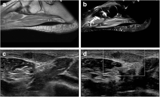
“You are shown a sagittal MRI of the foot in a 52-year-old man with diabetes. There are two nodules along the medial band of the plantar aponeurosis, low signal on T1 and intermediate-to-low on T2, blending with the fascia. He has similar nodules in the other foot.”
“You are shown MRI of a 44-year-old woman who had a 3 cm dorsal foot lump 'shelled out' at a local hospital eight months ago as a presumed ganglion. Histology reported a spindle cell lesion. She now has a nodular recurrence at the scar.”
Red flags mandating sarcoma referral
- Deep to the deep fascia
- Progressively enlarging, at any size
- Painful solid mass in a young adult
- Recurrence after previous excision
- 'Haematoma' persisting beyond six weeks
- Solid and enhancing on MRI with contrast
Site tells you the diagnosis
- Plantar aponeurosis, medial cord, multinodular — plantar fibromatosis
- 3rd then 2nd web space, plantar to intermetatarsal ligament — Morton's neuroma
- Dorsum, sinus tarsi, tendon sheath, anechoic — ganglion
- Along a named nerve, fusiform with tail sign — schwannoma
- Juxta-articular, small, painful, young adult — synovial sarcoma
- Along tendon sheath of a digit, blooming — tenosynovial giant cell tumour
Signal shortcuts
- Follows fat on all sequences and fully suppresses — lipoma
- Low on T1 and low on T2 — collagen, haemosiderin or urate
- Very bright T2, serpiginous, phleboliths — vascular malformation
- T1 brighter than muscle — melanin (clear cell sarcoma) or haemorrhage or fat
- Heterogeneous, necrotic, avidly enhancing — sarcoma until proven otherwise
- Bright T2 with NO internal enhancement — true cyst
Investigation order
- Weight-bearing radiographs of both feet
- Ultrasound with Doppler — cystic versus solid, foreign body, dynamic web space
- MRI with intravenous gadolinium for every solid or indeterminate mass
- CT for mineralisation characterisation; dual-energy CT for urate; CT chest for staging
- Image-guided core biopsy only at the sarcoma unit, tract in the line of resection
Never do
- Never shell out an undiagnosed solid mass
- Never use size less than 5 cm to exclude sarcoma in the foot
- Never biopsy before MRI and before sarcoma unit discussion
- Never rely on fine-needle aspiration for a primary sarcoma diagnosis
- Never accept 'clinical correlation' as the answer to an indeterminate enhancing mass
Evidence Base
Management and Outcome of Acral Soft-Tissue Sarcomas
- 63 acral soft tissue sarcomas presenting to one tertiary sarcoma service 2000-2016; 36 in the foot, 27 in the hand. The commonest foot subtype was SYNOVIAL SARCOMA (11 cases)
- IN 41 OF 63 (65 PER CENT) THE TUMOUR MEASURED LESS THAN 5 CM, with a median largest dimension of just 3 cm (range 2 to 6)
- 27 patients (43 PER CENT) had already undergone INADVERTENT EXCISION before referral to the sarcoma unit
- Those patients showed no significant difference in survival or local recurrence, but were significantly MORE LIKELY TO REQUIRE AMPUTATION (p equals 0.008)
- Primary surgery at the sarcoma unit was excision with limb salvage in 43 (68 per cent), partial digit or ray amputation in 14 (22 per cent) and more proximal amputation in six (10 per cent); after treating local recurrence the final rates were 24 per cent partial and 19 per cent proximal amputation
- Reassuringly, the authors also conclude that sarcoma excision WITH LIMB PRESERVATION does not increase the risk of local recurrence - limb salvage is not a compromise on oncological control
- Five-year survival was 82 per cent. Larger size (over 5 cm) predicted lower survival (p equals 0.04) and higher local recurrence (p equals 0.009). The authors' explicit conclusion: size 'should NOT be used as a diagnostic threshold for referral'
Synovial Sarcoma of the Hand and Foot: An Institutional Review
- 84 patients with primary hand (20) or FOOT (64) synovial sarcoma treated 1979-2019 at a single cancer centre
- 63 (75 per cent) presented with localised disease at a median age of 36; 21 (25 per cent) already had metastases at a median age of 30
- For localised disease, five-year progression-free survival was 82 per cent and overall survival 88 per cent, with LOCAL recurrence-free survival of 100 PER CENT
- SIZE STRATIFIES OUTCOME BELOW THE USUAL THRESHOLD: under 3.0 cm gave 95 per cent five-year PFS and 100 per cent OS, against 84 per cent PFS for 3.0-4.9 cm and 53 PER CENT for 5.0 cm or more (p equals 0.007)
- Tumours of 5.0 cm or more carried a hazard ratio for progression of 7.99 (95 per cent CI 1.68 to 37.91); the authors recommend considering systemic therapy at 3.0 cm and above
- Metastatic presentation was not uniformly fatal - five-year overall survival was 50 per cent in that group - and YOUNGER age (15-39 versus 40 and above) predicted better overall survival both in localised (p equals 0.04) and in metastatic disease (p equals 0.03)
Tumours of the Foot: A 10-Year Retrospective Analysis
- 195 foot tumours identified from 4,997 consecutive orthopaedic oncology records at one Malaysian centre over eleven years - foot tumours were 3.9 per cent of the unit's workload
- 148 were benign and 47 malignant: ABOUT A QUARTER OF FOOT TUMOURS REACHING AN ONCOLOGY UNIT WERE MALIGNANT
- 144 were soft tissue and 47 bone; soft tissue tumours occurred in an older population while bone tumours clustered in the second decade
- GANGLION was the commonest benign soft tissue tumour, and the commonest MALIGNANT soft tissue tumour was MALIGNANT MELANOMA - not a sarcoma
- On the bone side the answers differ: giant cell tumour was the commonest benign bone tumour and CHONDROSARCOMA the commonest malignant one
- Amputation rate 5.64 per cent, recurrence 1.54 per cent, mortality 3.08 per cent; mean MSTS score 79 per cent and TESS 76.2 per cent
Ultrasound Versus Magnetic Resonance Imaging for Morton Neuroma: Systematic Review and Meta-Analysis
- Systematic review by two independent reviewers of studies to April 2014; 277 identified, 14 included in the meta-analysis
- ALL included studies used SURGERY as the reference standard
- Pooled sensitivity: ultrasound 0.91 (95 per cent CI 0.83-0.96) and MRI 0.90 (0.82-0.96) - NO significant difference (Q test p equals 0.88)
- Pooled specificity: MRI 1.00 (0.73-1.00); ultrasound 0.854 with a very wide interval of 0.41 to 1.00
- The authors conclude ultrasound is as accurate as MRI and is likely the most cost-effective imaging method for this diagnosis
Recurrence Rate After Wide Resection of Plantar Fibromatosis: A Case Series and Systematic Literature Review
- 12 patients reviewed 2 to 13 years after wide resection, assessed clinically and with MRI for recurrence, plus a systematic review of the literature
- In the authors' own series 2 of 12 (17 per cent) recurred over a mean FOLLOW-UP of 7.8 years (range 2 to 13) - 7.8 years is the observation period, not the time to recurrence; median Foot Functional Index 1 (0-66) and AOFAS 95 (44-100)
- The systematic review pooled 6 studies, 109 feet in 92 patients
- RECURRENCE TRACKED THE WIDTH OF EXCISION: 67 PER CENT after local resection, 42 per cent after wide resection, and 27 PER CENT after fasciectomy
- The authors recommend wide resection or fasciectomy over local resection on the strength of that gradient