The Antigravity Engine and the Gatekeeper of the Popliteal Fossa
- Origin: soleal line and the middle third of the posterior tibia, the posterior head and upper quarter of the fibula, and the fibrous tendinous arch between them.
- The soleal arch is the tunnel through which the popliteal artery and tibial nerve pass into the deep posterior compartment - a genuine entrapment site.
- Innervated by the tibial nerve, roots S1 and S2, with separate branches from those to gastrocnemius.
- It is monoarticular, crossing only the ankle, which is the entire basis of the Silfverskiold test.
- Type I slow-twitch fibre predominance makes it a fatigue-resistant postural muscle, not a sprinter.
- “If dorsiflexion improves when the knee is flexed, the tight structure is gastrocnemius - because soleus does not cross the knee.
- “The soleus is the local muscle flap of choice for a middle-third tibial defect; medial gastrocnemius covers the proximal third.
- “Soleal venous sinuses are the commonest site of origin of calf deep vein thrombosis.
- “Functional popliteal artery entrapment in the athlete is often caused by a hypertrophied soleal arch rather than an anomalous gastrocnemius head.
Overview
Soleus is the broad, flat, multipennate muscle lying deep to the gastrocnemius in the superficial posterior compartment of the leg. Together the two form the triceps surae, converging on the tendo-Achilles. The single anatomical fact that governs everything about soleus is that it is monoarticular - it crosses the ankle only, and never the knee. Gastrocnemius crosses both.
It is a postural muscle. Standing quietly, the ground reaction force passes anterior to the ankle axis and creates a continuous dorsiflexion moment; soleus is the muscle that resists it. In walking it is the principal controller of tibial advance over the fixed foot through mid-stance, and it generates the majority of the plantarflexion power at push-off. It is also a pump: the venous sinusoids within its substance are squeezed with every contraction, and this is the dominant mechanism of venous return from the lower limb.
Soleus takes origin from two bones - the soleal line of the tibia and the posterior head and upper quarter of the fibula - and between these two bony origins there is a fibrous tendinous arch, the soleal arch (arcus tendineus musculi solei), spanning the gap.
- The popliteal artery and the tibial nerve pass deep to this arch to enter the deep posterior compartment, where the artery divides into anterior tibial and tibioperoneal trunk. The anterior tibial artery actually leaves the popliteal artery just above the arch and passes forward through the interosseous membrane.
- Because this is a fixed fibrous ring with no give, it is a genuine entrapment site:
- Popliteal artery entrapment syndrome (type V and functional forms) - the artery is compressed by the arch, classically in a young athlete with a hypertrophied calf.
- Tibial nerve entrapment at the soleal arch - a proximal, high tibial neuropathy producing calf pain, plantar paraesthesia and weakness of the deep compartment muscles including tibialis posterior and the long toe flexors. It is much rarer than distal tarsal tunnel syndrome and is routinely missed.
- Surgical consequence: any decompression of the proximal tibial nerve or popliteal artery in this region requires division of the soleal arch under direct vision, taking care of the vessels immediately deep to it.
GPSSuperficial Posterior Compartment
Hook:All three are tibial nerve, S1-S2. Only soleus is monoarticular - that is the whole basis of the Silfverskiold test.

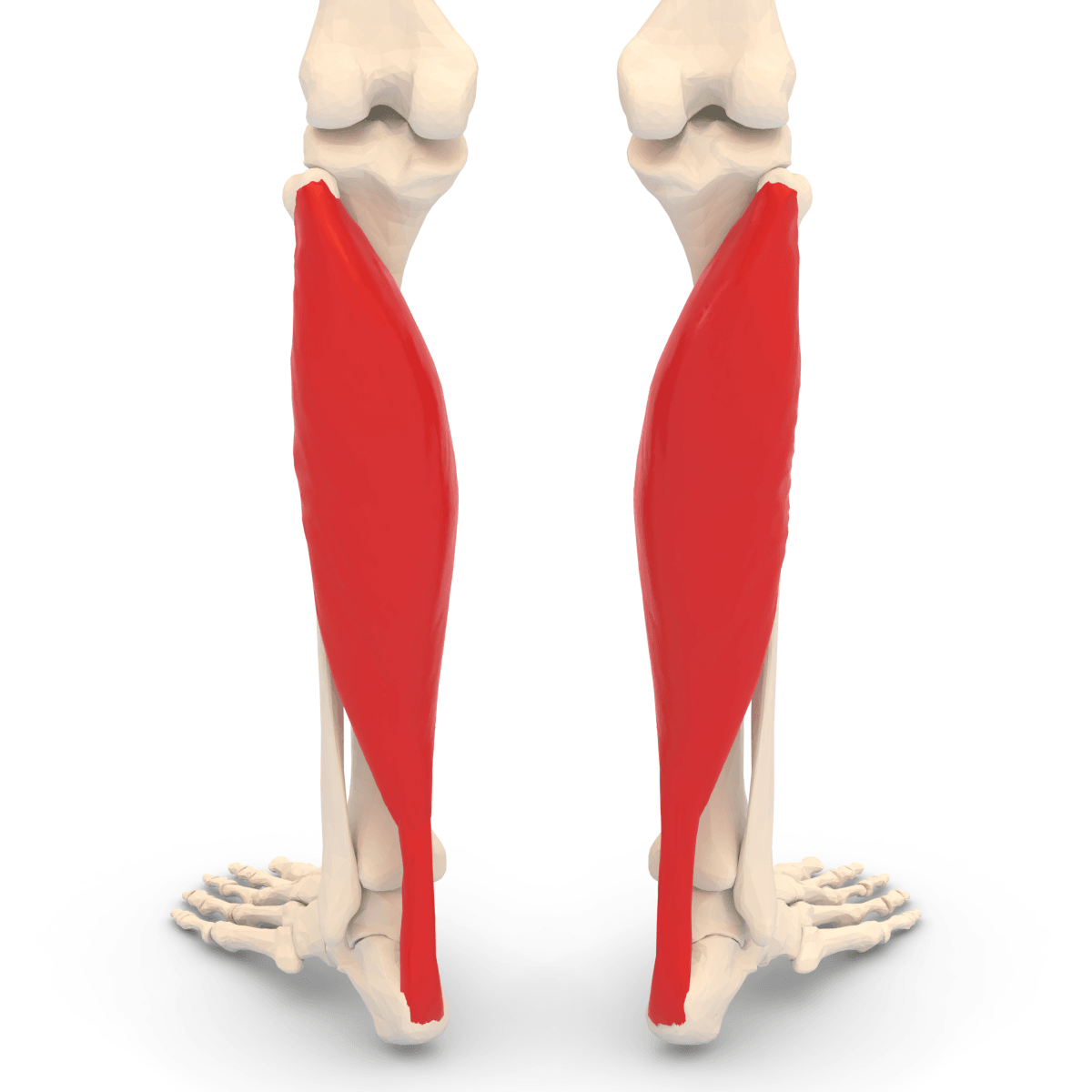
Attachments, Innervation and Relations
Origin
- Soleal line on the posterior surface of the tibia, and the middle third of the medial border of the tibia.
- Posterior aspect of the head of the fibula and the upper quarter of the posterior surface of the fibular shaft.
- The fibrous tendinous soleal arch bridging the two bony origins, deep to which the popliteal vessels and tibial nerve pass.
- The muscle is multipennate, with an extensive internal aponeurotic system. Its physiological cross-sectional area is very large relative to its fibre length - the classic architecture of a force-producing rather than an excursion-producing muscle.
Insertion
- Fibres converge onto a broad posterior aponeurosis which fuses with the deep surface of the gastrocnemius aponeurosis to form the tendo-Achilles.
- The soleal contribution enters the tendon more distally and more anteriorly (deep) than the gastrocnemius contribution, and the composite tendon spirals approximately 90 degrees as it descends, so that soleal fibres come to lie medially at the calcaneal insertion and gastrocnemius fibres laterally.
- Insertion onto the middle third of the posterior surface of the calcaneal tuberosity, distal to the retrocalcaneal bursa.
The Achilles spiral - why it matters
- The rotation concentrates stress in a region roughly 2 to 6 cm proximal to the insertion, which is both the least vascular zone and the site of the great majority of Achilles ruptures and of mid-substance tendinopathy.
- It also means that at open repair the fibre orientation must be respected; the tendon is not a simple parallel cable.
When dividing the soleal arch - during a proximal tibial nerve decompression, a popliteal artery entrapment release, or a proximally based soleus flap harvest - remember that the popliteal artery and vein and the tibial nerve lie directly beneath it. The arch must be divided from medial to lateral under direct vision, with a retractor or a finger protecting the vessels, and never with a blind sweep of scissors. Injury here is limb-threatening.
Action and Biomechanics
Primary action
- Ankle plantarflexion, and by virtue of its insertion medial to the subtalar axis after the Achilles spiral, a contribution to hindfoot inversion when the hindfoot is neutral or varus. Critically, when the hindfoot is already in valgus the Achilles vector passes lateral to the subtalar axis and the triceps surae becomes an evertor - a deforming force in the collapsing flatfoot.
- It does not act on the knee. Gastrocnemius flexes the knee; soleus does not.
Gastrocnemius versus soleus
- Gastrocnemius
- Knee and ankle (biarticular)
- Soleus
- Ankle only (monoarticular)
- Gastrocnemius
- Predominantly type II fast-twitch
- Soleus
- Predominantly type I slow-twitch
- Gastrocnemius
- Explosive power - jumping, sprinting
- Soleus
- Postural endurance and stance-phase control
- Gastrocnemius
- Longer fibres, greater excursion
- Soleus
- Multipennate, huge cross-sectional area, short fibres, high force
- Gastrocnemius
- Ankle plantarflexion with the knee extended
- Soleus
- Ankle plantarflexion with the knee flexed to 90 degrees
- Gastrocnemius
- Gastrocnemius recession (Strayer, Baumann, Vulpius)
- Soleus
- Requires tendo-Achilles lengthening
- Gastrocnemius
- Proximal third of the tibia
- Soleus
- Middle third of the tibia
- Gastrocnemius
- Medial head tear - tennis leg
- Soleus
- Rarely ruptures in isolation
Role in gait
- Loading response: relatively quiet; tibialis anterior is controlling the foot.
- Mid-stance: soleus becomes the dominant muscle in the limb. It contracts eccentrically to control the forward advance of the tibia over the fixed foot. Without it the tibia collapses forward into dorsiflexion and the patient adopts a crouch gait with a flexed knee - the exact deformity produced by an over-lengthened Achilles tendon.
- Terminal stance and pre-swing: it contracts concentrically, generating the largest single burst of positive power in the gait cycle and driving push-off.
- Swing: silent.
The soleus is the plantarflexion-knee extension couple. By restraining forward tibial advance in mid-stance it keeps the ground reaction force anterior to the knee, which generates an extension moment and lets the knee stay straight without quadriceps effort.
- Lengthen the Achilles excessively - most classically by performing a tendo-Achilles lengthening when the Silfverskiold test showed only an isolated gastrocnemius contracture - and the soleus becomes incompetent.
- The tibia now advances unchecked, the ground reaction force moves behind the knee, a flexion moment develops, and the patient must fire quadriceps continuously to stay upright.
- The result is a crouch gait: energetically expensive, progressive, associated with patellofemoral pain and patella alta, and extremely difficult to reverse.
This is the reason the Silfverskiold test is not an academic exercise. Once you have decided to lengthen, a positive Silfverskiold test means gastrocnemius recession, not tendo-Achilles lengthening.
But the test does not make that decision for you. DiGiovanni measured 34 patients with forefoot and midfoot pain against 34 matched asymptomatic controls: dorsiflexion of 10 degrees or less with the knee extended was present in 88 per cent of the patients - and in 44 per cent of the controls. Tighten the definition to 5 degrees and it is 65 versus 24 per cent. Between a quarter and nearly half of people with no foot symptoms whatever meet the usual definition of a tight gastrocnemius. So a positive test identifies a mechanical contributor to be weighed against the clinical picture, and never by itself an indication to operate. What DiGiovanni does establish is the mechanism: the deficit vanished when the knee was flexed to 90 degrees (17.9 against 22.3 degrees, p = 0.09, not significant), which is the experimental validation of Silfverskiold's principle in the non-spastic adult foot.
Force and architecture
- The triceps surae generates by far the largest plantarflexion torque of any muscle group about the ankle. Cadaveric measurement of relative muscle strength below the knee (Silver, de la Garza and Rang, 1985) found the plantarflexors of the ankle to be six times as strong as the dorsiflexors. Soleus contributes the majority of the static and endurance component.
- Its short fibres and huge physiological cross-sectional area mean high force but low excursion - the architecture of a postural strut.
- Type I fibre predominance (commonly quoted around 70 to 90 per cent in soleus, versus roughly half in gastrocnemius) makes it fatigue-resistant and explains its role in quiet standing.
The calf muscle pump
- Each soleus contraction compresses the intramuscular venous sinuses and propels blood proximally; competent valves in the deep and perforating veins prevent reflux.
- Ejection fraction of the calf pump in a normal limb is substantial, and the pump is the principal mechanism opposing the hydrostatic column in the upright human.
- Pump failure - from immobility, muscle weakness, ankle stiffness (which prevents the pump cycling) or valvular incompetence - produces venous hypertension, oedema, lipodermatosclerosis and venous ulceration in the gaiter area.
- Clinical translation: a stiff ankle is a failed calf pump. This is one of the underappreciated arguments for restoring ankle motion after trauma, and the reason ankle pump exercises are prescribed from day one after any lower-limb surgery.
Surface Anatomy and Examination
Palpation and inspection
- Soleus is palpable as the broad muscle mass either side of the Achilles in the distal third of the calf, extending further distally than gastrocnemius. Its medial and lateral borders emerge from beneath the gastrocnemius bellies roughly at the junction of the middle and distal thirds of the leg.
- The gastrocnemius bellies are the prominent bulges in the upper calf; the soleus is what you feel below and lateral to them.
- Asymmetry in calf circumference measured 10 cm below the tibial tuberosity is a crude but useful record of triceps surae wasting.
Named tests and signs
- How to perform
- Measure passive ankle dorsiflexion with the knee extended, then with the knee flexed to 90 degrees, holding the hindfoot in neutral or slight inversion and the subtalar joint locked
- Positive finding
- Dorsiflexion improves markedly with the knee flexed
- What it means
- Isolated gastrocnemius contracture - treat with recession
- False positives
- Allowing the hindfoot to evert or the midfoot to dorsiflex falsely increases the measured range
- How to perform
- Resisted plantarflexion with the knee flexed to 90 degrees (gastrocnemius slackened)
- Positive finding
- Weakness
- What it means
- Soleus weakness or S1 lesion
- False positives
- Pain inhibition, Achilles pathology
- How to perform
- Rise onto the toes on one leg, repeated up to 20 to 25 times
- Positive finding
- Reduced number, reduced height, loss of heel inversion
- What it means
- Triceps surae insufficiency or S1 radiculopathy; loss of inversion suggests tibialis posterior dysfunction
- False positives
- Balance failure, forefoot pain, ankle arthritis
- How to perform
- Tap the Achilles with the ankle relaxed in slight dorsiflexion
- Positive finding
- Absent or depressed
- What it means
- S1-S2 lesion
- False positives
- Age-related loss, peripheral neuropathy - always compare sides
- How to perform
- Prone with knees flexed to 90 degrees; squeeze the calf
- Positive finding
- No passive plantarflexion
- What it means
- Achilles tendon rupture
- False positives
- Intact plantaris or an incomplete tear may produce a false negative; always compare with the other side
- How to perform
- Palpate the pedal pulses during resisted plantarflexion and during passive maximal dorsiflexion, with ankle-brachial index or duplex
- Positive finding
- Pulse loss or a fall in ankle-brachial index on provocation
- What it means
- Popliteal artery entrapment, possibly at a hypertrophied soleal arch
- False positives
- Pulse obliteration occurs in a substantial minority of asymptomatic people - imaging correlation is essential
Getting the Silfverskiold test right
- Patient supine, knee fully extended.
- Lock the subtalar joint in neutral or slight inversion by grasping the heel - this is the step most commonly omitted, and without it the midfoot dorsiflexes and gives a falsely generous reading.
- Dorsiflex the ankle with a gentle, sustained force applied along the lateral border of the foot, not the forefoot, and record the angle.
- Flex the knee to 90 degrees and repeat.
- Interpretation: a marked increase in dorsiflexion with the knee flexed (commonly taken as an increase of 10 degrees or more, or a change from restricted to more than 10 degrees of dorsiflexion) indicates an isolated gastrocnemius contracture. No change means a combined gastrocsoleus contracture or a bony block.
Complications
- Mechanism
- Artery and vein lie immediately deep to the arch
- Prevention
- Divide the arch under direct vision with the vessels protected; never a blind sweep
- Management
- Immediate vascular repair or interposition vein graft; vascular surgical support
- Mechanism
- Forcing the arc of rotation to reach a distal-third defect
- Prevention
- Respect the middle-third territory; use free tissue transfer distally
- Management
- Debridement and secondary flap - usually a free flap
- Mechanism
- Over-zealous proximal dissection
- Prevention
- Identify and preserve the pedicle in the proximal 5 to 8 cm before dividing minor pedicles
- Management
- The flap is lost; plan alternative cover
- Mechanism
- Tight fascial closure of the donor compartment
- Prevention
- Leave the donor fascia open; close skin only
- Management
- Urgent fasciotomy
- Mechanism
- The nerve lies superficial in the posterior midline
- Prevention
- Posteromedial incision, blunt dissection, visualise the lateral aponeurotic edge
- Management
- Neuroma excision and burial if symptomatic
- Mechanism
- Tendo-Achilles lengthening performed for an isolated gastrocnemius contracture
- Prevention
- Silfverskiold test in every case; recession when the test is positive
- Management
- Extremely difficult to reverse - bracing, quadriceps strengthening, occasionally distal femoral extension osteotomy in CP
- Mechanism
- Loss of the plantarflexion-knee extension couple
- Prevention
- Measured lengthening, protect with a cast in the corrected but not over-corrected position
- Management
- Ground reaction AFO; salvage is difficult
- Mechanism
- Deep transverse fascia not divided
- Prevention
- Deliberately open the deep compartment along the posterior tibial border
- Management
- Late claw toes, cavovarus and plantar sensory loss requiring tendon release or transfer
- Mechanism
- Stasis in the valveless soleal sinuses during immobility
- Prevention
- Early mobilisation, ankle pump exercises, mechanical and chemical prophylaxis
- Management
- Anticoagulation per local protocol
- Mechanism
- Over-aggressive cuts in a degenerate tendon
- Prevention
- Limit the extent of each cut; test the correction incrementally
- Management
- Cast immobilisation or open repair
Clinical Relevance
Popliteal artery entrapment syndrome
- Presentation: a young, athletic patient - classically a runner or footballer with well-developed calves - with exercise-induced calf claudication, sometimes paraesthesia or coldness, relieved by rest. It is frequently misdiagnosed as chronic exertional compartment syndrome or medial tibial stress syndrome.
- Anatomical classification (types I to IV, with type V for venous involvement and a separate functional type). The types are conventionally credited to Love and Whelan, whose 1965 paper named the syndrome and described the variants; the numbering, type V and the functional category are later additions by subsequent authors, so do not attribute a specific type number to the 1965 citation.
- Type I: the popliteal artery courses medial to a normally positioned medial head of gastrocnemius.
- Type II: the medial head of gastrocnemius attaches abnormally laterally, displacing the artery medially.
- Type III: an accessory slip of gastrocnemius entraps the artery.
- Type IV: the artery lies deep to popliteus or a fibrous band.
- Type V: any of the above with popliteal vein involvement as well.
- Functional (type VI in some schemes): no anatomical anomaly - entrapment results from muscular hypertrophy, most often of the soleal arch or the medial gastrocnemius, compressing a normally positioned artery during exercise.
- The soleus link: the soleal arch is the specific structure implicated in many functional cases. A hypertrophied, fibrous arch compresses the artery in plantarflexion and in forced dorsiflexion.
- Investigation: duplex ultrasound with provocative manoeuvres, magnetic resonance angiography or CT angiography in neutral and in provoked positions, and ankle-brachial index before and after exercise. Beware the base rate: up to 50 per cent of normal limbs show transient popliteal compression at the extremes of plantarflexion or dorsiflexion (Levien 1999). Provoked compression is therefore close to a normal finding, and the diagnosis rests on provoked compression plus discrete, reproducible, exertional symptoms - never on the image alone.
- Consequences of delay: repeated compression causes intimal damage, post-stenotic aneurysm, thrombosis and distal embolisation - a limb-threatening progression in a young patient.
- Treatment, and it differs by type. For anatomical entrapment (types I to IV), operate at diagnosis rather than observing: in Levien's series 15 of 58 such limbs had already occluded by presentation. Release is through a posterior (S-shaped) or medial approach, dividing the offending muscle slip or the soleal arch. For functional entrapment, decompress only where the symptoms are discrete and typical - given the 50 per cent base rate of provoked compression in normals, a liberal indication here operates on people who do not have the disease. Where the artery is already degenerate or occluded, replace the segment with vein rather than patching it, because the histological damage extends beyond the visibly abnormal wall.
Tibial nerve entrapment at the soleal arch
- A proximal tibial neuropathy, far rarer and far more often missed than distal tarsal tunnel syndrome.
- Symptoms: deep calf pain worse with exercise, plantar paraesthesia, and weakness of the deep posterior compartment - tibialis posterior, flexor digitorum longus and flexor hallucis longus - producing weak inversion and weak toe flexion with a normal ankle jerk pattern and preserved gastrocnemius and soleus function if the branches to them arise proximal to the arch.
- Key discriminator from tarsal tunnel syndrome: in a soleal arch entrapment the Tinel sign is positive high in the proximal calf, not behind the medial malleolus, and the calf pain is prominent.
- Treatment: decompression by division of the soleal arch, taking great care of the popliteal vessels immediately deep to it.
Achilles pathology involving soleus
- Mid-substance Achilles tendinopathy and rupture occur in the hypovascular zone 2 to 6 cm above the insertion, corresponding to the region of the Achilles spiral where soleal and gastrocnemius fibres cross.
- Isolated soleus strain is a recognised but underdiagnosed injury in distance runners, presenting as diffuse deep calf pain that is worse with knee-flexed calf raises (soleus loaded) rather than the more acute, localised, knee-extended pain of a medial gastrocnemius tear (tennis leg).
- Accessory soleus muscle is an anatomical variant presenting as a soft-tissue mass in the posteromedial ankle, sometimes causing exertional pain or even a localised compartment syndrome; it is distinguished from a tumour by its muscle signal on MRI.
Deep vein thrombosis and the soleal sinuses
- The valveless soleal sinusoids are the commonest origin of calf deep vein thrombosis, and this is why calf DVT is the starting point of most propagating lower-limb thrombosis after immobilisation, surgery or long-haul travel.
- Prophylaxis logic: intermittent pneumatic compression, graduated compression stockings, early mobilisation and active ankle pumping all work by re-engaging the calf pump and emptying the sinuses.
- Clinical caveat: calf tenderness and swelling are unreliable; the diagnosis needs duplex ultrasound, guided by a pre-test probability score and D-dimer where appropriate.
Chronic venous insufficiency
- Failure of the calf pump - from valvular incompetence, muscle weakness or a stiff ankle - produces ambulatory venous hypertension, gaiter-area lipodermatosclerosis, haemosiderin staining and venous ulceration.
- Orthopaedic relevance: ankle stiffness after a pilon or plafond fracture, prolonged immobilisation, and post-thrombotic syndrome all degrade the pump. Restoring ankle motion and encouraging active plantarflexion is part of the vascular as well as the musculoskeletal rehabilitation.
Superficial posterior compartment syndrome
- Less common than the anterior compartment, but recognised, and the soleus is the bulk of the compartment. Signs are pain on passive ankle dorsiflexion, a tense calf and sural nerve paraesthesia.
- Do not forget the deep posterior compartment, which sits beneath the soleus behind the deep transverse fascia and is the compartment most frequently missed at fasciotomy. Its late sequel is claw toes with plantar sensory loss.
Surgical Relevance
The soleus flap - the workhorse of the middle third
The classic teaching
- Proximal third tibial defect: medial gastrocnemius flap (occasionally lateral gastrocnemius).
- Middle third tibial defect: soleus flap - usually a medial hemisoleus.
- Distal third tibial defect: free tissue transfer, or a local fasciocutaneous, perforator or reverse-flow flap. A distally based soleus flap is described but is unreliable and should not be offered as the answer for a distal-third defect in a viva.
- Modern practice increasingly uses perforator-based propeller flaps and free flaps even in the middle third, but the anatomical teaching about muscle territory remains the exam answer and the safe default where microsurgery is unavailable.
Why soleus works as a flap
- Mathes and Nahai type II: a dominant proximal pedicle from the posterior tibial artery (medial half) or the peroneal artery (lateral half), with minor segmental pedicles distally. The minor pedicles can be divided and the muscle rotated on its dominant pedicle.
- Muscle provides vascularised cover over exposed bone and metalwork, obliterates dead space, and delivers antibiotic and cellular defence to a contaminated bed - all superior to a skin graft alone.
Gastrocnemius recession versus tendo-Achilles lengthening
- Level
- Proximal, intramuscular
- What is lengthened
- Gastrocnemius aponeurosis within the muscle belly, deep to gastrocnemius
- Indication
- Isolated gastrocnemius contracture, particularly in children
- Principal risk
- Under-correction; small gain per cut
- Level
- Distal gastrocnemius aponeurosis
- What is lengthened
- Gastrocnemius aponeurosis divided and allowed to recede off the soleal aponeurosis
- Indication
- The standard adult gastrocnemius recession for a positive Silfverskiold test
- Principal risk
- Sural nerve injury - it lies immediately superficial in the midline
- Level
- Gastrocsoleus aponeurosis
- What is lengthened
- Inverted V or tongue-in-groove of the combined aponeurosis
- Indication
- Mild combined contracture
- Principal risk
- Over-lengthening if extended too far distally
- Level
- Tendo-Achilles
- What is lengthened
- The whole tendon, including the soleal contribution
- Indication
- True combined gastrocsoleus contracture with a negative Silfverskiold test
- Principal risk
- Over-lengthening producing calcaneus gait and crouch; complete rupture
- Level
- Tendo-Achilles
- What is lengthened
- The whole tendon under direct control
- Indication
- Severe fixed equinus, revision surgery
- Principal risk
- Wound problems, over-lengthening, adhesion
- The decision rule is simple and absolute: a positive Silfverskiold test means gastrocnemius recession. A negative test - no improvement with the knee flexed - means the soleus is also tight and a tendo-Achilles lengthening is required. Performing a tendo-Achilles lengthening for an isolated gastrocnemius contracture is the classic error that produces a crouch gait.
- Sural nerve safety in a Strayer recession: the sural nerve and short saphenous vein lie in the posterior midline superficial to the gastrocnemius aponeurosis, passing from between the gastrocnemius heads proximally to the lateral side distally. Use a posteromedial longitudinal incision, identify the plane bluntly, and visualise the lateral edge of the aponeurosis before cutting across it.
Other surgical contexts
- Posteromedial approach to the tibia: the interval passes between the soleus (retracted posteriorly) and the deep flexors. Elevating soleus from the posteromedial tibial border gives access to the posterior tibial cortex for plating or for bone grafting a nonunion.
- Posteromedial fasciotomy: the incision lies 1 to 2 cm posterior to the posteromedial tibial border. The superficial posterior compartment is opened first, then the deep transverse fascia must be deliberately divided along the posterior tibial border to open the deep posterior compartment. Skipping this second step is the classic incomplete fasciotomy.
- Achilles surgery: in chronic rupture with a gap, a gastrocnemius turn-down (Bosworth) or V-Y lengthening uses the proximal aponeurosis; the soleus contributes muscle bulk that can be advanced but is not a graft source.
- Bone grafting and nonunion: the posteromedial soleus can be mobilised to provide a vascularised bed over a grafted middle-third tibial nonunion, and this is often as valuable as the graft itself.
Guidelines, Registries & Global Practice
Anatomical variation across populations
- The soleal arch varies from a thin, membranous band to a thick, tough tendinous structure. A thick fibrous arch in a hypertrophied calf is the substrate for functional popliteal artery entrapment, which is why the condition clusters in athletic populations and in military recruits.
- Accessory soleus is a described variant, presenting as a posteromedial ankle mass, occasionally with exertional pain. Reported prevalence in cadaveric and imaging series is low but not negligible, and it is more often recognised now because of the volume of ankle MRI performed.
- Plantaris - the third muscle of the superficial compartment - is absent in a reported 7 to 20 per cent of limbs depending on the series and population. Its long slender tendon is a recognised autograft donor for hand and foot reconstruction where it is present.
- The relative contribution of gastrocnemius and soleus to Achilles fibre orientation varies, and the degree of Achilles spiral is not constant.
Side-by-side guidance
- Position relevant to soleus
- Joint orthoplastic management of open tibial fractures, with definitive soft tissue cover ideally within 72 hours; local muscle flap or free flap chosen by defect location and the zone of injury.
- Position relevant to soleus
- Teaches soleus as the standard local flap for middle-third tibial cover and emphasises leaving the donor fascia open; emphasises deliberate division of the deep transverse fascia in posteromedial fasciotomy.
- Position relevant to soleus
- Support the Silfverskiold test as a routine component of foot and ankle assessment and gastrocnemius recession as the treatment for isolated gastrocnemius contracture.
- Position relevant to soleus
- Strongly discourage isolated percutaneous tendo-Achilles lengthening for a positive Silfverskiold test because of the risk of crouch gait; recommend multilevel assessment before single-level surgery.
- Position relevant to soleus
- Recommend provocative imaging plus symptom correlation before operating for popliteal artery entrapment, since provoked compression is common in asymptomatic limbs.
- Position relevant to soleus
- Recommend mechanical prophylaxis with intermittent pneumatic compression and early mobilisation alongside risk-stratified pharmacological prophylaxis in lower-limb surgery.
Resource-dependent practice
- Well-resourced settings: microsurgical free tissue transfer is available for any defect, provoked magnetic resonance angiography is used for entrapment, and instrumented gait analysis informs cerebral palsy surgery.
- Limited-resource settings: the soleus flap is one of the most valuable operations in the trauma surgeon repertoire, because it requires no microsurgery, no implants and no special equipment, and delivers definitive vascularised cover to an open tibial fracture. Its anatomical territory and its limitations must be known precisely. The Silfverskiold test costs nothing and prevents the single most damaging error in equinus surgery.
- Popliteal artery entrapment is under-diagnosed globally because the workup requires provoked imaging; a high index of suspicion in the young athlete with exercise-limited calf pain is the practical substitute.
Registry and outcome signals
- National open fracture audits consistently associate early definitive soft tissue cover with lower deep infection rates and better union, supporting the orthoplastic pathway irrespective of whether the cover is local or free.
- Cerebral palsy outcome series consistently report that isolated tendo-Achilles lengthening in ambulant children is associated with progression to crouch gait, and this observation has shifted practice worldwide toward selective gastrocnemius recession and multilevel surgery.
MCQ Practice Points
Q: Why does knee flexion change ankle dorsiflexion in a gastrocnemius contracture but not in a soleus contracture? A: Soleus crosses only the ankle. Gastrocnemius crosses the knee and the ankle, so flexing the knee slackens it. This is the entire basis of the Silfverskiold test.
Q: What passes deep to the soleal arch? A: The popliteal artery and vein and the tibial nerve. The anterior tibial artery has already branched off just proximal to the arch.
Q: What is the innervation of soleus? A: The tibial nerve, roots S1 and S2, with separate proximal and distal branches - the distal branch entering the deep surface of the muscle.
Q: Which local muscle flap covers the middle third of the tibia? A: Soleus (usually a medial hemisoleus). Medial gastrocnemius covers the proximal third; the distal third needs free tissue transfer or a local fasciocutaneous or perforator flap.
Q: What is the vascular pattern of soleus? A: Type II - one dominant proximal pedicle plus minor segmental pedicles. The medial half is supplied by the posterior tibial artery and the lateral half by the peroneal artery.
Q: Which fibre type predominates in soleus and why does it matter? A: Type I slow-twitch, making it fatigue-resistant and suited to postural and stance-phase work, in contrast to the type II predominance of gastrocnemius.
Q: Where do most calf deep vein thromboses originate? A: In the valveless soleal venous sinuses, which pool blood during immobility - the anatomical rationale for calf compression and ankle pump exercises.
Q: Why does over-lengthening the Achilles cause a crouch gait? A: It destroys the plantarflexion-knee extension couple. An incompetent soleus lets the tibia advance, moving the ground reaction force behind the knee and creating a flexion moment the quadriceps must resist continuously.
Q: Where do soleal fibres lie at the calcaneal insertion? A: Medially. The Achilles spirals roughly 90 degrees as it descends, so soleal fibres become medial and gastrocnemius fibres lateral. The spiral concentrates stress 2 to 6 cm above the insertion - the rupture and tendinopathy zone.
Q: What is the classic omission in a posteromedial fasciotomy? A: Failure to divide the deep transverse fascia and open the deep posterior compartment beneath the soleus. The late result is claw toes, cavovarus and plantar sensory loss.
Exam Viva Scenarios
Practise clinical reasoning and management decisions out loud
“A 23-year-old competitive middle-distance runner describes cramping pain in the right calf coming on reproducibly after about eight minutes of running and settling within two minutes of stopping. He has heavily muscled calves. At rest his pedal pulses are normal and his neurological examination is normal. He has been treated for medial tibial stress syndrome without benefit. What is your differential and how do you proceed?”
“A 41-year-old motorcyclist has a Gustilo IIIB open tibial shaft fracture with a 6 cm by 4 cm soft tissue defect over the middle third of the tibia. The bone is exposed and the fracture has been stabilised with an intramedullary nail after thorough debridement. There is no microsurgical service in your hospital tonight. How do you plan soft tissue cover?”
“A 7-year-old boy with spastic diplegic cerebral palsy, GMFCS level II, walks on his toes. Passive ankle dorsiflexion is minus 10 degrees with the knee extended and plus 5 degrees with the knee flexed to 90 degrees, with the hindfoot held in neutral. His knees are straight in stance. What is the diagnosis and what would you do?”
Anatomy
- Origin: soleal line and middle third of posterior tibia, fibular head and upper quarter of fibula, plus the soleal arch
- Insertion: Achilles into the middle third of the posterior calcaneus - soleal fibres end medially after the 90-degree spiral
- Nerve: tibial, S1-S2, proximal and distal branches
- Artery: posterior tibial (medial half) and peroneal (lateral half) - Mathes and Nahai type II
- Monoarticular - crosses the ankle only
The Soleal Arch
- Fibrous arch between the tibial and fibular origins
- Popliteal artery and vein and tibial nerve pass deep to it
- Anterior tibial artery branches just proximal to the arch
- Site of popliteal artery entrapment and proximal tibial neuropathy
- Divide only under direct vision
Function
- Dominant plantarflexor through stance; eccentric control of tibial advance
- Plantarflexion-knee extension couple keeps the knee straight
- Type I slow-twitch, fatigue-resistant, postural
- Calf pump - soleal sinuses drive venous return
Silfverskiold
- Dorsiflexion improves with knee flexed = isolated gastrocnemius = recession
- No change = combined gastrocsoleus = tendo-Achilles lengthening
- Lock the hindfoot in neutral or the midfoot substitutes
- Over-lengthening causes crouch gait - very hard to reverse
Flap
- Middle third of tibia - medial hemisoleus, proximally based
- Preserve the dominant pedicle in the proximal 5-8 cm
- Split along the central raphe; release from the Achilles
- Leave the donor fascia open
- Do not force it into the distal third - use a free flap
Evidence Base
Reduction of the Uncrossed Two-Joint Muscles of the Leg to One-Joint Muscles in Spastic Conditions
- Recognised that a biarticular gastrocnemius and a monoarticular soleus can be distinguished by comparing dorsiflexion with the knee extended and flexed
- Proposed selective surgical treatment of gastrocnemius rather than the whole tendo-Achilles in spastic equinus
- Established the anatomical logic that underpins gastrocnemius recession
- The eponymous test remains in daily use a century later
Isolated Gastrocnemius Tightness
- 34 consecutive patients with metatarsalgia or midfoot/forefoot pain against 34 age, weight and sex-matched asymptomatic controls, measured with a purpose-built electrogoniometer
- KNEE EXTENDED: mean maximal dorsiflexion 4.5 degrees in patients versus 13.1 degrees in controls (p less than 0.001)
- KNEE FLEXED 90 DEGREES: 17.9 versus 22.3 degrees - and this difference was NOT significant (p = 0.09), which is the finding that localises the problem to gastrocnemius
- Defining gastrocnemius contracture as dorsiflexion of 5 degrees or less: 65 per cent of patients - but ALSO 24 PER CENT OF ASYMPTOMATIC CONTROLS
- Relaxing the definition to 10 degrees or less: 88 per cent of patients and 44 PER CENT OF CONTROLS
- Gastrocsoleus contracture (10 degrees or less with the knee flexed) in 29 per cent of patients and 15 per cent of controls
The Myth of Muscle Balance - Relative Strengths and Excursions of Normal Muscles About the Foot and Ankle
- Muscle fibre lengths and muscle weights below the knee were measured in the lower limbs of FIVE cadavers to derive relative strength and excursion
- The plantarflexors of the ankle were found to be SIX times as strong as the dorsiflexors
- The authors discarded the concept of muscle balance in tendon transfer surgery and proposed that TASK APPROPRIATENESS should be the guide
- Because fibre length and excursion are constantly related, contracture is accompanied by decreased excursion - and tendon lengthening improves the DEFORMITY but does NOT improve the decreased active range of movement