The Muscle the Posterior Interosseous Nerve Lives Inside
- Two heads: a SUPERFICIAL head from the lateral epicondyle, radial collateral and annular ligaments, and a DEEP head from the SUPINATOR CREST and fossa of the ULNA. The posterior interosseous nerve passes BETWEEN them.
- Innervated by the POSTERIOR INTEROSSEOUS NERVE (C5, C6) — the branches arise proximal to or at the arcade of Frohse, which is why supinator function is preserved in a posterior interosseous nerve palsy at the arcade.
- The ARCADE OF FROHSE is the tendinous proximal margin of the superficial head. Spinner showed it is membranous in the fetus and becomes tendinous with age and use, explaining why compression is an acquired adult problem.
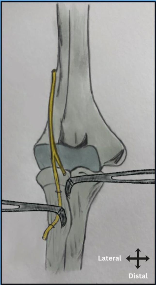
- The posterior interosseous nerve crosses the radial shaft a mean of 5.2 cm distal to the radiocapitellar joint in PRONATION (minimum 3.8 cm), falling to a mean 3.3 cm in SUPINATION (minimum 2.2 cm).
- Radial tunnel syndrome is a PAIN syndrome without motor loss; posterior interosseous nerve palsy is a MOTOR syndrome (finger drop) usually without pain. Same nerve, same anatomical sites, opposite presentations.
- “In a posterior interosseous nerve palsy the wrist still extends but in RADIAL DEVIATION, because extensor carpi radialis longus (radial nerve proper, proximal to the division) works while extensor carpi ulnaris does not.
- “Sensation is NORMAL in a posterior interosseous nerve palsy — the superficial radial nerve branches off before the supinator. Numbness in the dorsal first web means a more proximal radial nerve lesion.
- “Pronate for a posterolateral or Thompson approach and supinate for a volar Henry approach — the directions are opposite because you approach from opposite sides.
- “The supinator motor branches are a recognised nerve transfer donor: Bertelli transferred them to the posterior interosseous nerve to restore thumb and finger extension in tetraplegia with strong wrist extensors.
Overview
Supinator is a broad, flat muscle that wraps around the proximal third of the radius like a sleeve. As a supinator of the forearm it is secondary to the biceps. Its entire surgical significance derives from one relationship: the posterior interosseous nerve passes through it, between its two heads.
That single fact generates:
- The arcade of Frohse, the commonest site of posterior interosseous nerve compression and the anatomical origin of both radial tunnel syndrome and posterior interosseous nerve palsy.
- The safe zone — the distance from the radiocapitellar joint within which the proximal radius can be exposed or plated without endangering the nerve, and the reason forearm rotation is a deliberate intra-operative manoeuvre.
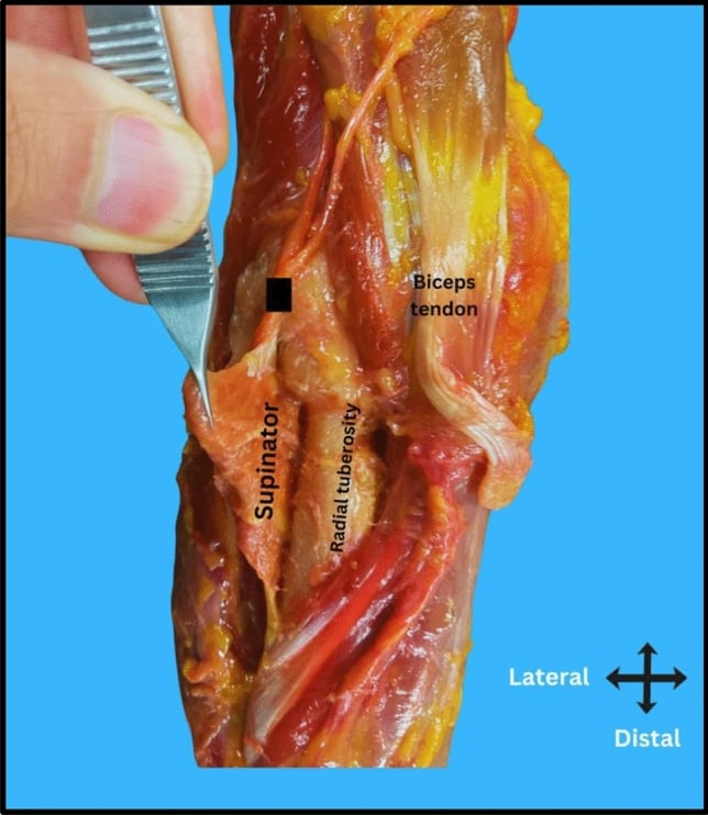
- The subperiosteal elevation technique that makes a volar Henry approach to the proximal radius safe.
This is the single highest-yield concept on this page and the one candidates most often get backwards.
Why the nerve moves. The posterior interosseous nerve is fixed within the substance of the supinator, and the supinator is wrapped around the radius. Rotate the radius and the supinator rotates with it, carrying the nerve.
The measured effect (Diliberti and colleagues, 32 cadaveric specimens, posterolateral approach between anconeus and extensor carpi ulnaris):
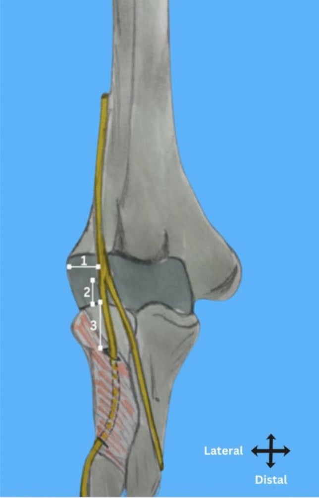
- Pronation: safe zone from the capitellum averaged 52.0 plus or minus 7.8 mm, with a minimum of 38 mm. Nerve-to-shaft angle 27.8 plus or minus 6.7 degrees.
- Supination: safe zone fell to a mean of 33.4 plus or minus 5.7 mm, as little as 22 mm. Nerve-to-shaft angle 47.4 plus or minus 6.8 degrees.
- Pronation therefore buys approximately 2 cm of safe zone from a posterolateral direction.
The rule, stated in both directions:
- PRONATE for a posterolateral (Kocher) or dorsal (Thompson) approach. Pronation swings the nerve anteriorly and medially, away from the dorsal and lateral surface you are working on.
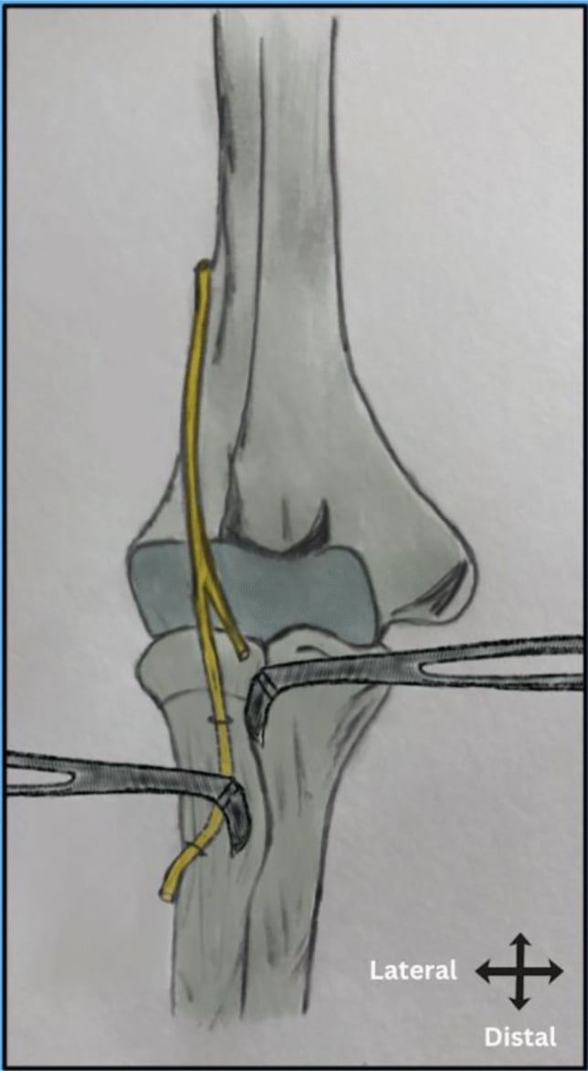
- SUPINATE for the volar (Henry) approach. Supination swings the nerve posteriorly and laterally, away from the volar surface, and brings the supinator insertion anteriorly so it can be elevated subperiosteally off the radius with the nerve protected inside it.
They are opposite because you approach from opposite sides. The nerve is not "safer in pronation" in the abstract — it is safer in pronation from behind, and safer in supination from in front.
FREASRadial Tunnel Compression Sites
Hook:Five sites, proximal to distal, and the release must address all of them — inadequate decompression is the documented cause of failure.
Attachments, Innervation and Relations
Two Heads
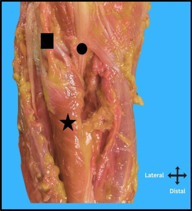
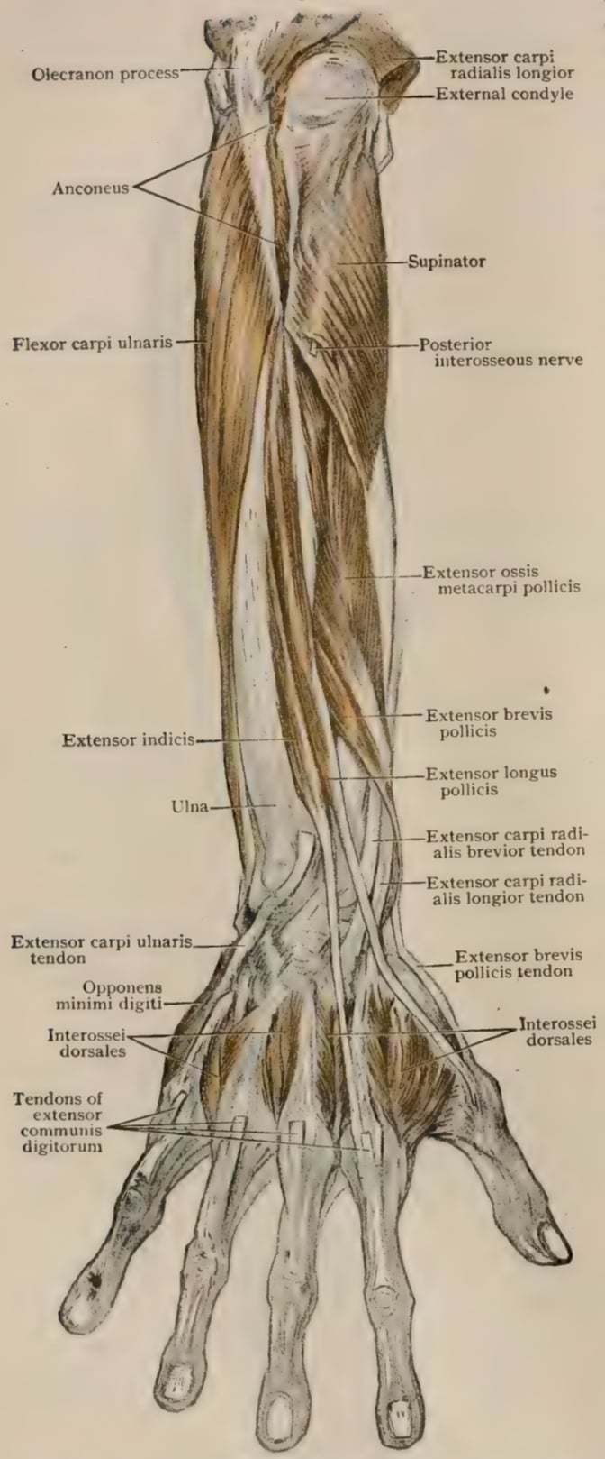
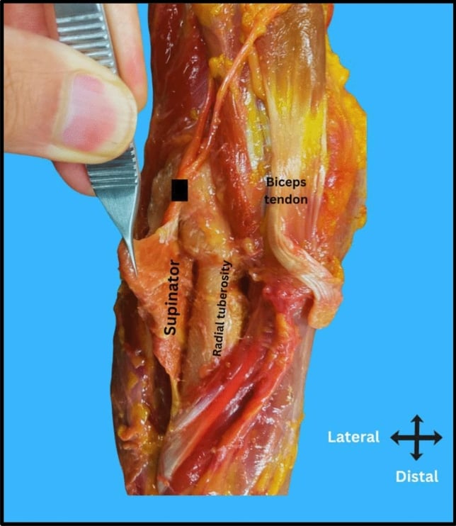
The supinator has a superficial (humeral) head and a deep (ulnar) head, and the posterior interosseous nerve passes between them.
- Lateral epicondyle of the humerus (deep to the common extensor origin).
- Radial collateral ligament of the elbow.
- Annular ligament of the proximal radioulnar joint.
- Its proximal margin is the ARCADE OF FROHSE — a tendinous arch over the posterior interosseous nerve.
- Supinator crest of the ulna — a bony ridge running distally from the posterior aspect of the lateral side of the olecranon, and the insertion point of the lateral ulnar collateral ligament.
- Supinator fossa, the depression anterior to the crest.
- The posterior part of the annular ligament and the interosseous membrane region.
Insertion
- The two heads converge and wrap around the radius to insert on the lateral, posterior and anterior surfaces of the proximal third of the radius, from just distal to the radial tuberosity down to the insertion of pronator teres.
- The insertion is circumferential over the proximal third — which is exactly why the muscle can be elevated subperiosteally as a sleeve, carrying the nerve safely within it.
Bony Landmarks to Quote
- Supinator crest of the ulna — also the insertion of the lateral ulnar collateral ligament, so a fracture or dissection here has ligamentous as well as muscular consequences.
- Radial tuberosity — the proximal limit of the supinator insertion, and the landmark for distal biceps repair.
- The radiocapitellar joint — the fixed proximal reference from which all posterior interosseous nerve safe-zone distances are measured.
Action and Biomechanics
Primary Action
Supination of the forearm — unwrapping itself from the radius and rotating the radius laterally.
Supinator versus Biceps
- Supinator
- Posterior interosseous (radial) C5, C6
- Biceps brachii
- Musculocutaneous C5, C6
- Supinator
- Weaker; the workhorse for slow, unresisted supination
- Biceps brachii
- Roughly two to three times more powerful; the prime supinator against resistance
- Supinator
- Works at ALL elbow positions, including full extension
- Biceps brachii
- Most efficient with the elbow at 90 degrees; poor in extension
- Supinator
- NONE — it does not meaningfully flex the elbow
- Biceps brachii
- Powerful elbow flexor as well
- Supinator
- Recruited first for slow, low-load supination
- Biceps brachii
- Recruited with speed and resistance
The key functional point: supinator alone handles slow, unresisted supination, and it does so at any elbow angle. Biceps is recruited for fast or resisted supination, and only performs well with the elbow flexed. This is why:
- Testing supination with the elbow EXTENDED minimises the biceps contribution and preferentially loads the supinator — the basis of one of the provocative tests for radial tunnel syndrome.
- A distal biceps rupture costs 40 to 50 per cent of supination power but leaves slow unresisted supination essentially intact, because supinator remains.
- A screwdriver is turned with biceps; the hand is turned palm-up to receive change with supinator.
The Anatomy of Supination
- Supination is rotation of the radius around the ulna, occurring at the proximal and distal radioulnar joints simultaneously.
- The axis passes from the centre of the radial head proximally to the fovea of the ulnar head distally.
- Normal arc: roughly 85 degrees of supination and 75 degrees of pronation from neutral, though functional range is smaller.
- Functional range (Morrey): approximately 50 degrees of pronation and 50 degrees of supination suffices for most activities of daily living, which is the figure to quote when discussing acceptable outcomes after forearm trauma or synostosis.
What Happens When It Fails
- Isolated supinator paralysis is functionally minor: biceps compensates for resisted supination, and the patient loses endurance in slow unresisted supination rather than absolute power.
- A posterior interosseous nerve palsy at the arcade spares the supinator entirely, so supination is normal while the fingers drop — a striking and diagnostically useful dissociation.
- Supinator contracture or scarring after proximal radial fracture or surgery contributes to loss of pronation and, together with heterotopic bone in the interosseous space, to proximal radioulnar synostosis.
Surface Anatomy and Examination
Locating the Radial Tunnel
- The point of maximal tenderness in radial tunnel syndrome is 4 to 5 cm distal to the lateral epicondyle, over the supinator in the mobile wad.
- The point of maximal tenderness in lateral epicondylitis is at or within 1 cm of the lateral epicondyle, over the extensor carpi radialis brevis origin.
- That 4 cm difference is the single most useful bedside discriminator, and it is the first thing to elicit in any patient with lateral elbow pain that has not responded to treatment.
Clinical Tests and What They Mean
- How to perform
- Palpate the lateral epicondyle, then 4 to 5 cm distal over the supinator in the mobile wad
- Positive finding
- Maximal tenderness 4 to 5 cm DISTAL to the epicondyle
- What it means
- Radial tunnel syndrome rather than lateral epicondylitis
- False positives
- The two coexist in a meaningful minority — tenderness at both sites does not exclude either
- How to perform
- Elbow extended, forearm pronated, wrist neutral; resist extension of the middle finger at the metacarpophalangeal joint
- Positive finding
- Pain in the proximal forearm over the supinator
- What it means
- Tension of the tendinous ECRB medial edge over the PIN
- False positives
- Also positive in lateral epicondylitis, since it loads ECRB — poor specificity
- How to perform
- Elbow fully extended, forearm pronated; resist supination
- Positive finding
- Pain in the proximal forearm
- What it means
- Supinator loading with the nerve in its substance; elbow extension minimises the biceps contribution
- False positives
- Pain from a distal biceps or radiocapitellar problem
- How to perform
- Ask the patient to extend the wrist against gravity
- Positive finding
- Extension PRESENT but in RADIAL DEVIATION
- What it means
- PIN palsy — ECRL working (radial nerve proper), ECU paralysed
- False positives
- Tenodesis effect from finger flexion mimics active extension
- How to perform
- Light touch over the dorsal first web space
- Positive finding
- NORMAL in PIN palsy; reduced in a proximal radial nerve lesion
- What it means
- Localises the lesion above or below the radial nerve division
- False positives
- Overlap with the lateral antebrachial cutaneous territory at the margins
- How to perform
- Inject local anaesthetic into the radial tunnel over the supinator
- Positive finding
- Temporary abolition of pain, accompanied by a transient PIN palsy confirming correct placement
- What it means
- Supports a diagnosis of radial tunnel syndrome
- False positives
- Diffusion to the lateral epicondyle can relieve epicondylitis pain too — inject a small volume accurately
The Central Discrimination
- Radial tunnel syndrome
- PAIN — deep, aching, proximal forearm
- Posterior interosseous nerve palsy
- WEAKNESS — finger drop
- Lateral epicondylitis
- Pain at the lateral epicondyle
- Radial tunnel syndrome
- NONE
- Posterior interosseous nerve palsy
- Finger and thumb extension lost
- Lateral epicondylitis
- None
- Radial tunnel syndrome
- None
- Posterior interosseous nerve palsy
- NONE (superficial radial spared)
- Lateral epicondylitis
- None
- Radial tunnel syndrome
- 4 to 5 cm distal to the epicondyle
- Posterior interosseous nerve palsy
- May be none
- Lateral epicondylitis
- At or within 1 cm of the epicondyle
- Radial tunnel syndrome
- Normal
- Posterior interosseous nerve palsy
- Present but in RADIAL DEVIATION
- Lateral epicondylitis
- Normal but painful
- Radial tunnel syndrome
- Usually NORMAL — a clinical diagnosis
- Posterior interosseous nerve palsy
- Abnormal, confirming the lesion
- Lateral epicondylitis
- Normal
- Radial tunnel syndrome
- Pain abolished
- Posterior interosseous nerve palsy
- No change in weakness
- Lateral epicondylitis
- Usually unchanged if injected accurately in the tunnel
Imaging and Electrodiagnostics
- Radial tunnel syndrome is a clinical diagnosis. Nerve conduction studies and electromyography are characteristically normal, which does not exclude it — this frequently causes diagnostic paralysis and is worth stating explicitly in a viva.
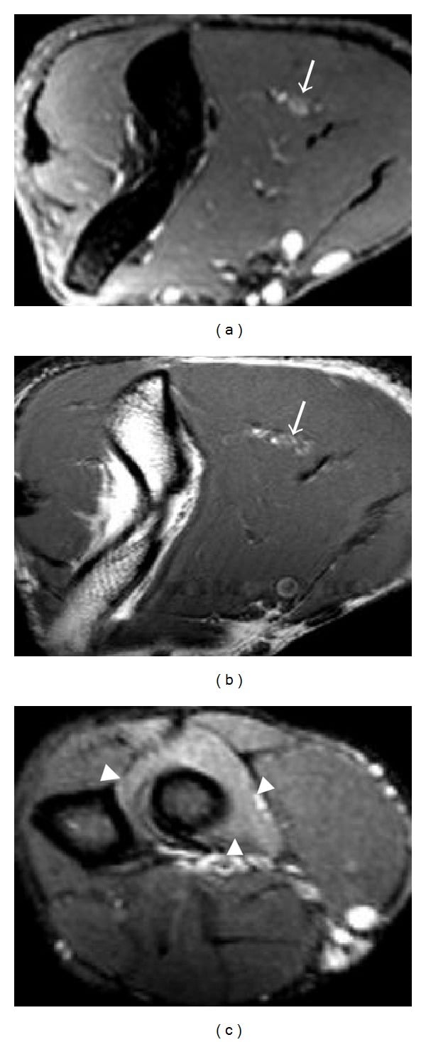
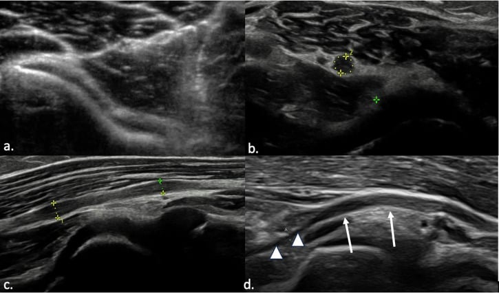
- Posterior interosseous nerve palsy should be confirmed electrodiagnostically at around 3 to 4 weeks, and MRI or high-resolution ultrasound may show denervation oedema in the posterior compartment muscles with supinator spared, or a compressive lesion such as a lipoma, ganglion or synovitis.
- In rheumatoid arthritis, a posterior interosseous nerve palsy from radiocapitellar synovitis must be distinguished from extensor tendon rupture at the wrist. The tenodesis test separates them: passively flexing the wrist produces finger extension if the tendons are intact (a nerve problem) but not if they are ruptured.
Complications
Denervation and Donor Morbidity
- Harvesting one or two supinator motor branches for transfer leaves the muscle partly innervated and biceps as the prime supinator; donor morbidity is minimal and this is the basis of the Bertelli transfer.
- Complete supinator denervation produces loss of endurance in slow unresisted supination rather than absolute weakness; it is not functionally disabling.
- Transecting the supinator muscle belly in an approach, rather than elevating it, both denervates and devascularises portions of it and unnecessarily exposes the nerve — it should not be done.
Unpredictable Outcomes of Radial Tunnel Release
- Patients must be counselled that radial tunnel decompression has less predictable results than carpal or cubital tunnel release, because the diagnosis is clinical, electrodiagnostics are normal, and the pain frequently has a multifactorial component including coexistent lateral epicondylitis and central sensitisation.
- A diagnostic local anaesthetic block that abolishes the pain is the best available predictor of a good result and should be obtained before offering surgery.
Stiffness and Loss of Rotation
- Any surgery at the proximal radius risks loss of forearm rotation from supinator scarring, capsular contracture and heterotopic bone. Early active pronation and supination is the single most important preventive measure.
Clinical Relevance
Definition
Compression of the posterior interosseous nerve within the radial tunnel producing pain without motor deficit. It is a contentious diagnosis precisely because it has no objective findings.
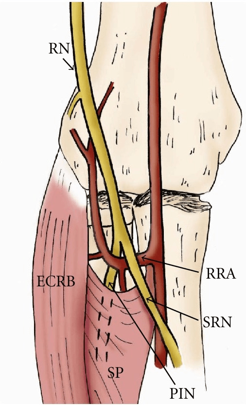
The Five Compression Sites (Proximal to Distal)
- Fibrous bands anterior to the radiocapitellar joint.
- Leash of Henry — the radial recurrent vessels crossing the nerve.
- Tendinous medial edge of extensor carpi radialis brevis.
- Arcade of Frohse — the tendinous proximal margin of the superficial head of supinator. The commonest site.
- Distal edge of the supinator.
Presentation
- Deep, aching pain in the proximal extensor forearm, worse with repetitive pronation and supination.
- Night pain is characteristic and helps distinguish it from lateral epicondylitis.
- Maximal tenderness 4 to 5 cm distal to the lateral epicondyle.
- No weakness, no sensory loss, normal electrodiagnostics.
The Coexistence Problem
Radial tunnel syndrome and lateral epicondylitis coexist in a meaningful minority of patients. A lateral epicondylitis that fails to respond to appropriate treatment should prompt reassessment for a proximal nerve component — and equally, a supposed radial tunnel that improves with an epicondylar injection was probably epicondylitis.
Management
- Non-operative first, and for a prolonged period: activity modification, avoidance of repetitive forearm rotation, splinting the wrist in extension with the elbow flexed and the forearm supinated (which relaxes the supinator), and physiotherapy including nerve gliding.
- Surgical decompression is reserved for persistent symptoms after an adequate non-operative trial, and outcomes are less predictable than for carpal or cubital tunnel decompression — patients must be counselled accordingly.
- The technical imperative is completeness. Hartz and colleagues, in their pronator teres syndrome series, identified inadequate decompression or misdiagnosis as the causes of failure, and the same holds here. All five sites must be released.
- Approach: either the Thompson (dorsal, extensor digitorum communis / extensor carpi radialis brevis) interval, or the brachioradialis-splitting or anterolateral (Henry-type) approach. The anterolateral approach gives better access to the proximal sites (fibrous bands and the leash of Henry); the Thompson approach gives better access to the arcade and the distal supinator edge.
Surgical Relevance
The Four Ways In and What Each Does to the Supinator
- Interval
- Brachioradialis (radial) / flexor carpi radialis (median)
- Forearm position
- SUPINATE for the proximal third
- Supinator handling
- Elevated SUBPERIOSTEALLY off the radius from its insertion, with the nerve protected inside it
- PIN risk
- Low IF supinated and elevated subperiosteally; high if the muscle is cut across
- Interval
- Extensor digitorum communis / extensor carpi radialis brevis
- Forearm position
- PRONATE
- Supinator handling
- Split in the line of its fibres, or the nerve identified within it and protected
- PIN risk
- Moderate — the nerve is directly in the field; some surgeons identify it routinely
- Interval
- Anconeus (radial) / extensor carpi ulnaris (PIN)
- Forearm position
- PRONATE
- Supinator handling
- Not usually violated; the approach stops proximal to it
- PIN risk
- Low if the dissection stays within 3.8 cm of the radiocapitellar joint in pronation
- Interval
- Extensor digitorum communis / extensor carpi radialis brevis, more anterior
- Forearm position
- PRONATE
- Supinator handling
- Proximal edge encountered
- PIN risk
- Higher — the nerve is closer to this more anterior interval
The Volar Henry Manoeuvre in Detail
This is the technique most often asked for verbatim:
- Develop the interval between brachioradialis (laterally, taking the superficial radial nerve with it) and flexor carpi radialis (medially, taking the radial artery with it).
- Ligate the leash of Henry — the radial recurrent vessels crossing the field at the level of the radial neck.
- FULLY SUPINATE the forearm. This brings the supinator insertion anteriorly into view and rotates the posterior interosseous nerve, held within the muscle, posteriorly and laterally away from you.
- Elevate the supinator subperiosteally off the radius, starting at its insertion and working proximally. The nerve travels with the muscle, protected within its substance. At no point is the muscle transected.
- This exposes the proximal radius safely.
Why this works: you are never dissecting toward the nerve. You are lifting the whole muscular sleeve, nerve included, off the bone in one layer.
The Thompson Approach
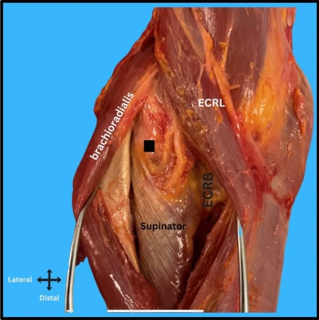
- Interval: extensor digitorum communis (posterior interosseous nerve) and extensor carpi radialis brevis (variable innervation) — not a strictly reliable internervous plane, which is one of its criticisms.
- PRONATE the forearm to move the nerve away.
- The nerve is encountered within the supinator; many surgeons formally identify it, particularly if plating is planned or the exposure needs to extend proximally.
- Safe zone: at least 3.8 cm distal to the radiocapitellar joint in pronation, and often more, but this is the figure to work to.
Guidelines, Registries & Global Practice
Anatomical Variation
- The arcade of Frohse is not present in everyone as a tendinous structure. Spinner's central observation is developmental: it is membranous in the fetus and becomes tendinous with age and use. Reported prevalence of a distinctly tendinous arcade in adult cadaveric series varies widely, from roughly a third to nearly all specimens depending on how strictly it is defined — a genuine source of confusion in the literature and a reason the reported prevalence figures should be quoted with caution.
- The extensor carpi radialis brevis innervation is variable — from the radial nerve proper, from the posterior interosseous nerve, or from the bifurcation itself. This is why the degree of preserved wrist extension in a posterior interosseous nerve palsy varies between patients.
- A single-headed supinator, or accessory slips, are described but uncommon.
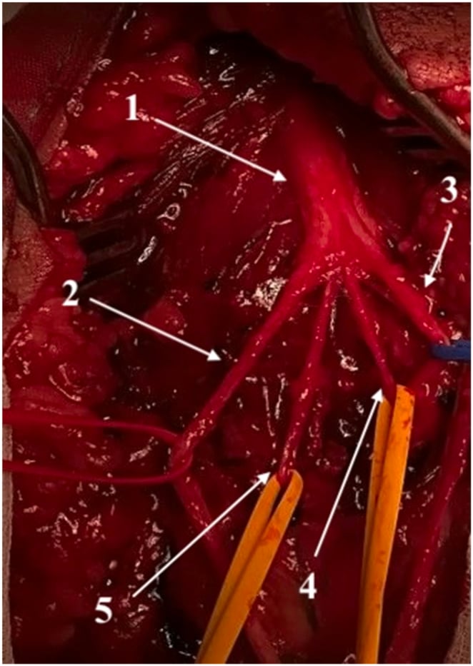
- The supinator motor branches were found as a consistent pair (one to each head) near the arcade of Frohse in cadaveric study, carrying 70 per cent of the myelinated fibres of the posterior interosseous nerve — the anatomical basis for the direct coaptation used in nerve transfer.
Side-by-Side Guidance
- Position relevant to supinator and the PIN
- Describes the volar Henry approach with explicit instruction to SUPINATE for the proximal third and elevate the supinator subperiosteally from its insertion, and the dorsal Thompson approach with instruction to PRONATE.
- Position relevant to supinator and the PIN
- No formal guideline. Consensus favours a prolonged non-operative trial, a confirmatory diagnostic block before surgery, complete release of all five sites, and explicit counselling that outcomes are less predictable than for carpal or cubital tunnel release.
- Position relevant to supinator and the PIN
- A spontaneous posterior interosseous nerve palsy warrants imaging to exclude a space-occupying lesion before attribution to the arcade of Frohse.
- Position relevant to supinator and the PIN
- Supinator branch to posterior interosseous nerve transfer is an accepted option in patients with strong wrist extensors, exploiting the short reinnervation distance; it competes with, rather than replaces, tendon transfer.
Global Practice Differences
- Nerve transfer for radial and posterior interosseous nerve palsy requires microsurgical infrastructure, early referral and electrodiagnostic support. In settings where patients present late, tendon transfer remains the appropriate and reliable answer — and for an isolated posterior interosseous nerve palsy that reconstruction is simpler than for a complete radial palsy, because wrist extension is already preserved.
- Radial tunnel decompression is performed far less often in some health systems than others, reflecting genuine international disagreement about whether the entity is over-diagnosed. Candidates should be able to argue both sides: the diagnosis rests on a symptom with no objective correlate, but the anatomical substrate is well described and a subset of patients clearly benefit.
- Access to MRI for a spontaneous posterior interosseous nerve palsy is not universal. Where it is unavailable, high-resolution ultrasound is an excellent and widely available substitute for detecting a lipoma or ganglion in the radial tunnel, and should be the default first-line investigation in those settings.
Registry and Outcome Signals
- Neither radial tunnel release nor posterior interosseous nerve decompression is captured in national registries; the evidence is institutional series and small comparative studies. Reported success rates for radial tunnel release vary widely, which is itself the most honest thing to say about the operation.
- Nerve versus tendon transfer for radial nerve palsy has been compared in a retrospective cohort showing both improve function significantly, with better grip strength after nerve transfer but no difference in pinch strength or patient-reported outcome — supporting a decision driven by time from injury and patient tolerance for a longer recovery rather than by a claim of superiority.
Rehabilitation Consensus
- After any proximal radius surgery, early active pronation and supination is the priority; forearm rotation is the function most readily and most permanently lost.
- After nerve transfer, motor re-education exploits the synergy between the donor and recipient action — patients learn to initiate extension by attempting supination, then progressively dissociate the two.
MCQ Practice Points
Q: What are the two heads of supinator and what runs between them? A: A superficial head from the lateral epicondyle, radial collateral and annular ligaments, and a deep head from the SUPINATOR CREST of the ulna. The POSTERIOR INTEROSSEOUS NERVE passes between them.
Q: What is the arcade of Frohse? A: The tendinous proximal margin of the superficial head of supinator — the commonest site of posterior interosseous nerve compression. It is membranous in the fetus and becomes tendinous with age.
Q: How far distal to the radiocapitellar joint does the PIN cross the radial shaft? A: A mean of 52 mm in PRONATION (minimum 38 mm) and 33 mm in SUPINATION (minimum 22 mm) from a posterolateral direction.
Q: Which forearm position for which approach? A: PRONATE for dorsal/Thompson and posterolateral/Kocher. SUPINATE for volar Henry. Opposite, because you approach from opposite sides.
Q: Why is supinator function preserved in a PIN palsy at the arcade of Frohse? A: Its motor branches arise PROXIMAL to or at the arcade, before the nerve enters the muscle.
Q: Why does the wrist deviate radially in a PIN palsy? A: Extensor carpi radialis longus (radial nerve proper) is working while extensor carpi ulnaris (PIN) is paralysed.
Q: Is sensation affected in a PIN palsy? A: NO. The superficial radial nerve branches off before the supinator. Dorsal first web numbness indicates a more proximal radial nerve lesion.
Q: How do radial tunnel syndrome and PIN palsy differ? A: Radial tunnel syndrome is PAIN without motor loss; PIN palsy is MOTOR loss usually without pain. Same nerve, same sites, opposite presentations.
Q: Where is the point of maximal tenderness in radial tunnel syndrome? A: 4 to 5 cm DISTAL to the lateral epicondyle over the supinator — compared with at or within 1 cm of the epicondyle in lateral epicondylitis.
Q: When is supinator the main supinator and when is biceps? A: Supinator handles SLOW, UNRESISTED supination at any elbow angle; biceps is recruited for FAST or RESISTED supination and works best with the elbow flexed.
Q: Why are the supinator motor branches expendable donors? A: They carry C5 and C6 fibres and biceps duplicates the function, so sacrificing them does not abolish supination — the basis for coapting them directly to the posterior interosseous nerve.
Clinical Imaging
Imaging Atlas
Exam Viva Scenarios
Practise clinical reasoning and management decisions out loud
“You need to plate a fracture of the proximal third of the radius. Talk me through your approach and, specifically, what you do with the forearm and why.”
“A 42-year-old joiner has had lateral elbow pain for 14 months. He has had physiotherapy, a brace and two corticosteroid injections into the lateral epicondyle with only transient benefit. He describes a deep ache in the forearm and is woken by it at night. Examination shows full power in all muscle groups and normal sensation. What is going on?”
“A 55-year-old woman presents with a three-week history of progressive inability to straighten her fingers. There was no injury. On examination she cannot extend the metacarpophalangeal joints of any finger or the thumb. Wrist extension is present but the wrist deviates radially. Sensation is entirely normal. What is the lesion, and what is your next step?”
Anatomy
- Superficial head: lateral epicondyle, radial collateral and annular ligaments
- Deep head: SUPINATOR CREST and fossa of the ulna
- PIN passes BETWEEN the two heads
- Insertion: wraps the proximal third of the radius, lateral, posterior and anterior
Innervation
- Posterior interosseous nerve C5, C6
- Branches arise PROXIMAL to or at the arcade of Frohse
- Therefore SPARED in a PIN palsy at the arcade
- Two to four branches - expendable nerve transfer donors
Radial Tunnel - Five Sites
- 1. Fibrous bands anterior to the radiocapitellar joint
- 2. Leash of Henry (radial recurrent vessels)
- 3. Tendinous medial edge of ECRB
- 4. ARCADE OF FROHSE - commonest site
- 5. Distal edge of supinator
Safe Zone
- PRONATION: mean 52 mm, minimum 38 mm from the capitellum
- SUPINATION: mean 33 mm, minimum 22 mm
- PRONATE for Thompson/Kocher; SUPINATE for volar Henry
- Elevate supinator SUBPERIOSTEALLY as a sleeve with the nerve inside
Clinical
- Radial tunnel = PAIN, no motor loss, normal EMG, tender 4-5 cm distal to the epicondyle
- PIN palsy = finger drop, wrist extends in RADIAL DEVIATION, sensation NORMAL
- Spontaneous PIN palsy = image for a mass
- Rheumatoid: tenodesis test distinguishes nerve palsy from tendon rupture
Evidence Base
The Arcade of Frohse and Its Relationship to Posterior Interosseous Nerve Paralysis
- The classic reference defining the arcade of Frohse and its relationship to posterior interosseous nerve paralysis
- No abstract is indexed for this 1968 paper; the claims below are what the title and MeSH indexing support, plus the conclusion universally attributed to it
- Indexed MeSH covers forearm anatomy and innervation, FETAL anatomy, and paralysis of peripheral nervous system origin — consistent with a study comparing fetal and adult specimens
- The conclusion attributed to it throughout the subsequent literature is that the arcade is MEMBRANOUS in the fetus and becomes TENDINOUS with age and use, making compression an acquired adult problem rather than a congenital one
- It established the arcade as the principal site of posterior interosseous nerve compression, and the anatomical distinction from a more proximal radial nerve lesion in which brachioradialis is also affected
Anatomical Considerations Regarding the Posterior Interosseous Nerve During Posterolateral Approaches to the Proximal Radius
- Thirty-two cadaveric specimens dissected through the posterolateral interval between anconeus and extensor carpi ulnaris, with the posterior interosseous nerve exposed
- Pronation allowed safe exposure of at least the proximal 38 mm of the lateral radius, with a mean proximal safe zone of 52.0 plus or minus 7.8 mm
- Supination reduced the safe zone to as little as 22 mm, with a mean of 33.4 plus or minus 5.7 mm
- The angle formed by the nerve and the radial shaft averaged 47.4 plus or minus 6.8 degrees in supination, decreasing to 27.8 plus or minus 6.7 degrees in pronation
- The authors concluded that approaching the lateral aspect of the proximal radius is safest in pronation
Transfer of Supinator Motor Branches to the Posterior Interosseous Nerve to Reconstruct Thumb and Finger Extension in Tetraplegia
- Case report: a patient with tetraplegia and paralysis of thumb and finger extension underwent bilateral transfer of supinator motor branches to the posterior interosseous nerve
- Surgery was performed 7 months after the spinal cord injury
- At 6 months postoperatively, with the wrist in neutral, extension of the thumb and fingers was almost full bilaterally
- The authors concluded that in tetraplegic patients with STRONG WRIST EXTENSORS, supinator motor branch transfer is a promising alternative for reconstructing thumb and finger extension
- Level 5 evidence: a single patient treated bilaterally, not a comparative series
Transfer of Supinator Motor Branches to the Posterior Interosseous Nerve in C7-T1 Brachial Plexus Palsy
- Four adult patients with C7-T1 root lesions operated between 5 and 7 months after injury; shoulder, elbow and wrist motion preserved but complete palsy of finger motion
- Supinator function is preserved in these lesions because its innervation stems from the C6 root
- Finger flexion was reconstructed by brachialis transfer; finger and thumb extension by transferring supinator motor branches to the posterior interosseous nerve
- Performed through an incision over the proximal third of the radius, dissecting between extensor carpi radialis brevis and extensor digitorum communis
- At 12 months all four patients could open the hand and fully extend the metacarpophalangeal joints; thumb abduction improved from 0 to 5 cm from the lateral aspect of the index finger
Radial Nerve Palsy: Nerve Transfer Versus Tendon Transfer to Restore Function
- Retrospective comparison of 30 patients treated with tendon transfers and 16 with nerve transfers for isolated radial nerve injury
- Fifteen of the 16 nerve transfer patients also received a pronator teres to extensor carpi radialis brevis tendon transfer for immediate wrist extension
- Both groups improved significantly in grip and pinch strength, DASH and quality-of-life scores after surgery
- Postoperative grip strength was significantly higher in the nerve transfer group; pinch strength did NOT differ, and neither did DASH or quality-of-life scores - on the patient-reported measures the two operations were equivalent
- Nerve transfer patients were younger, presented earlier and had longer follow-up
The Pronator Teres Syndrome: Compressive Neuropathy of the Median Nerve
- Thirty-nine patients with a clinical diagnosis of pronator teres syndrome seen over seven years
- Electrophysiological testing showed abnormalities in only a few patients, and localisation was rarely possible
- Surgical exploration of 36 forearms in 32 of the 39 patients showed intramuscular tendinous bands in pronator teres and indentation of the flexor superficialis muscle belly in most forearms, with occasional vascular and muscular abnormalities
- Intra-operative recordings improved shortly after release in only 6 of the 10 forearms tested - even direct measurement at the time of decompression did not confirm the lesion in every case
- Of 36 operations, 28 gave good or excellent results, five fair, and three unchanged
- The causes of failure were INADEQUATE DECOMPRESSION or MISDIAGNOSIS