The Muscle the Median Nerve Passes Through
- Two heads: a HUMERAL (superficial) head from the medial supracondylar ridge and common flexor origin, and an ULNAR (deep) head from the medial side of the CORONOID PROCESS. The MEDIAN NERVE passes between them; the ULNAR ARTERY passes DEEP to both.
- Insertion: the middle third of the LATERAL surface of the radius, at the point of maximum convexity — the pronator tuberosity.
- Innervated by the MEDIAN NERVE (C6, C7), with branches arising PROXIMAL to the muscle. Pronator teres function is therefore PRESERVED in a pronator syndrome caused by compression between its own heads.
- Pronator syndrome is a PAIN and PARAESTHESIA syndrome with proximal forearm tenderness and NO thenar wasting; anterior interosseous nerve syndrome is a PURE MOTOR syndrome with NO sensory loss.
- Pronator teres to extensor carpi radialis brevis is the standard wrist extension transfer in radial nerve palsy, and is used as an immediate internal splint even when a nerve transfer is planned.
- “Pronator syndrome is distinguished from carpal tunnel syndrome by involvement of the PALMAR CUTANEOUS BRANCH territory (the thenar eminence skin), which arises proximal to the carpal tunnel and is therefore SPARED in carpal tunnel syndrome.
- “In pronator syndrome, night symptoms are UNCOMMON — the opposite of carpal tunnel syndrome, where they are the rule.
- “Hartz and colleagues found intramuscular tendinous bands in the pronator in most of 36 explored forearms; inadequate decompression and misdiagnosis were the two causes of failure.
- “The ligament of Struthers arises from a supracondylar process present in roughly 1 per cent of people, and is the ONLY cause of proximal median compression visible on a plain radiograph.
Overview
Pronator teres runs obliquely across the proximal forearm from the medial epicondylar region to the lateral radius, forming the medial boundary of the cubital fossa. It is the primary pronator of the forearm, and three of its features drive its surgical importance:
- The median nerve passes between its two heads, making it the third of the four classical sites of proximal median nerve compression and the muscle after which pronator syndrome is named.
- Its insertion on the lateral radius is released in the middle-third window of the volar Henry approach and is a deforming force in proximal radial shaft fractures.
- It is expendable and synergistic with wrist extension, making it the standard donor for the pronator teres to extensor carpi radialis brevis transfer that restores wrist extension in radial nerve palsy.
Three median nerve syndromes, and examiners will hand you all three in a differential. Learn them as a triangle of positive and negative findings.
- Pronator syndrome
- Yes — including the thenar eminence skin (palmar cutaneous branch)
- Anterior interosseous nerve syndrome
- NONE — pure motor
- Carpal tunnel syndrome
- Yes — but the thenar eminence skin is SPARED
- Pronator syndrome
- Usually none or subtle; forearm ache and hand weakness reported
- Anterior interosseous nerve syndrome
- FPL, FDP to index (and middle), pronator quadratus — cannot make an OK sign
- Carpal tunnel syndrome
- Thenar wasting and weak abduction in advanced cases
- Pronator syndrome
- Uncommon
- Anterior interosseous nerve syndrome
- Absent
- Carpal tunnel syndrome
- Characteristic — the classic presentation
- Pronator syndrome
- Proximal forearm
- Anterior interosseous nerve syndrome
- Usually none
- Carpal tunnel syndrome
- At the wrist
- Pronator syndrome
- Resisted pronation with the elbow extended; resisted elbow flexion and supination; resisted long finger FDS
- Anterior interosseous nerve syndrome
- Often preceded by severe pain then painless weakness
- Carpal tunnel syndrome
- Phalen and Durkan tests
- Pronator syndrome
- Frequently NORMAL
- Anterior interosseous nerve syndrome
- Abnormal in FPL, FDP index, pronator quadratus
- Carpal tunnel syndrome
- Abnormal — the diagnostic standard
The single most useful discriminator between pronator syndrome and carpal tunnel syndrome is the palmar cutaneous branch of the median nerve. It arises from the median nerve roughly 5 to 8 cm proximal to the wrist crease and passes superficial to the flexor retinaculum, not through the carpal tunnel. It supplies the skin over the thenar eminence and the radial palm.
- Carpal tunnel syndrome: the compression is distal to the branch, so thenar eminence skin sensation is NORMAL.
- Pronator syndrome: the compression is proximal to the branch, so thenar eminence skin sensation IS affected.
And why pronator teres itself works in pronator syndrome: the motor branches to pronator teres arise from the median nerve proximal to the muscle, before the nerve dives between the two heads. Compression between the heads is therefore distal to its own innervation.
SLAPProximal Median Nerve Compression Sites
Hook:Each site has its OWN provocative test — the test that reproduces the symptoms tells you which site to release.
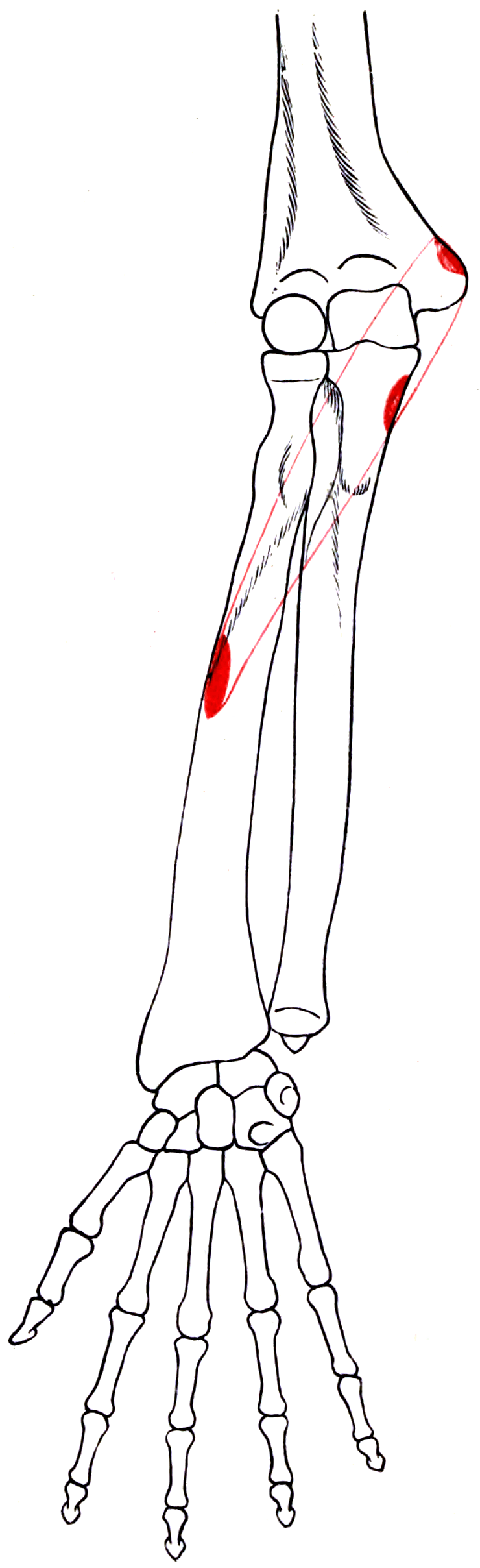
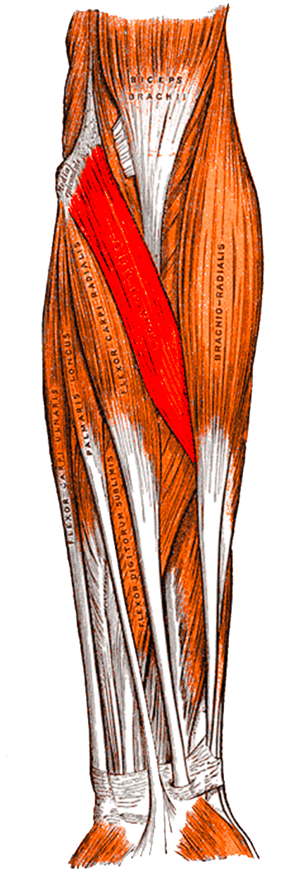
Attachments, Innervation and Relations
Two Heads
- The medial supracondylar ridge of the humerus, just above the medial epicondyle.
- The common flexor origin on the medial epicondyle.
- The medial intermuscular septum and the deep antebrachial fascia.
- It is the larger of the two heads and forms most of the muscle bulk.
- The medial side of the coronoid process of the ulna, just distal to the sublime tubercle.
- It is the smaller head, and it is absent in roughly 5 to 10 per cent of limbs — a variation with direct clinical relevance, since when the deep head is absent the median nerve passes deep to the whole muscle and compression at this site cannot occur in the classical manner.
Insertion
- The middle third of the lateral surface of the radius, at the point of maximum convexity of the radial bow — sometimes called the pronator tuberosity or the pronator teres tubercle.
- The insertion is a broad, flat, roughened area roughly 4 to 5 cm long, immediately distal to the supinator insertion and proximal to the middle of the bone.
- Its position at the apex of the radial bow gives it a good pronation moment arm and makes it a deforming force in fractures at that level.
Bony Landmarks to Quote
- Humeral head origin: medial supracondylar ridge and medial epicondyle.
- Ulnar head origin: medial coronoid process, just distal to the sublime tubercle (which is where the anterior band of the medial collateral ligament inserts).
- Insertion: lateral radius at the apex of the radial bow, approximately the mid-shaft.
Action and Biomechanics
Primary Action
Pronation of the forearm — pronator teres is the primary pronator, working at all speeds and against resistance.
Secondary Action
Weak elbow flexion. Because it crosses the elbow from the medial supracondylar ridge, it contributes a small flexion moment, but it is never the limiting factor.
Pronator Teres versus Pronator Quadratus
- Pronator teres
- Median (main trunk) C6, C7
- Pronator quadratus
- ANTERIOR INTEROSSEOUS nerve C7, C8
- Pronator teres
- Proximal forearm, crossing obliquely
- Pronator quadratus
- Distal forearm, transverse, deep
- Pronator teres
- PRIMARY pronator; recruited for fast and resisted pronation
- Pronator quadratus
- Initiates and sustains SLOW, UNRESISTED pronation; active throughout
- Pronator teres
- Weak flexion
- Pronator quadratus
- None — does not cross the elbow
- Pronator teres
- Its contribution falls with the elbow EXTENDED, since the muscle is lengthened across two joints
- Pronator quadratus
- Unaffected by elbow position
- Pronator teres
- None beyond pronation and weak flexion
- Pronator quadratus
- STABILISES the distal radioulnar joint by compressing the radius against the ulna
The clinical test that follows: to isolate pronator quadratus, test resisted pronation with the elbow FULLY FLEXED. Elbow flexion slackens pronator teres and minimises its contribution, leaving pronator quadratus as the main pronator being tested. This is the standard way to detect an anterior interosseous nerve lesion clinically.
Conversely, resisted pronation with the elbow EXTENDED maximally loads pronator teres and is one of the provocative tests for pronator syndrome.
The Radial Bow
- Pronator teres inserts at the apex of the radial bow, the point of maximum lateral convexity of the radius.
- The radial bow is essential to forearm rotation: it provides the space for the radius to rotate around the ulna. Loss of the bow after a malreduced forearm fracture directly reduces pronation and supination.
- Restoring the radial bow is one of the explicit goals of forearm fracture fixation, and pronator teres is the muscle that pulls against it.
What Happens When It Fails
- Loss of pronator teres alone (as after harvest for transfer) is usually described as costing little, because pronator quadratus continues to pronate. It has been measured and the cost is larger than that. Skie tested six cadavers and six patients and found a statistically significant loss of pronation strength of 24 to 44 per cent depending on the method, with the cadaveric arm confirming reduced force and range after simulating the transfer. Patients still function, and the transfer remains the standard — but "expendable" should mean the loss is acceptable, not there is no loss. It matters most where pronation is load-bearing (manual work, crutch or wheelchair use) and where the transfer is being done as an internal splint while awaiting nerve recovery, which is the situation the authors specifically flag.
- Loss of both pronators (a high median nerve lesion) abolishes active pronation; the patient compensates by shoulder abduction and internal rotation to bring the palm downward, a highly visible substitution.
- Pronator teres contracture contributes to a fixed pronation deformity in cerebral palsy and after forearm trauma; pronator teres rerouting (transferring the tendon around the radius so it becomes a supinator) is a described treatment in cerebral palsy.
Surface Anatomy and Examination
Palpation
- With the elbow flexed to about 90 degrees and the forearm supinated, ask the patient to pronate against resistance. The pronator teres stands out as an oblique cord running from the medial epicondylar region across the proximal forearm toward the lateral radius.
- It forms the medial edge of the cubital fossa, and the brachial artery pulse and the median nerve are found just medial to the biceps tendon and just lateral to the muscle's proximal edge.
- Tenderness over the proximal muscle belly, roughly 4 to 6 cm distal to the medial epicondyle, is the key finding in pronator syndrome.
Clinical Tests and What They Mean
- How to perform
- Elbow fully extended, forearm supinated; resist pronation
- Positive finding
- Reproduction of forearm pain and paraesthesia
- What it means
- Compression BETWEEN THE TWO HEADS of pronator teres
- False positives
- Medial epicondylitis; the test also loads the common flexor origin
- How to perform
- Elbow at 120 to 135 degrees, forearm supinated; resist flexion
- Positive finding
- Reproduction of symptoms
- What it means
- Compression under the LACERTUS FIBROSUS, which tightens in this position
- False positives
- Distal biceps pathology
- How to perform
- Resist proximal interphalangeal flexion of the middle finger with the other digits held extended
- Positive finding
- Reproduction of symptoms
- What it means
- Compression at the FDS proximal arch (sublimis bridge)
- False positives
- Also loads FDS itself, causing muscular pain
- How to perform
- As described for a supracondylar process
- Positive finding
- Reproduction of symptoms
- What it means
- Compression by a LIGAMENT OF STRUTHERS — obtain a radiograph looking for a supracondylar process
- False positives
- Uncommon; do not over-diagnose
- How to perform
- Light touch over the skin of the thenar eminence, NOT the thumb pulp
- Positive finding
- REDUCED
- What it means
- Palmar cutaneous branch involvement, so the lesion is PROXIMAL to the carpal tunnel
- False positives
- Requires careful technique; test the thenar SKIN, not the digits
- How to perform
- Ask the patient to make a circle with thumb and index finger
- Positive finding
- A flat, pinched, triangular pinch instead of a round O
- What it means
- Anterior interosseous nerve palsy — FPL and FDP index cannot flex the terminal joints
- False positives
- Attritional tendon rupture in rheumatoid disease produces the same sign
- How to perform
- Elbow fully flexed to slacken pronator teres; resist pronation
- Positive finding
- Weakness
- What it means
- Pronator quadratus deficit — an anterior interosseous nerve lesion
- False positives
- Pain inhibition
Distinguishing Pronator Syndrome from Carpal Tunnel Syndrome
- Pronator syndrome
- AFFECTED (palmar cutaneous branch involved)
- Carpal tunnel syndrome
- SPARED (branch arises proximal to the tunnel)
- Pronator syndrome
- Uncommon
- Carpal tunnel syndrome
- CHARACTERISTIC
- Pronator syndrome
- Proximal volar forearm, 4 to 6 cm distal to the medial epicondyle
- Carpal tunnel syndrome
- None, or at the wrist
- Pronator syndrome
- Proximal forearm
- Carpal tunnel syndrome
- At the wrist
- Pronator syndrome
- Negative
- Carpal tunnel syndrome
- Positive
- Pronator syndrome
- Frequently NORMAL
- Carpal tunnel syndrome
- Abnormal — the diagnostic standard
- Pronator syndrome
- Resisted pronation, resisted elbow flexion and supination, resisted FDS
- Carpal tunnel syndrome
- Wrist position and compression
Electrodiagnostics: Manage Expectations
Hartz and colleagues, in the definitive clinical series of 39 patients, found that electrophysiological testing showed abnormalities in only a few patients, and localisation of the abnormality was possible only rarely. This is the honest position: pronator syndrome is a clinical diagnosis, and a normal study does not exclude it. Electrodiagnostics are performed principally to exclude carpal tunnel syndrome and cervical radiculopathy, not to confirm the pronator.
Complications
Donor-Site Morbidity
- Loss of pronator teres costs measurable pronation power and endurance, but pronator quadratus maintains functional pronation and patients rarely complain. It is the accepted trade-off.
- Do not harvest pronator teres in a patient who also has an anterior interosseous nerve lesion, since pronator quadratus would then also be out and the patient would lose active pronation entirely.
- In cerebral palsy, over-correction of a pronation deformity by rerouting can produce a fixed supination deformity, which is functionally worse than the pronation it replaced. Patient selection and careful tensioning are essential.
Medial Antebrachial Cutaneous Neuroma
- The medial antebrachial cutaneous nerve crosses the antecubital fossa subcutaneously and is at risk in any medial or anterior elbow incision. A painful neuroma at this site is a recognised and disabling complication.
- Avoidance: identify and protect the nerve in the subcutaneous plane; spread rather than cut.
Adhesions and Loss of Rotation
- Any surgery in the proximal forearm risks adhesions and loss of forearm rotation. Early active pronation and supination, guided by hand therapy, is the single most important preventive measure.
- After a pronator teres transfer, the tendon is passed subcutaneously around the radius; a rough tunnel or a sharp angle of pull produces adhesions and a poor result.
Clinical Relevance
Presentation
Hartz and colleagues described the classical picture from 39 patients over seven years:
- Aching discomfort in the forearm
- Weakness in the hand
- Numbness in the thumb and index finger
- Cyclic stress usually brought on the symptoms — repetitive pronation and supination at work or in sport.
- The distinctive physical finding was tenderness over the proximal part of the pronator teres, aggravated by resisted pronation of the forearm, flexion of the elbow, and occasionally by resisted contraction of the flexor superficialis of the long finger.
The Four Compression Sites and Their Provocative Tests
- Structure
- Ligament of Struthers, from a supracondylar process to the medial epicondyle
- Provocative test
- Resisted elbow flexion at about 120 degrees with the forearm pronated
- Prevalence
- Supracondylar process in roughly 1 per cent of people; the ONLY site visible on a plain radiograph
- Structure
- Lacertus fibrosus (bicipital aponeurosis)
- Provocative test
- Resisted elbow flexion with the forearm SUPINATED
- Prevalence
- Common contributor
- Structure
- BETWEEN THE TWO HEADS of pronator teres (tendinous bands)
- Provocative test
- Resisted pronation with the elbow EXTENDED
- Prevalence
- Tendinous bands found in MOST explored forearms in the Hartz series
- Structure
- Proximal fibrous arch of flexor digitorum superficialis (sublimis bridge)
- Provocative test
- Resisted long finger FDS contraction
- Prevalence
- Indentation of the FDS muscle belly seen in most explored forearms
Surgical Findings and Results (Hartz and colleagues)
- 36 forearms in 32 patients explored.
- Intramuscular tendinous bands in the pronator found in most.
- Indentation of the flexor superficialis muscle belly in most.
- Vascular and muscular abnormalities occasionally.
- Intra-operative recordings showed improvement shortly after release in 6 of the 10 forearms tested.
- Results: 28 good or excellent, 5 fair, 3 unchanged.
- The causes of failure were INADEQUATE DECOMPRESSION or MISDIAGNOSIS.
Management
- Non-operative first, and for a prolonged period: activity modification to avoid repetitive pronation and supination, splinting, and physiotherapy with nerve gliding.
- Surgical decompression if symptoms persist: through a lazy-S incision across the antecubital fossa, all four sites are released — the ligament of Struthers and any supracondylar process, the lacertus fibrosus, the tendinous bands and deep head of pronator teres, and the flexor digitorum superficialis arch.
- The lesson from the Hartz data: release everything. Partial decompression is one of the two documented reasons the operation fails.
Surgical Relevance
The Standard Wrist Extension Transfer for Radial Nerve Palsy
Pronator teres to extensor carpi radialis brevis is the first transfer named in every radial nerve palsy reconstruction, and it appears in essentially every published set.
Why Pronator Teres Is the Right Donor
- Expendable — but not free. Pronator quadratus continues to pronate and patients function well, which is why the transfer is standard. The measured cost is nonetheless real: Skie found a statistically significant 24 to 44 per cent loss of pronation strength in six patients, matched by reduced force and range in a cadaveric simulation of the same transfer. Consent for it, and weigh it in a manual worker or anyone who loads the forearm in pronation.
- Median-innervated, therefore unaffected by the radial nerve lesion.
- Synergistic: pronation and wrist extension occur together in normal grasp, so motor re-education is straightforward — the patient is taught to think about pronating to extend the wrist, then progressively dissociates the two.
- Adequate excursion and power: wrist extension requires only about 3 cm of excursion, which pronator teres comfortably provides. It loses roughly one MRC grade on transfer but the resulting wrist extension is functional.
- Straight line of pull once the tendon is passed around the radius.
Why ECRB Rather Than ECRL
- Extensor carpi radialis brevis inserts on the base of the THIRD metacarpal, in line with the axis of the forearm, so it produces central, balanced wrist extension.
- Extensor carpi radialis longus inserts on the base of the SECOND metacarpal and produces radially deviated wrist extension.
- Transferring into extensor carpi radialis brevis alone gives a balanced wrist. Some surgeons weave the transfer through both tendons; the risk is radial deviation if extensor carpi radialis longus dominates.
Technique
- The pronator teres tendon is harvested with a long strip of periosteum from its radial insertion, to lengthen it and provide robust tissue for the weave.
- It is passed around the radius subcutaneously, from volar to dorsal, superficial to brachioradialis and extensor carpi radialis longus, to reach extensor carpi radialis brevis.
- Pulvertaft weave into extensor carpi radialis brevis, tensioned with the wrist in about 30 to 45 degrees of extension and the elbow flexed.
- Tension check: with the wrist in the set position, the transfer should be taut; the tenodesis effect should be preserved.
The Standard Radial Nerve Palsy Transfer Set
- Standard transfer
- PRONATOR TERES to extensor carpi radialis brevis
- Alternative
- None widely used — PT is the near-universal choice
- Comment
- Synergistic, expendable, excursion-matched
- Standard transfer
- Flexor carpi ulnaris to extensor digitorum communis
- Alternative
- Flexor carpi radialis to EDC; flexor digitorum superficialis to EDC
- Comment
- FCU sacrifices the main wrist flexor and ulnar deviator; FCR preserves it. Raskin and Wilgis showed no significant functional impairment after FCU transfer at a mean 8 years
- Standard transfer
- Palmaris longus to a rerouted extensor pollicis longus
- Alternative
- FDS ring to EPL if palmaris longus is absent (about 15 per cent)
- Comment
- Rerouting the EPL out of the third compartment improves the vector to combine extension with abduction
As an Internal Splint Alongside Nerve Transfer
Patterson and colleagues reported that 15 of 16 patients undergoing nerve transfer for radial nerve palsy also received a pronator teres to extensor carpi radialis brevis tendon transfer at the same time. The rationale is elegant: the tendon transfer provides immediate wrist extension while the nerve transfer reinnervates the finger extensors over months. The tendon transfer is an internal splint. In that series the nerve transfer group achieved significantly higher grip strength, with no difference in pinch strength or patient-reported outcome. Read the comparison carefully before quoting it as nerve-beats-tendon. Because 15 of the 16 also had the tendon transfer, the study compares tendon transfer alone against tendon transfer plus nerve transfer — which is an addition, not an alternative. The groups also differed significantly in the three variables that most affect a strength result: the nerve transfer patients were younger, operated sooner after injury, and followed for longer. The defensible reading is the authors' own — both approaches improve pain, function and satisfaction, and the choice turns on presentation timing and on whether the patient can wait months for reinnervation.
Guidelines, Registries & Global Practice
Anatomical Variation
- The ulnar (deep) head is absent in roughly 5 to 10 per cent of limbs. When it is absent, the median nerve passes deep to the whole muscle, and compression at this site cannot occur in the classical manner — a variation with direct diagnostic and surgical relevance.
- A supracondylar process of the humerus, from which the ligament of Struthers arises, is present in approximately 1 per cent of people, with reported prevalence varying by population. It is a vestigial structure and is the only one of the four compression sites visible on a plain radiograph — which is why a lateral humeral radiograph is worth obtaining when a proximal median compression is suspected.
- Gantzer muscle, an accessory head of flexor pollicis longus arising from the medial epicondyle or coronoid, is present in a substantial minority of limbs and is a described cause of anterior interosseous nerve compression.
- Palmaris longus is absent in roughly 15 per cent of limbs, with marked population variation (lower prevalence of absence in some Asian populations, higher in some European series). Its presence must be confirmed before planning a transfer that depends on it.
- Martin-Gruber anastomosis — a median-to-ulnar communication in the forearm — is present in roughly 15 to 20 per cent of limbs and produces atypical patterns of weakness and confusing nerve conduction studies.
Side-by-Side Guidance
- Position relevant to pronator teres
- Describes the volar Henry approach with release of the pronator teres insertion for the middle third with the forearm pronated, and subperiosteal elevation of the supinator for the proximal third with the forearm supinated.
- Position relevant to pronator teres
- No formal guideline. Consensus favours a prolonged non-operative trial, exclusion of carpal tunnel syndrome and cervical radiculopathy, and systematic release of all four sites if surgery is undertaken.
- Position relevant to pronator teres
- Near-universal agreement that pronator teres to extensor carpi radialis brevis is the wrist extension transfer; disagreement persists over flexor carpi ulnaris versus flexor carpi radialis as the finger extension donor.
- Position relevant to pronator teres
- Early presentation with a reconstructable nerve favours nerve transfer, often with a pronator teres to extensor carpi radialis brevis tendon transfer as an internal splint; late presentation favours tendon transfer alone.
Global Practice Differences
- The FCU versus FCR debate for finger extension is genuinely unsettled. Raskin and Wilgis showed that despite the anatomic loss, wrist function was not significantly impaired after flexor carpi ulnaris transfer at a mean of eight years, with work simulation indistinguishable from controls — which supports the traditional FCU-based set. Others prefer flexor carpi radialis to preserve the main wrist flexor and ulnar deviator, particularly in manual workers. Both are defensible.
- Nerve transfer requires microsurgical infrastructure and early referral. In settings where patients present late — which is most of the world for humeral shaft fractures with nerve injury — the tendon transfer set is the appropriate, reliable and equipment-light answer, requiring no implants and no specialised instruments.
- Pronator syndrome is diagnosed at very different rates in different health systems, reflecting the same tension as radial tunnel syndrome: a symptom-based diagnosis with no objective correlate. Candidates should be able to argue both that the anatomical substrate is well described and that the entity is easily over-diagnosed.
Registry and Outcome Signals
- Neither pronator decompression nor tendon transfer is captured in national registries; the evidence base is institutional series, and the Hartz series of 39 patients remains a defining reference despite its age.
- The best comparative data on nerve versus tendon transfer show both improve function significantly, with better grip strength after nerve transfer but no difference in pinch strength or patient-reported outcome — supporting a decision driven by time from injury rather than by a claim of superiority.
Rehabilitation Consensus
- After tendon transfer, contemporary practice favours early protected active motion under supervised hand therapy over prolonged immobilisation, to reduce adhesions and speed motor relearning.
- Motor re-education for a pronator teres transfer exploits the natural synergy: patients are taught to initiate wrist extension by attempting pronation, then progressively dissociate the two movements. Synergistic transfers are re-educated faster than non-synergistic ones, which is one of the reasons pronator teres is preferred.
MCQ Practice Points
Q: What are the two heads of pronator teres and what passes between them? A: A humeral (superficial) head from the medial supracondylar ridge and common flexor origin, and an ulnar (deep) head from the medial CORONOID. The MEDIAN NERVE passes between them.
Q: What is the relationship of the ulnar artery to pronator teres? A: It passes DEEP to BOTH heads. The nerve goes between; the artery goes under.
Q: Where does pronator teres insert? A: The middle third of the LATERAL surface of the radius, at the apex of the radial bow (the pronator tuberosity).
Q: How does the palmar cutaneous branch distinguish pronator syndrome from carpal tunnel syndrome? A: It arises 5 to 8 cm proximal to the wrist and passes SUPERFICIAL to the flexor retinaculum. Thenar eminence skin sensation is therefore affected in pronator syndrome and SPARED in carpal tunnel syndrome.
Q: Are night symptoms typical of pronator syndrome? A: NO — they are uncommon. Night symptoms are characteristic of carpal tunnel syndrome.
Q: Name the four sites of proximal median nerve compression. A: Ligament of Struthers, lacertus fibrosus, between the two heads of pronator teres, and the proximal fibrous arch of flexor digitorum superficialis.
Q: What is the sensory deficit in anterior interosseous nerve syndrome? A: NONE. It is a pure motor syndrome — flexor pollicis longus, flexor digitorum profundus to index (and middle), and pronator quadratus.
Q: How do you test pronator quadratus in isolation? A: Resisted pronation with the elbow FULLY FLEXED, which slackens pronator teres and minimises its contribution.
Q: Why is pronator teres the donor of choice for wrist extension in radial nerve palsy? A: Expendable (pronator quadratus remains), median-innervated, SYNERGISTIC with wrist extension, and excursion-matched (wrist extension needs only about 3 cm).
Q: Why transfer into extensor carpi radialis brevis rather than longus? A: ECRB inserts on the base of the THIRD metacarpal, in line with the forearm axis, giving balanced extension. ECRL inserts on the second and causes radial deviation.
Q: How does pronator teres determine reduction position in a radial shaft fracture? A: Proximal to its insertion, the proximal fragment is SUPINATED (supinator and biceps unopposed) — fix in supination. Distal to its insertion, the proximal fragment is NEUTRAL — fix in neutral.
Q: What forearm position for each window of the Henry approach? A: SUPINATE for the supinator (proximal third), PRONATE for the pronator teres (middle third). Rotate to bring each insertion anteriorly into view.
Exam Viva Scenarios
Practise clinical reasoning and management decisions out loud
“A 38-year-old assembly-line worker has six months of aching in the right proximal forearm and numbness in the thumb and index finger. She was referred with a diagnosis of carpal tunnel syndrome, but nerve conduction studies are normal and a night splint has not helped. What is your differential and how do you sort it out?”
“A 45-year-old man has a complete radial nerve palsy 14 months after an open humeral shaft fracture. Exploration at the time showed the nerve to be transected and it was grafted, but there has been no recovery. What do you offer him and why is pronator teres involved?”
“You are fixing an isolated radial shaft fracture. How does the level of the fracture relative to pronator teres change your reduction, and how do you deal with the muscle in the approach?”
Anatomy
- Humeral head: medial supracondylar ridge and common flexor origin
- Ulnar head: medial CORONOID (absent in 5-10 per cent)
- MEDIAN NERVE passes BETWEEN the heads; ULNAR ARTERY passes DEEP to both
- Insertion: lateral radius at the apex of the radial bow (middle third)
Innervation
- Median nerve C6, C7
- Branches arise PROXIMAL to the muscle - so PT works in pronator syndrome
- AIN arises from the DORSAL median nerve 5-8 cm distal to the lateral epicondyle
- Palmar cutaneous branch: 5-8 cm proximal to the wrist, SUPERFICIAL to the retinaculum
Four Compression Sites
- 1. Ligament of Struthers (supracondylar process, about 1 per cent) - the only one on a radiograph
- 2. Lacertus fibrosus - resisted elbow flexion with supination
- 3. Between the two PT heads - resisted pronation with the elbow EXTENDED
- 4. FDS proximal arch - resisted long finger FDS
Diagnosis
- Pronator syndrome: thenar SKIN affected, night symptoms uncommon, EMG often NORMAL
- Carpal tunnel: thenar skin SPARED, night symptoms characteristic, EMG abnormal
- AIN syndrome: PURE MOTOR, no sensory loss, cannot make a round OK sign
- Isolate pronator quadratus by resisted pronation with the elbow FULLY FLEXED
Transfer and Approach
- PT to ECRB: expendable, synergistic, excursion-matched (wrist needs about 3 cm)
- ECRB (3rd metacarpal) not ECRL (2nd) - avoids radial deviation
- Full set: PT to ECRB, FCU or FCR to EDC, PL to rerouted EPL
- Henry: SUPINATE for the supinator, PRONATE for the pronator teres
Evidence Base
The Pronator Teres Syndrome: Compressive Neuropathy of the Median Nerve
- Thirty-nine patients with a clinical diagnosis of pronator teres syndrome seen over a seven-year period
- Typical complaints were aching forearm discomfort, hand weakness and numbness in the thumb and index finger, usually brought on by cyclic stress
- The distinctive physical finding was tenderness over the proximal pronator teres, aggravated by resisted pronation, elbow flexion and occasionally resisted long finger flexor superficialis contraction
- Electrophysiological testing showed abnormalities in only a few patients, and localisation of the abnormality was possible only rarely
- Surgical exploration of 36 forearms showed intramuscular tendinous bands in the pronator and indentation of the flexor superficialis muscle belly in most; 28 of 36 operations gave good or excellent results, 5 fair and 3 unchanged, with inadequate decompression or misdiagnosis the causes of failure
Radial Nerve Palsy: Nerve Transfer Versus Tendon Transfer to Restore Function
- Retrospective review of 30 patients with isolated radial nerve injury treated with tendon transfers and 16 treated with nerve transfers
- Fifteen of the 16 nerve transfer patients also received a concomitant PRONATOR TERES to EXTENSOR CARPI RADIALIS BREVIS tendon transfer to provide immediate wrist extension
- Both groups showed significant improvement in grip and pinch strength, DASH scores and quality-of-life scores after surgery
- Postoperative grip strength was significantly higher in the nerve transfer group; pinch strength did not differ
- Nerve transfer patients were younger, presented earlier and had longer follow-up; both approaches gave improved pain, function and satisfaction
Flexor Carpi Ulnaris Transfer for Radial Nerve Palsy: Functional Testing of Long-Term Results
- Six patients with complete irreparable radial nerve palsy treated with the STANDARD transfer set: PRONATOR TERES to extensor carpi radialis brevis, flexor carpi ulnaris to extensor digitorum communis, and palmaris longus to a rerouted extensor pollicis longus
- Mean follow-up 8 years (range 3 to 15), compared against 10 matched volunteer controls
- A functional range of motion was maintained in all patients and wrist power was sufficient for all activities of daily living
- Work simulation testing (hammering, sawing, tightening screws, using pliers) showed no significant difference from controls
- Despite the anatomic loss of flexor carpi ulnaris, wrist function was not significantly impaired
Functional Deficit After Transfer of the Pronator Teres for Acquired Radial Nerve Palsy
- Sets out the four criteria a motor donor must meet: adequate strength, correct line of pull, synergistic action, and NO UNACCEPTABLE FUNCTIONAL LOSS - then tests the fourth for the commonest donor in the upper limb
- Six cadavers and six patients studied, biomechanically and clinically
- Cadaveric simulation of the pronator teres to extensor carpi radialis brevis transfer showed decreased range of motion and decreased force developed under similar load
- Clinically there was a STATISTICALLY SIGNIFICANT LOSS OF PRONATION STRENGTH OF 24 TO 44 PER CENT, depending on the testing method
- The authors flag this as important where the transfer is performed as an internal splint WHILE AWAITING radial nerve recovery - the patient may recover the radial nerve and keep the pronation deficit
The Arcade of Frohse and Its Relationship to Posterior Interosseous Nerve Paralysis
- The classic reference defining the arcade of Frohse and its relationship to posterior interosseous nerve paralysis
- No abstract is indexed for this 1968 paper; the claims below rest on the title, the MeSH indexing (forearm anatomy and innervation, FETAL anatomy, paralysis of peripheral nervous system origin) and the conclusion universally attributed to it
- That conclusion is that the arcade is membranous in the fetus and becomes tendinous with age and use, making compression an acquired adult problem
- It established the arcade as the principal site of posterior interosseous nerve compression and the framework for distinguishing it from a more proximal radial nerve lesion
Anatomical Considerations Regarding the Posterior Interosseous Nerve During Posterolateral Approaches to the Proximal Radius
- Thirty-two cadaveric specimens dissected to quantify the safe zone along the proximal radius
- Pronation allowed safe exposure of at least the proximal 38 mm of the lateral radius, mean 52.0 plus or minus 7.8 mm
- Supination reduced the safe zone to as little as 22 mm, mean 33.4 plus or minus 5.7 mm
- The nerve-to-shaft angle averaged 47.4 degrees in supination, falling to 27.8 degrees in pronation
- Approaching the lateral proximal radius is safest in pronation
Vulnerability of the Posterior Interosseous Nerve During Proximal Radius Exposures
- THREE approaches compared on 30 cadaveric upper limbs: the modified anterior Henry approach, Thompson's posterior approach, and a modified Gordon-Boyd approach
- Structures were localised, marked and imaged radiographically, and plates and screws were then applied through each approach to define their relationship to the nerve
- The ANTERIOR approach was judged relatively safe and extensible both proximally and distally - with the caution that SCREW placement can still injure the nerve
- Thompson's posterior approach, although favoured because the radius is superficial dorsally, CARRIES THE RISK of posterior interosseous nerve injury when the most proximal radius is exposed
- The Gordon-Boyd approach exposes both bones but requires excessive muscle stripping