A measured difference in limb length is a sign, not a diagnosis — the job is to say whether it is congenital, acquired shortening, or overgrowth, and whether the growth plate is still working
- Apparent discrepancy from pelvic obliquity, adduction/abduction contracture or scoliosis is far commoner than true bone shortening — exclude it before imaging.
- Discrepancy is a moving target: what matters is not today's number but the projected discrepancy at skeletal maturity, which needs serial measurement plus bone age.
- Hemihypertrophy (true overgrowth) mandates abdominal surveillance for Wilms tumour and hepatoblastoma — this is the systemic must-not-miss.
- Any unilateral shortening with a painful, warm or stiff joint is septic arthritis sequelae or Perthes until proven otherwise.
- A discrepancy of less than 2 cm at maturity is generally not treated; 2-5 cm is epiphysiodesis territory; greater than 5 cm needs lengthening or prosthetic fitting.
- Look at the whole limb: femoral, tibial and foot height contributions must be separated because the treatment site differs.
- “Ask for the block test first — it converts a vague complaint into a number and separates true from apparent.
- “Congenital causes shorten proportionally (a constant percentage); acquired growth arrest produces an accelerating absolute difference.
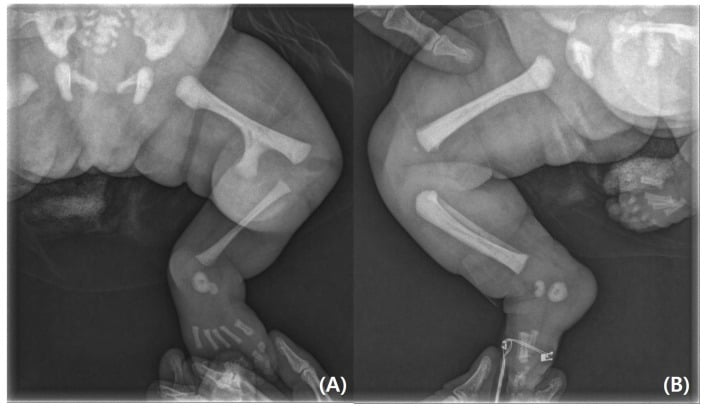
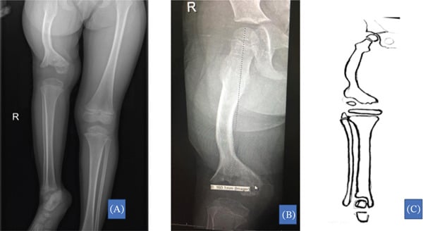
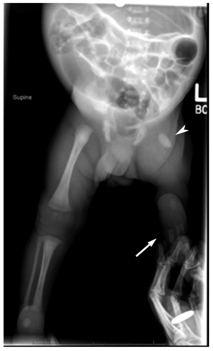
- “Femoral shortening with a bulbous, laterally-facing hip and limited abduction is proximal focal femoral deficiency, not DDH.
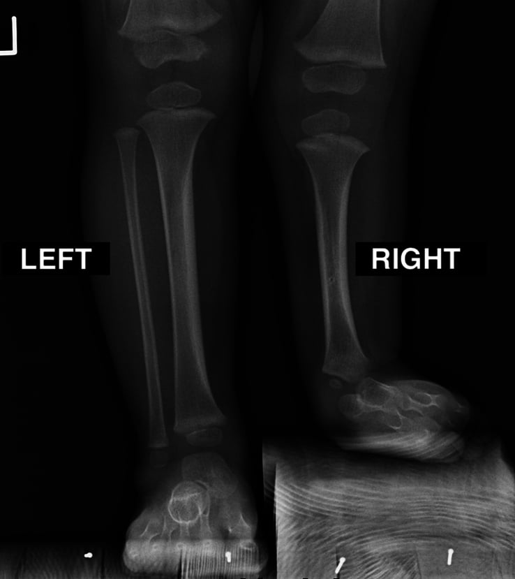
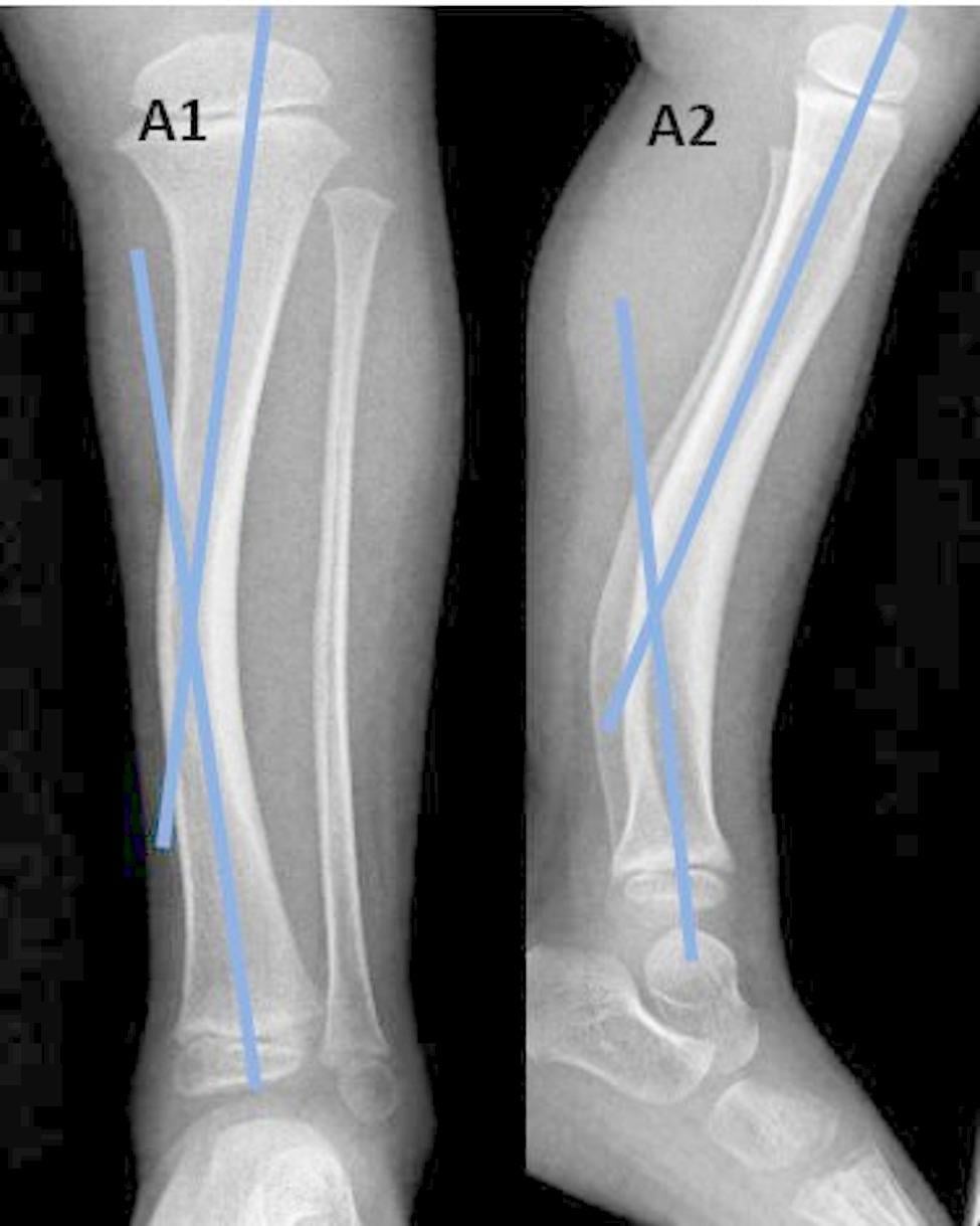
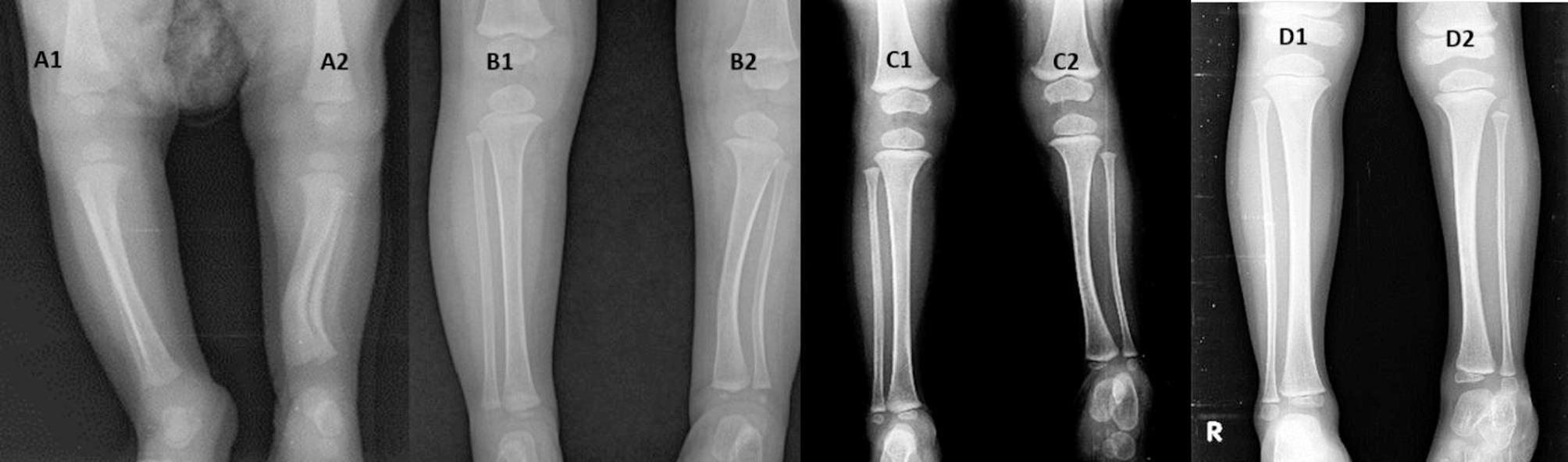
- “In fibular hemimelia the leg is short AND the foot has fewer rays with an equinovalgus ankle — check the foot, not just the leg.
- “Neuromuscular limbs are short because they are unloaded — the shortening is diffuse, thin and osteopenic, not focal.
Umbilicus-to-medial-malleolus measures apparent length and includes pelvic obliquity. True length is ASIS to ipsilateral medial malleolus. A fixed adduction contracture makes a normal limb look short; abduction contracture makes it look long.
A 2 cm discrepancy in a 4-year-old with a complete physeal arrest will become far greater; the same 2 cm in a 14-year-old girl at Risser 4 will barely change. Always pair the measurement with bone age and remaining growth.
Isolated hemihyperplasia and Beckwith-Wiedemann spectrum carry an embryonal tumour risk. The correct answer is three-monthly surveillance - but the two tests run for DIFFERENT durations, which is the detail that scores: abdominal ultrasound to age 7 (Wilms tumour) and serum alpha-fetoprotein to age 4 (hepatoblastoma). Refer to a clinical geneticist, because current consensus stratifies by molecular subtype rather than screening everyone identically. Not just shoe raises.
Total limb length includes calcaneal and talar height. A short limb with a normal scanogram of femur and tibia may be a hindfoot problem, or the knee may be held flexed by a contracture. Measure segments separately.
Recognising the Pattern
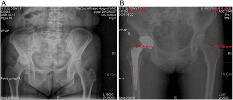
Definition. Limb length discrepancy (LLD) is a difference in the length of the lower limbs measured from a fixed pelvic landmark to the ground, expressed in centimetres and attributed to a segment: femur, tibia, or foot.
Confirming it is genuinely present.
- Clinical block test — the patient stands with knees straight and blocks of known thickness are placed under the short side until the iliac crests are level. The block height equals the functional discrepancy. This is the single most reproducible bedside measurement and includes foot height.
- True length — ASIS to ipsilateral medial malleolus with the pelvis square and both limbs in identical positions. Compare sides.
- Apparent length — xiphisternum or umbilicus to medial malleolus. A difference in apparent length with equal true lengths means pelvic obliquity from contracture or scoliosis.
- Segment localisation — Galeazzi test: supine, hips and knees flexed, feet flat. A lower knee indicates tibial shortening; a knee that is more posterior indicates femoral shortening.
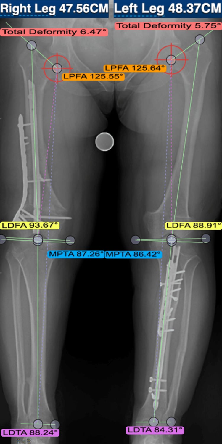
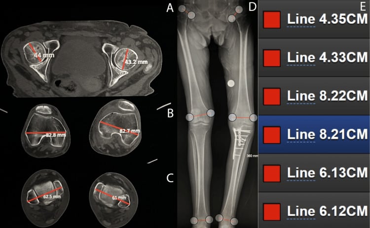
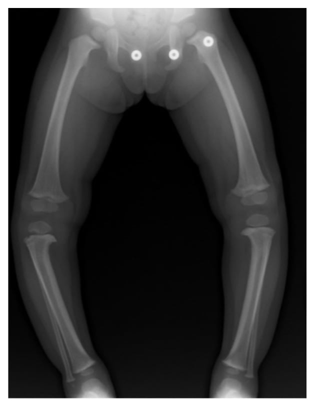
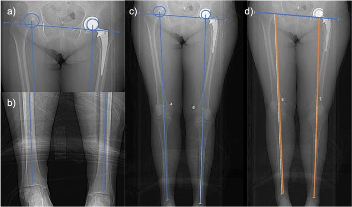
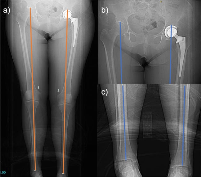
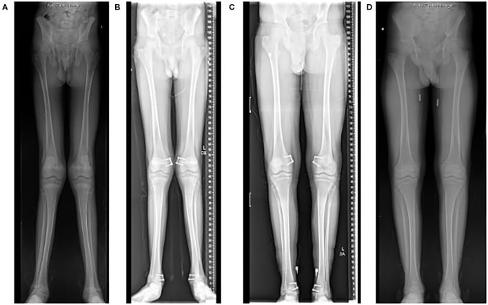
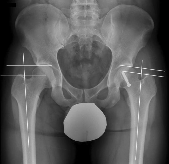
- Radiographic confirmation — standing full-length AP of both limbs (EOS or a calibrated scanogram) with a ruler in the field, patient weight-bearing and patellae forward, with a block under the short side to level the pelvis.
Saying it out loud in a viva. "This is a standing full-length AP radiograph of both lower limbs in a skeletally immature patient. There is a limb length discrepancy of approximately X centimetres, short on the [side]. Measuring segments, the femoral difference is X and the tibial difference is Y, so the shortening is predominantly [femoral/tibial]. The mechanical axis passes [medial/lateral] to the knee centre. I would want the clinical block test, a bone age film, and serial measurements before committing to a projection."
Mimics and false positives.
- Pelvic obliquity from a fixed adduction contracture (cerebral palsy, post-septic hip) — corrects on the block test only partially and true lengths are equal.
- Scoliosis with a lumbosacral hemivertebra — the pelvis is obliquely set within the spine.
- Knee or hip flexion contracture — shortens the limb functionally without bone shortening; check with the limbs extended.
- Radiographic magnification and malrotation — an externally rotated femur projects shorter; an uncalibrated film exaggerates. Always ask for the ruler.
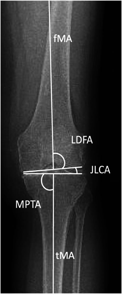
- Genu valgum or varum — angular deformity shortens the limb geometrically; the bone lengths may be equal.
- Equinus contracture — a fixed equinus foot lengthens the functional limb and may mask a true shortening.
Next Investigation
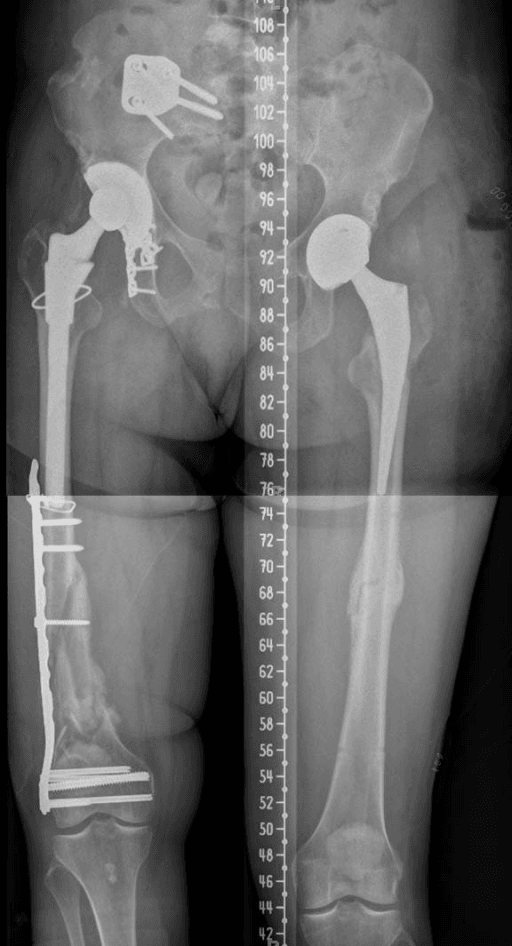
Radiation matters: a full-length scanogram carries a real gonadal dose and these children are measured repeatedly for a decade. Use EOS where available, shield, and do not order films at intervals shorter than 6 months without a reason.
The Differential
- Typical age / setting
- Any age, months to years after a documented injury
- Discriminating feature
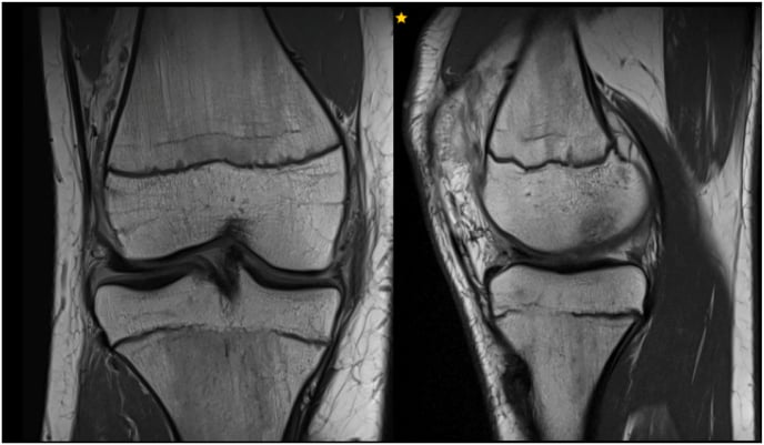
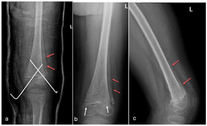
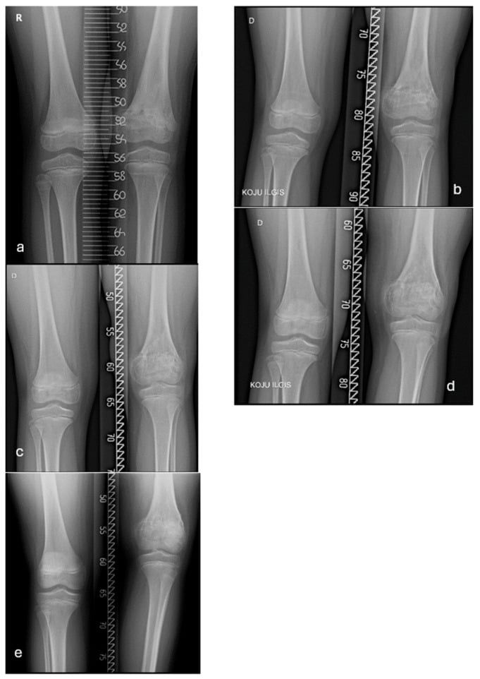
- Progressive shortening WITH angular deformity converging toward the bar; Park-Harris growth arrest line is oblique and converges on the bar rather than parallel to the physis
- What confirms it
- MRI physeal mapping (fat-suppressed 3D) or CT to size and locate the bar as a percentage of physeal area
- Typical age / setting
- Sequel of neonatal or infant sepsis, presenting at ages 2-10
- Discriminating feature
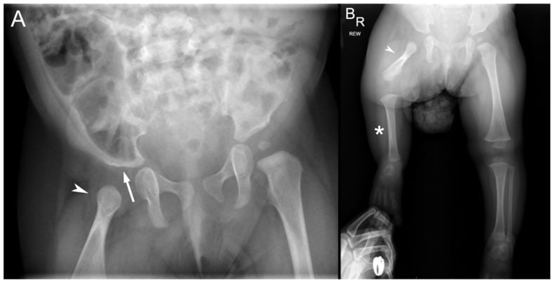
- Severe shortening far out of proportion to the history, often with femoral head loss and a stiff painless hip; frequently a history of NICU line sepsis
- What confirms it
- Radiographs showing absent or deformed capital femoral epiphysis; MRI for residual physis
- Typical age / setting
- Recognised in infancy or early childhood
- Discriminating feature
- The LONG side is abnormal — asymmetry involves soft tissue, hemiface, tongue or trunk, not just the limb; asymmetry is proportional and non-progressive in ratio
- What confirms it
- Clinical asymmetry across body segments plus tumour surveillance ultrasound; genetic testing for 11p15 imprinting defect
- Typical age / setting
- Ages 8-18 with pain, or a survivor of childhood cancer
- Discriminating feature
- Night pain, a metaphyseal lesion with periosteal reaction, or shortening dating precisely from radiotherapy or endoprosthesis
- What confirms it
- MRI of the whole bone; oncological history and radiotherapy field records
- Typical age / setting
- Well child, any age, discrepancy typically less than 2 cm
- Discriminating feature
- Diagnosis of exclusion: no deformity, no dysmorphism, symmetric segments, ratio of shortening constant on serial measurement
- What confirms it
- Serial scanograms plus bone age showing a stable percentage difference; no imaging abnormality
- Typical age / setting
- Infant to walking age; unilateral dislocation
- Discriminating feature
- Apparent rather than true shortening, with limited abduction, asymmetric thigh creases and a positive Galeazzi at the KNEE level
- What confirms it
- Ultrasound before 4-6 months; AP pelvis thereafter with broken Shenton line and proximal femoral migration
- Typical age / setting
- Boys aged 4-8, painful limp
- Discriminating feature
- Modest shortening (usually less than 2 cm) with coxa magna, coxa breva and loss of hip internal rotation and abduction
- What confirms it
- AP and frog-lateral pelvis showing fragmentation and subchondral fracture; MRI if early
- Typical age / setting
- Spastic hemiplegia, walking age onwards
- Discriminating feature
- The affected limb is short, THIN and osteopenic overall, with equinus and reduced foot size — disuse affects the whole limb, not one physis
- What confirms it
- Neurological examination; radiographs show slender diaphyses and generalised osteopenia on the affected side
- Typical age / setting
- Recognised at birth or in infancy
- Discriminating feature
- Short tibia with anteromedial bowing and a skin dimple, absent or short fibula, equinovalgus ball-and-socket ankle and missing lateral rays of the foot
- What confirms it
- AP and lateral tibia and foot radiographs counting rays and assessing the ankle mortise
- Typical age / setting
- Birth
- Discriminating feature
- The whole shortening is femoral, the thigh is short bulbous and held in flexion, abduction and external rotation, and the knee often has cruciate deficiency
- What confirms it
- AP pelvis and femur assessing acetabulum and proximal femoral segment; Aitken classification; MRI in infants for cartilaginous continuity
- Typical age / setting
- Ages 2-10, in the 1-2 years after a healed femoral shaft fracture
- Discriminating feature
- The previously FRACTURED side is longer, typically by up to about 1-2 cm, and the overgrowth plateaus by about 2 years post-injury
- What confirms it
- Serial scanograms showing overgrowth that stabilises; no physeal abnormality
- Typical age / setting
- Childhood, with a port-wine stain or varicosities
- Discriminating feature
- Overgrowth of the limb with a cutaneous vascular stain and lateral venous anomaly; warmth and bruit in high-flow lesions
- What confirms it
- Duplex ultrasound and contrast MR angiography of the limb
- Typical age / setting
- Girls aged 2-8 with a chronically swollen knee
- Discriminating feature
- The AFFECTED side is LONGER early (chronic synovitis hyperaemia accelerates the physis), then shorter if the physis closes prematurely; look for accompanying uveitis
- What confirms it
- Knee radiographs showing epiphyseal overgrowth and squared patella; MRI synovitis; ANA and slit lamp
- Typical age / setting
- Childhood, often with multiple lesions
- Discriminating feature
- Discrepancy accompanied by a matrix-containing or expansile lesion abutting the physis, or anterolateral tibial bowing in NF1; often bilateral but asymmetric
- What confirms it
- Skeletal survey; characteristic lucent columns (Ollier), sessile exostoses, or ground-glass matrix
Abdominal tumour incidence in syndromic and isolated hemihyperplasia — the basis for surveillance
- Isolated (idiopathic) hemihyperplasia carried a 1.2% abdominal tumour incidence (3 of 250 children: one adrenal carcinoma, two Wilms tumours) - real, but lower than historically quoted.
- Syndromic hemihypertrophy carried a higher risk (1 of 10 with Beckwith-Wiedemann syndrome developed a hepatoblastoma, 10%) - so the syndromic child and the isolated overgrowth are not the same risk.
- Retrospective review recommending geneticist referral to stratify which children benefit most from surveillance - supports surveillance while noting the isolated-overgrowth risk is lower than classic figures.
Leg Length Discrepancy After Femoral Shaft Fractures in Children - Review After Skeletal Maturity
- Thirty patients reviewed and radiographed AT SKELETAL MATURITY after an isolated closed femoral shaft fracture in childhood treated conservatively
- Where the fracture occurred between ages 7 and 13, the limb overgrew by ABOUT 1 CM - and did so regardless of sex, upper-limb dominance, age, fracture site or fracture configuration
- Excessive fracture overlap AT THE TIME OF INJURY increased overgrowth; overlap at union did not
- Angulation remodelled in children injured under 10 years, but in older children it sometimes ADDED to shortening rather than correcting
- The practical target: aim for about 1 cm of overlap at union in the 7-to-13-year-old, and correct angular deformity in children over 10 - this keeps the maximum discrepancy at maturity to about 1 cm
Multiplier Method for Predicting Limb-Length Discrepancy
- The premise, and the reason the method works only where it works: in CONGENITAL and developmental discrepancy the short limb grows at a rate PROPORTIONAL to the normal limb, so the discrepancy stays a constant fraction of limb length
- The multiplier is mature length divided by length at each age; it proved EQUIVALENT ACROSS ALL PERCENTILE GROUPS, varying only with age and sex
- For a congenital discrepancy the mature value is simply current discrepancy multiplied by the age- and sex-specific multiplier - obtainable from a SINGLE measurement, with no graphs
- For progressive developmental (non-congenital) discrepancy the formula differs: current discrepancy plus growth inhibition times growth remaining - two different formulae, and using the wrong one is the error
- Multipliers derived from radiographic, clinical and anthropological databases were similar despite differences in race, ethnicity and generation; predictions correlated well with the Moseley method and with actual outcomes in lengthening and epiphysiodesis groups
Narrowing It Down
- 1Step 1 — Is it true or apparent?
Do the block test standing, then measure true (ASIS to medial malleolus) against apparent (umbilicus to medial malleolus) lengths supine with the pelvis squared.
Equal true lengths with unequal apparent lengths means pelvic obliquity from a hip contracture or spinal deformity — treat the contracture or the curve, not the bone. Only a TRUE discrepancy proceeds to scanography.
- 2Step 2 — Which segment, and does the foot contribute?
Perform the Galeazzi test to localise femur versus tibia, then compare shoe sizes and calcaneal height.
Femoral-only shortening points to PFFD, DDH, Perthes or a distal femoral physeal bar. Tibial-only shortening points to fibular or tibial hemimelia, proximal tibial arrest, or Blount disease. Multi-segment shortening with a small foot suggests a neuromuscular or global congenital cause.
- 3Step 3 — Congenital or acquired?
Date the onset from the history: present since birth, or following a documented fracture, infection, tumour treatment or arthritis.
Congenital discrepancy grows as a CONSTANT PERCENTAGE of the normal side, so it projects from a single measurement by the multiplier method. Acquired discrepancy accelerates in absolute terms and demands physeal imaging and serial measurement.
- 4Step 4 — Is the short side abnormal, or is the long side abnormal?
Measure each limb against the child's own height percentile rather than only against each other, and decide which limb is the outlier.
If the LONG limb is the outlier, think hemihypertrophy, vascular malformation, JIA hyperaemia or post-fracture overgrowth — and in hemihypertrophy this is the point at which tumour surveillance is triggered, before any orthopaedic plan. If the SHORT limb is the outlier, think physeal arrest, congenital deficiency or neuromuscular disuse.
- 5Step 5 — Is there associated deformity or joint disease?
Look specifically for angular deformity that worsens with growth, for a stiff, painful or unstable hip, for anterolateral tibial bowing, and for a ball-and-socket ankle.
Progressive angulation indicates an asymmetric (peripheral) physeal bar. A stiff hip suggests Perthes, post-septic sequelae or slipped epiphysis. Anterolateral bowing with a short leg raises NF1 and congenital pseudarthrosis. A ball-and-socket ankle is strongly associated with longstanding fibular deficiency or tarsal coalition.
- 6Step 6 — Are there systemic or syndromic features?
Examine the skin, face, abdomen and neurology, not just the legs.
Cafe-au-lait macules and axillary freckling (NF1), macroglossia and omphalocele (Beckwith-Wiedemann), port-wine stain and varicosities (Klippel-Trenaunay), chronic knee swelling and uveitis (JIA), or spasticity and equinus (cerebral palsy). Each redirects the workup away from isolated orthopaedics, and two of them are urgent.
- 7Step 7 — What will the discrepancy be at maturity?
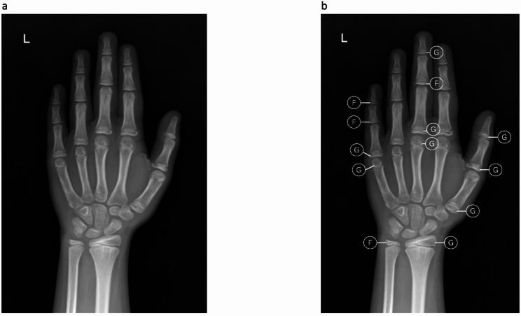
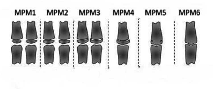
Obtain a bone age (Greulich and Pyle, left hand) and at least two scanograms 6-12 months apart, then project: multiplier method for a congenital discrepancy from a single visit, Moseley straight-line graph or Green-Anderson charts for acquired arrest.
Only the PROJECTED MATURE discrepancy dictates treatment, never the current one: observation and a shoe raise below about 2 cm, contralateral epiphysiodesis for roughly 2-5 cm, lengthening or amputation with prosthetic fitting above about 5-6 cm.
MCQ Practice Points
Q: A child has equal true limb lengths (ASIS to medial malleolus) but a 3 cm apparent difference. What is the mechanism? A: Pelvic obliquity, most often from a fixed hip adduction contracture on the apparently short side (or abduction contracture on the opposite side), or from a structural spinal curve. The treatment is directed at the contracture or the spine, not at the bone length.
Q: Which method best predicts mature discrepancy from a single clinic visit in a congenital case? A: The multiplier method. Congenital discrepancy remains a constant percentage of the growing limb, so current discrepancy multiplied by an age- and sex-specific factor gives the predicted mature value. Acquired arrests are better handled with the Moseley straight-line graph or Green-Anderson charts because the percentage is not constant.
Q: A 5-year-old had a mid-shaft femoral fracture at age 3 and now has a 1 cm LONGER injured limb. Management? A: Reassurance and observation. Hyperaemic overgrowth after paediatric femoral shaft fracture is well recognised, is usually modest, and plateaus by about 2 years post-injury. It is one reason for accepting mild shortening at the time of fracture treatment in young children.
Q: Which physeal bar is potentially amenable to resection? A: A bar involving less than roughly half the physeal area, in a child with at least 2 years or 2 cm of growth remaining, with correctable or minimal associated deformity. Larger bars, near-mature children and post-infective bars have poor results, and completion of the arrest with subsequent length equalisation is preferred.
Q: Which limb is abnormal in a child with a chronically swollen knee from oligoarticular JIA? A: Early, the affected limb is LONGER because chronic synovial hyperaemia accelerates the adjacent physis, with epiphyseal overgrowth and a squared patella on radiographs. Late, premature physeal closure can reverse this into shortening. Concurrent ophthalmology referral for asymptomatic uveitis is essential.
Exam Viva Scenarios
Practise clinical reasoning and management decisions out loud
“You are shown a standing full-length AP radiograph of a 9-year-old boy. The right leg is 2.5 cm shorter and there is a 12 degree valgus deformity at the right knee. Two years earlier he sustained a proximal tibial metaphyseal fracture treated in a cast.”
“You are shown an AP pelvis and femora of a 14-month-old with a markedly short right thigh. The right proximal femur is short and bulbous, the acetabulum is shallow, and there appears to be no continuity between the femoral head and shaft.”
“You are shown clinical photographs and a scanogram of a 3-year-old girl whose left leg is 1.5 cm longer than the right. The left arm and left side of the face also appear larger. She is otherwise well.”
Confirm the sign
- Block test standing = functional discrepancy including the foot
- True length = ASIS to ipsilateral medial malleolus
- Apparent length = umbilicus to medial malleolus (includes pelvic obliquity)
- Galeazzi localises femur versus tibia
- Imaging = standing full-length AP with ruler, patellae forward, pelvis levelled
Must-not-miss causes
- Post-traumatic physeal bar — progressive shortening with angulation
- Post-septic physeal destruction — history of neonatal sepsis, stiff hip
- Hemihypertrophy — the LONG side is abnormal; screen for Wilms and hepatoblastoma
- Neoplasia or post-irradiation arrest — night pain, oncology history
Short side abnormal
- Congenital: PFFD, congenital short femur, fibular or tibial hemimelia
- Acquired arrest: fracture, infection, irradiation, Blount disease
- Hip disease: DDH, Perthes, SUFE sequelae
- Neuromuscular disuse: hemiplegic CP — short, thin, osteopenic, small foot
Long side abnormal
- Hemihypertrophy / Beckwith-Wiedemann
- Vascular malformation: Klippel-Trenaunay (low flow), Parkes-Weber (high flow)
- JIA hyperaemia around the knee
- Post-fracture femoral overgrowth (plateaus by about 2 years)
Prediction and thresholds
- Bone age plus 2 or more serial scanograms before any decision
- Multiplier method for congenital; Moseley or Green-Anderson for acquired
- Less than 2 cm projected: observe, shoe raise if symptomatic
- Roughly 2-5 cm: contralateral epiphysiodesis, timed by bone age
- Greater than 5-6 cm: lengthening (external fixator or intramedullary) or amputation with prosthesis
Answer-completing extras
- Always state which segment is short and whether the foot contributes
- Always mention remaining growth, not just today's number
- Always screen hemihypertrophy for abdominal tumours
- Minimise cumulative radiation — EOS where available