The Nerve That Lifts the Foot
- The common peroneal nerve divides into deep and superficial branches at the fibular neck, within the substance of peroneus longus.
- The deep peroneal nerve supplies the entire anterior compartment: tibialis anterior, extensor hallucis longus, extensor digitorum longus and peroneus tertius, plus extensor digitorum brevis in the foot.
- Its only cutaneous territory is the first dorsal web space - a patch the size of a coin that carries enormous diagnostic weight.
- Loss of dorsiflexion WITH preserved eversion localises the lesion to the deep peroneal nerve, not to the common peroneal nerve.
- Decompress a compartment when the differential pressure - diastolic minus compartment pressure - stays below 30 mmHg for more than two hours. The duration is part of the threshold, not a refinement of it.
- “First dorsal web space sensation is the earliest reliable clinical sign in anterior compartment syndrome of the leg.
- “An accessory peroneal nerve from the superficial peroneal nerve supplies extensor digitorum brevis in a substantial minority and can mask electrodiagnostic findings.
- “The anterocentral ankle arthroscopy portal is avoided precisely because the nerve and artery run beneath it.
- “Weak inversion with foot drop means L5 root, not peroneal nerve - tibialis posterior is L5 but travels in the tibial nerve.
Test the first web space.
- The deep peroneal nerve traverses the anterior compartment and is the first structure to fail.
- Altered sensation in the first dorsal web precedes weakness, pulselessness and every other classic sign.
- Pain out of proportion, pain on passive toe plantarflexion, and a numb first web mean fasciotomy, not observation.
- Differential pressure below 30 mmHg sustained beyond two hours is the objective threshold.
Test eversion, not just dorsiflexion.
- Dorsiflexion lost with eversion PRESERVED - deep peroneal nerve.
- Both lost, with lateral leg numbness - common peroneal nerve at the fibular neck.
- Both lost plus weak inversion and hip abduction - L5 root.
- Remove the dressing, flex the knee, and re-examine before ordering anything.
- What the Nerve Is Doing
- Common peroneal nerve on the medial border of biceps femoris
- Structures Alongside
- Biceps femoris tendon, lateral head of gastrocnemius
- What Puts It At Risk
- Knee dislocation, posterolateral corner injury
- What the Nerve Is Doing
- Winds subcutaneously around the neck and divides in peroneus longus
- Structures Alongside
- Fibular neck, peroneus longus arcade
- What Puts It At Risk
- Fibular neck fracture, plaster, positioning, proximal fibular surgery
- What the Nerve Is Doing
- Pierces the anterior intermuscular septum into the anterior compartment
- Structures Alongside
- Anterior tibial artery joins it
- What Puts It At Risk
- Compartment syndrome, proximal tibial osteotomy, plating
- What the Nerve Is Doing
- On the interosseous membrane, between tibialis anterior and extensor hallucis longus
- Structures Alongside
- Anterior tibial artery and venae comitantes
- What Puts It At Risk
- Anterior compartment syndrome, tibial nailing retractors
- What the Nerve Is Doing
- Beneath the inferior extensor retinaculum with the artery
- Structures Alongside
- EHL and EDL tendons, dorsalis pedis artery
- What Puts It At Risk
- Anterior tarsal tunnel syndrome, ankle arthroscopy, anterior approach
- What the Nerve Is Doing
- Lateral branch to EDB; medial branch to the first web
- Structures Alongside
- Dorsalis pedis, first and second metatarsals
- What Puts It At Risk
- Lisfranc surgery, dorsal midfoot approaches, tight laces
TEDPAAnterior Compartment Contents
Hook:Four muscles and one neurovascular bundle in the tightest compartment in the leg.
EVERSION DECIDESLocalising a Foot Drop
Hook:Work outward from the foot: eversion, inversion, hip. Each step moves the lesion more proximal.
ROOF, FLOOR, FOURAnterior Tarsal Tunnel
Hook:A retinaculum for a roof, a joint for a floor, and a nerve squeezed between.
Overview
The deep peroneal nerve - the deep fibular nerve in current terminology - is one of the two terminal branches of the common peroneal nerve. It carries fibres from L4, L5 and S1, predominantly L5, and it is the nerve of the anterior compartment of the leg and of ankle and toe dorsiflexion.
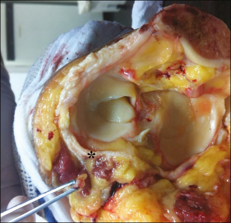
Its parent, the common peroneal nerve, arises from the sciatic nerve in the distal thigh, runs along the medial border of the biceps femoris tendon, crosses the lateral head of gastrocnemius, and then does the single most surgically consequential thing any peripheral nerve does: it winds subcutaneously around the neck of the fibula, tethered against bone with almost no soft tissue cover. It divides there, within the substance of peroneus longus, into the deep and superficial peroneal nerves.
Two clinical consequences follow immediately, and they are the reason this nerve is examined so heavily:
- A short, fixed, superficial segment against bone makes the nerve uniquely vulnerable to compression, traction and direct injury - from a plaster, a positioning error, a knee dislocation, a fibular neck fracture or a surgeon's retractor.
- A tiny sensory territory in the middle of a vulnerable compartment makes the first dorsal web space the most efficient sensory test in the lower limb.
The deep peroneal nerve runs the entire length of the anterior compartment, from the proximal intermuscular septum to the ankle, closely applied to the interosseous membrane. It is the most pressure-sensitive structure in that compartment, and its only cutaneous output is the first dorsal web space.
The result is that altered sensation between the hallux and second toe is the earliest reliable clinical sign of anterior compartment syndrome of the leg - earlier than weakness, far earlier than pulselessness, and detectable in a conscious patient with a single question. It costs nothing to test and it must be tested and documented in every tibial fracture, every high-energy leg injury, every tight cast and every post-fasciotomy review.
The corollary matters just as much: in an obtunded, intubated or regionally blocked patient this sign is unavailable, and objective compartment pressure measurement becomes mandatory rather than optional.
Course, Motor Supply and Relations
Origin and root value
- Terminal branch of the common peroneal nerve, itself the smaller terminal division of the sciatic nerve (L4, L5, S1, S2).
- Deep peroneal nerve root value: L4, L5 and S1, predominantly L5.
- The common peroneal nerve gives, before it divides, the recurrent articular branch to the knee and contributes the lateral sural cutaneous nerve to the calf. Both leave above the fibular neck, which is why they can be spared in a distal lesion.
The fibular neck
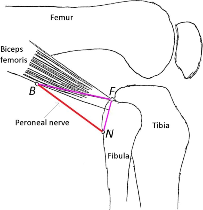
- The common peroneal nerve passes from the popliteal fossa along the medial border of the biceps femoris tendon, crosses the lateral head of gastrocnemius, and winds around the neck of the fibula approximately 2 to 4 cm distal to the tip of the fibular head.
- Here it lies subcutaneously, directly on periosteum, covered only by skin, fat and deep fascia. It is palpable and rollable in most people.
- It then passes deep to the arcade of the peroneus longus - the fibrous proximal edge of the muscle - and divides within the substance of the muscle into the deep and superficial peroneal nerves.
Anterior compartment course
- The deep peroneal nerve pierces the anterior intermuscular septum to enter the anterior compartment.
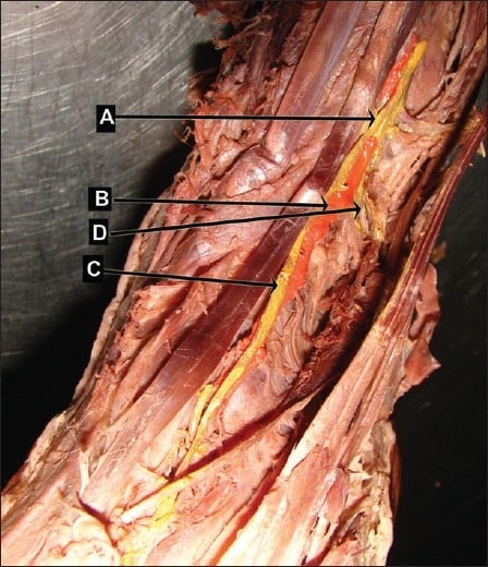
- It runs distally on the anterior surface of the interosseous membrane, initially between tibialis anterior and extensor digitorum longus, and in the distal leg between tibialis anterior and extensor hallucis longus.
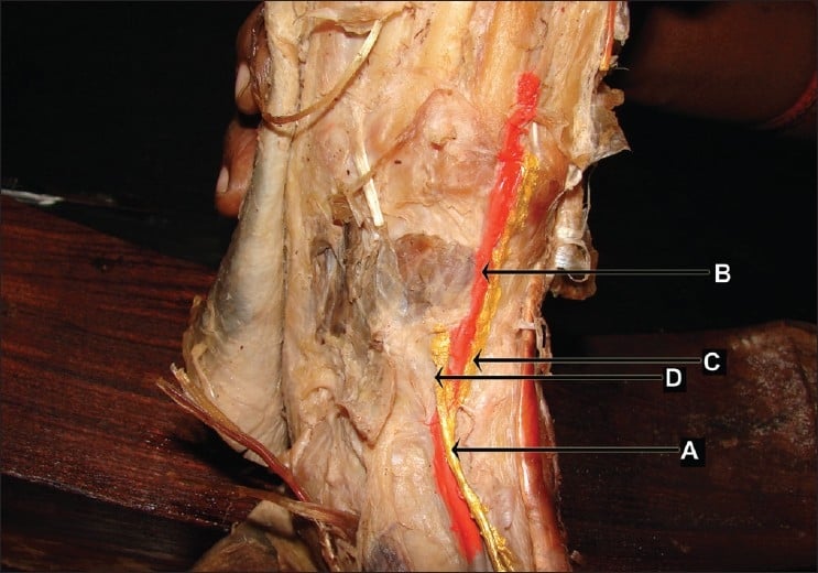
- It is joined by the anterior tibial artery shortly after entering the compartment. The nerve and artery travel together for the whole length of the compartment, the nerve crossing from lateral, to anterior, and back to the lateral side of the artery.
At the ankle
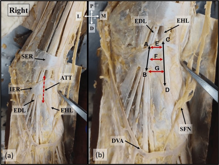
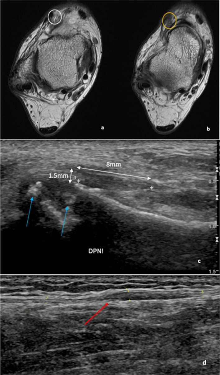
- The nerve passes beneath the inferior extensor retinaculum with the artery, in the anterior tarsal tunnel.
- At the level of the ankle joint it lies between the tendons of extensor hallucis longus and extensor digitorum longus, immediately lateral to the artery, which becomes the dorsalis pedis.
Terminal division
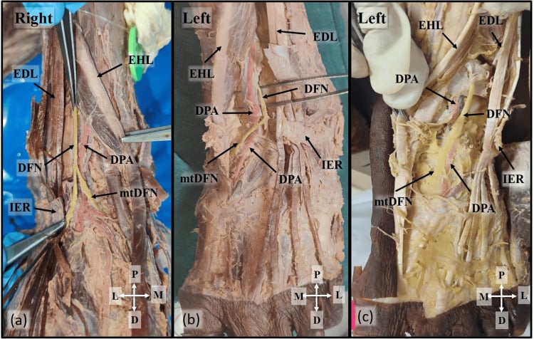
- Lateral terminal branch: passes laterally deep to extensor digitorum brevis, expands into a small ganglion-like swelling, and supplies extensor digitorum brevis and extensor hallucis brevis, with articular twigs to the tarsal and tarsometatarsal joints.
- Medial terminal branch: continues with the dorsalis pedis artery onto the dorsum of the foot, passes over the first intermetatarsal space, and divides into the dorsal digital nerves of the first web space - supplying the adjacent sides of the hallux and second toe. It frequently gives a small motor twig to the first dorsal interosseous.
The parent nerve is developed in common peroneal nerve anatomy, its sibling in superficial peroneal nerve anatomy, and the emergency it detects in compartment syndrome.
Surface Anatomy and Examination
Palpating the nerve
- Find the fibular head as a subcutaneous prominence below and behind the lateral tibial plateau.
- Move 2 to 4 cm distally and slightly anteriorly onto the neck of the fibula. In most people the common peroneal nerve can be rolled under the fingertip as a firm cord.
- Percussion here produces a Tinel sign radiating into the lateral leg and dorsum of the foot if the nerve is irritable.
- The anterior tarsal tunnel is found by palpating between the extensor hallucis longus and extensor digitorum longus tendons at the level of the ankle joint, just lateral to the dorsalis pedis pulse.
Motor examination
- Ankle dorsiflexion against resistance - tibialis anterior.
- Hallux dorsiflexion against resistance - extensor hallucis longus. Often the first to weaken.
- Lesser toe extension - extensor digitorum longus.
- Eversion against resistance - peroneus longus and brevis, superficial peroneal nerve. This is the discriminator.
- Inversion in plantarflexion - tibialis posterior, tibial nerve but L5 root. Weakness here points to a root lesion.
- Hip abduction - gluteus medius, L5 through the superior gluteal nerve. Weakness confirms a root lesion.
- Gait: a high-stepping steppage gait with a foot slap on heel strike.
- Wasting: flattening of the anterior compartment against the lateral tibial border; wasting of extensor digitorum brevis over the dorsolateral midfoot.
- Deformity: an established palsy produces an equinus contracture; check passive dorsiflexion with the knee flexed and extended.
- Skin: trophic change or an ulcer over the dorsum in a chronic palsy.
Named tests and manoeuvres
- How to Perform
- Light touch and pinprick between the hallux and second toe, compared with the other foot
- Positive Finding
- Reduced or altered sensation
- What It Means / False Positives
- Earliest sign in anterior compartment syndrome; abolished by regional block
- How to Perform
- Passively plantarflex the toes and ankle to stretch the anterior compartment
- Positive Finding
- Severe pain out of proportion
- What It Means / False Positives
- The most sensitive clinical sign of compartment syndrome; unreliable in a blocked or obtunded patient
- How to Perform
- Percuss 2-4 cm distal to the fibular head
- Positive Finding
- Paraesthesia radiating into the dorsum of the foot
- What It Means / False Positives
- Localises an irritable common peroneal nerve; mildly uncomfortable in normal limbs, so compare sides
- How to Perform
- Resist eversion with the foot in plantarflexion
- Positive Finding
- Weakness indicates superficial peroneal involvement
- What It Means / False Positives
- Preserved eversion with foot drop localises to the deep peroneal nerve
- How to Perform
- Resist extension of the great toe
- Positive Finding
- Weakness before ankle dorsiflexion weakens
- What It Means / False Positives
- Detects partial or evolving lesions early
- How to Perform
- Percuss between EHL and EDL at the ankle
- Positive Finding
- Paraesthesia into the first web space
- What It Means / False Positives
- Supports anterior tarsal tunnel syndrome
- How to Perform
- Measure compartment pressure and subtract from the diastolic blood pressure
- Positive Finding
- Differential pressure below 30 mmHg sustained for more than 2 hours
- What It Means / False Positives
- The objective decompression threshold; a single dip is not the trigger and absolute pressures alone over-diagnose
- Conduction block across the fibular head on peroneal motor studies is the classic finding in a compressive lesion at the neck and, when present, predicts a good prognosis.
- Needle electromyography distinguishes the levels: denervation in tibialis anterior and extensor hallucis longus with a normal peroneus longus points to the deep peroneal nerve; involvement of peroneus longus as well points to the common peroneal nerve; involvement of the short head of biceps femoris points above the fibular neck; and involvement of gluteus medius or tibialis posterior points to the L5 root.
- In anterior tarsal tunnel syndrome the abnormalities are confined to extensor digitorum brevis, with prolonged peroneal distal motor latency and reduced amplitude.
- Two pitfalls in interpreting extensor digitorum brevis findings were specifically highlighted in the original clinical descriptions: an accessory peroneal nerve can preserve the response and mask the diagnosis, and fibrillation potentials in extensor digitorum brevis are often dismissed as shoe-wear artefact while prolonged latencies are dismissed as a cool limb. Warm the foot and interpret in context.
- Baseline studies are most informative at 3 to 4 weeks after an acute injury, once Wallerian degeneration has declared itself.
Complications
- Mechanism
- Pressure on the fibular neck in lithotomy or lateral decubitus
- How to Avoid It
- Pad the fibular neck, re-check after every position change
- What to Do
- Remove pressure, splint at neutral, electromyography at 3-4 weeks
- Mechanism
- First web sensation not tested, or masked by a regional block
- How to Avoid It
- Test and document before analgesia; measure differential pressure when unreliable
- What to Do
- Emergency four-compartment fasciotomy
- Mechanism
- Osteotomy performed too close to the fibular neck
- How to Avoid It
- Osteotomise the middle third, at least 10 cm below the fibular head
- What to Do
- Explore and repair or graft if divided
- Mechanism
- Correction of a severe valgus and flexion deformity lengthens the nerve
- How to Avoid It
- Anticipate in high-risk deformity; avoid tight dressings
- What to Do
- Remove dressings, flex the knee, re-examine, splint at neutral
- Mechanism
- Articular branch to the superior tibiofibular joint not divided
- How to Avoid It
- Divide the articular branch and address the joint at the index operation
- What to Do
- Revision with articular branch division and joint resection or arthrodesis
- Mechanism
- Sustained retraction on the neurovascular bundle
- How to Avoid It
- Release retraction periodically; retract the bundle with EHL as a unit
- What to Do
- Usually recovers; reassure and document
- Mechanism
- Palsy managed without a splint
- How to Avoid It
- Ankle-foot orthosis at neutral from day one
- What to Do
- Serial casting or tendo-Achilles lengthening at reconstruction
- Mechanism
- Wrong diagnosis, or compression at a distal site left unreleased
- How to Avoid It
- Localise with a Tinel sign and a diagnostic block first
- What to Do
- Re-examine for a distal extensor hallucis brevis entrapment
Clinical Relevance
Why this nerve dominates the diagnosis
The anterior compartment of the leg is the compartment most commonly affected by acute compartment syndrome, and the deep peroneal nerve runs its entire length. Nerve tissue is more sensitive to ischaemia than muscle, so the nerve declares the problem first.
Clinical sequence
- Pain out of proportion to the injury, poorly relieved by opioid analgesia.
- Pain on passive stretch - passive plantarflexion of the toes and ankle.
- Paraesthesia in the first dorsal web space - the first objective sign, and the one that is specific to this compartment.
- Weakness of hallux and then ankle dorsiflexion.
- Pallor, paralysis and pulselessness are late and unreliable - a normal dorsalis pedis pulse never excludes the diagnosis.
The objective threshold
- McQueen and Court-Brown monitored anterior compartment pressure continuously for 24 hours in 116 tibial diaphyseal fractures. Using an absolute pressure of 30 mmHg as a threshold would have committed 43 per cent of patients to fasciotomy, and 40 mmHg would still have led to 23 per cent unnecessary fasciotomies.
- Using a differential pressure - diastolic blood pressure minus compartment pressure - of less than 30 mmHg identified all three genuine compartment syndromes with no missed cases.
- Practical rule: measure the differential pressure, not the absolute pressure, and decompress when it is below 30 mmHg for more than two hours. The duration is part of the threshold and is routinely dropped: McQueen and Duckworth's later review states the criterion as "the differential pressure being less than 30 mmHg for more than 2 h", and it is that sustained reading which carries a sensitivity of 94 per cent and a specificity of 98 per cent, against a sensitivity of only 13 to 64 per cent for clinical findings. A single transient dip below 30 is not the trigger; a single reading above it is not reassurance either. Measure within 5 cm of the fracture, where pressures are highest.
Fasciotomy and the nerve
- The standard two-incision, four-compartment fasciotomy uses an anterolateral incision, roughly 2 cm anterior to the fibular shaft, to open the anterior and lateral compartments.
- The superficial peroneal nerve lies in the lateral compartment and is the nerve at risk from this incision; the deep peroneal nerve lies deeper, on the interosseous membrane, and is at risk from over-enthusiastic deep dissection.
- Release the anterior compartment fascia along its whole length with long scissors, staying just anterior to the intermuscular septum, and identify the septum before releasing the lateral compartment.
- Document first web sensation and dorsiflexion power after fasciotomy as the new baseline.
Chronic exertional compartment syndrome
- Exercise-induced anterior compartment pain with transient first web paraesthesia and foot drop that resolves with rest is the classic history in a runner.
- Diagnosis is by post-exercise compartment pressure measurement; treatment is fasciotomy or fasciectomy of the affected compartments.
- Do not confuse it with a medial tibial stress syndrome or a stress fracture, neither of which produces a neurological symptom.
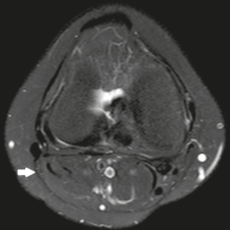
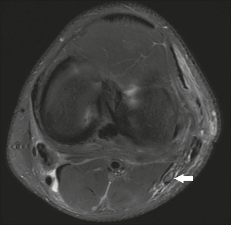
Surgical Relevance
- Fibular neck: there is no safe zone. The nerve lies subcutaneously on the neck, roughly 2 to 4 cm distal to the tip of the fibular head. Any work on the proximal fibula requires the nerve to be identified.
- Fibular osteotomy for a proximal tibial osteotomy: perform it in the middle third of the shaft, at least 10 cm distal to the fibular head, keeping well proximal to the syndesmosis so that ankle stability is preserved. The alternatives - fibular head excision or proximal tibiofibular joint release - both require direct visualisation of the nerve.
- Lateral tibial plateau plating: the nerve is behind the fibular neck; keep dissection anterior to the fibular head and never place a retractor blindly around the posterolateral proximal tibia.
- Anterior compartment fasciotomy: make the lateral incision approximately 2 cm anterior to the fibular shaft, identify the anterior intermuscular septum, and release the anterior compartment just anterior to it. The superficial peroneal nerve lies immediately behind the septum in the lateral compartment.
- Anterior ankle approach: work medial to extensor hallucis longus and retract the tendon and the neurovascular bundle laterally as a unit.
- Ankle arthroscopy: never use an anterocentral portal.
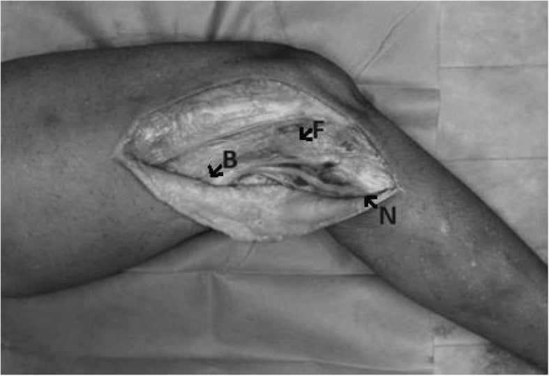
Exposing the nerve at the fibular neck
- Position: lateral decubitus or supine with a bump, knee flexed to relax the nerve.
- Incision: curvilinear, centred over the fibular head, extending posterior to the biceps femoris tendon proximally and distally over the neck.
- Landmark: identify the biceps femoris tendon and follow its medial border distally. The common peroneal nerve is always on the medial side of this tendon - this is the single most reliable way to find it.
- Trace the nerve distally over the neck, divide the arcade of peroneus longus, and follow the division into the deep and superficial branches.
- Do not skeletonise the nerve or strip it circumferentially over a long segment; it depends on its longitudinal epineurial vessels.
Decompression for compressive neuropathy
- Release the deep fascia over the nerve, the arcade of the peroneus longus, and any fibrous band at the entry into the anterior compartment.
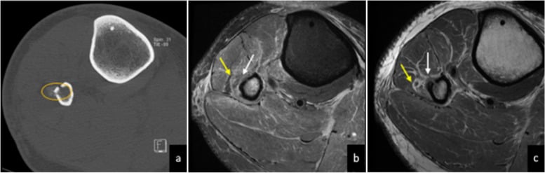
- Look for an intraneural ganglion if the palsy was spontaneous - a fusiform swelling of the nerve, sometimes with visible cystic fluid. Decompress the cyst, divide the articular branch to the superior tibiofibular joint, and address the joint, or the ganglion recurs.
- Postoperative: early mobilisation, an ankle-foot orthosis until dorsiflexion returns, and serial examination.
Prevention checklist for the whole team
- Pad the fibular neck of the dependent leg in every lateral case and every lithotomy case, and re-check after position changes.
- Document the pre-operative neurological status of both legs before any knee or proximal tibial procedure.
- Do not allow a circumferential dressing or cast to sit tightly over the fibular neck or the dorsum of the foot.
- Examine and document first web sensation and hallux dorsiflexion in recovery, before regional analgesia wears off, and again the next morning.
- Treat a new deficit as a mechanical problem until proven otherwise - remove dressings, flex the knee, and re-examine, rather than ordering an investigation first.
Guidelines, Registries & Global Practice
Anatomical variation across populations
- The level at which the common peroneal nerve divides varies; in some limbs it divides above the fibular neck and in others within the peroneus longus substance, which changes what a surgeon expects to see at exposure.
- Peroneus tertius is absent in a meaningful proportion of limbs, with reported absence rates varying widely across populations. Its absence does not produce a deficit and it should not be searched for as a landmark.
- The accessory peroneal nerve is reported in roughly a fifth to a quarter of limbs in electrodiagnostic and cadaveric series, with prevalence differing between populations. It is the single most important normal variant for interpreting a peroneal study.
- Anomalous fibrous bands at the peroneus longus arcade and at the entry to the anterior compartment are common and are a recognised cause of a spontaneous compressive neuropathy.
Differences in described technique
- Emphasis
- Identify and protect the common peroneal nerve for any proximal fibular or lateral proximal tibial procedure
- Practical Point
- The nerve is found on the medial border of the biceps femoris tendon - the most reliable landmark
- Emphasis
- Serial clinical assessment plus differential pressure monitoring in obtunded or blocked patients
- Practical Point
- Documented first web sensation on every review of a tibial fracture
- Emphasis
- Low threshold for four-compartment fasciotomy in high-energy tibial injury
- Practical Point
- A palpable pulse never excludes the diagnosis
- Emphasis
- Non-operative management of anterior tarsal tunnel syndrome first, with localisation before release
- Practical Point
- Address dorsal osteophytes and footwear, not just the retinaculum
- Emphasis
- Anticipate peroneal palsy after correction of severe valgus with flexion contracture
- Practical Point
- Remove dressings and flex the knee as the first response to a new deficit
Registry, service and resource considerations
- Arthroplasty registries record revision, not nerve injury. Peroneal palsy after total knee arthroplasty therefore appears far less often in registry data than in clinical practice, and its true incidence comes from institutional series.
- Compartment syndrome is a recognised source of litigation in every health system, and in every system the defence is the same: documented serial neurovascular examination, an explicit record of first web space sensation, a clear escalation plan, and prompt decompression once the threshold is reached.
- Regional anaesthesia policy varies internationally in high-risk tibial fractures. The unifying principle is that a dense block removes the earliest clinical sign, so where a block is used, objective pressure monitoring should be available.
- High-resource settings have continuous compartment pressure monitoring, high-resolution nerve ultrasound to detect an intraneural ganglion before exploration, and access to nerve transfer techniques for irrecoverable palsy.
- Limited-resource settings lose nothing on the diagnostic side. Testing the first dorsal web space, resisted hallux dorsiflexion and passive stretch pain requires no equipment and detects the emergency. The practical constraint is theatre access for timely fasciotomy, which makes early recognition and early escalation even more important.
- Orthotic provision is the real global inequity in this nerve. A well-fitted ankle-foot orthosis converts a disabling foot drop into a functional limb, and where orthotic services are limited, tendon transfer becomes a more attractive definitive option than it would be elsewhere.
MCQ Practice Points
Q: Where does the common peroneal nerve divide into deep and superficial branches? A: At the fibular neck, within the substance of peroneus longus, having passed beneath the arcade of that muscle roughly 2 to 4 cm distal to the tip of the fibular head.
Q: What is the entire cutaneous territory of the deep peroneal nerve? A: The first dorsal web space and the adjacent sides of the hallux and second toe. Nothing else.
Q: Which intrinsic muscles of the foot are supplied by the peroneal nerve? A: Extensor digitorum brevis and extensor hallucis brevis only, through the lateral terminal branch of the deep peroneal nerve. Every other intrinsic foot muscle is tibial.
Q: A patient has foot drop with preserved eversion. Where is the lesion? A: The deep peroneal nerve. Peroneus longus and brevis are supplied by the superficial peroneal nerve, so preserved eversion excludes a common peroneal lesion.
Q: What is the pressure threshold for fasciotomy? A: A differential pressure - diastolic blood pressure minus compartment pressure - of less than 30 mmHg. Absolute pressure thresholds of 30 or 40 mmHg lead to a high rate of unnecessary fasciotomy.
Q: What is the earliest reliable objective sign of anterior compartment syndrome of the leg? A: Altered sensation in the first dorsal web space, because the deep peroneal nerve traverses the whole compartment and nerve tissue fails before muscle.
Q: Why might extensor digitorum brevis responses be normal in genuine anterior tarsal tunnel syndrome? A: An accessory peroneal nerve arising from the superficial peroneal nerve passes behind the lateral malleolus to supply extensor digitorum brevis, bypassing the tunnel entirely.
Q: Where should a fibular osteotomy be performed during a lateral closing wedge proximal tibial osteotomy? A: In the middle third of the fibular shaft, at least 10 cm distal to the fibular head, keeping well proximal to the syndesmosis. The alternatives are fibular head excision or proximal tibiofibular joint release, both with the nerve visualised.
Q: Why does an intraneural ganglion of the common peroneal nerve recur after simple decompression? A: Because the cyst tracks along the articular branch from the superior tibiofibular joint. The articular branch must be divided and the joint addressed.
Q: Why is the anterocentral portal avoided in ankle arthroscopy? A: Because the deep peroneal nerve and the anterior tibial artery lie directly beneath it, between the extensor hallucis longus and extensor digitorum longus tendons.
Exam Viva Scenarios
Practise clinical reasoning and management decisions out loud
“A 22-year-old motorcyclist has a closed mid-shaft tibial fracture treated in a backslab six hours ago. He is requesting increasing analgesia. The foot is warm with a palpable dorsalis pedis pulse. What do you do?”
“A 68-year-old woman had a total knee arthroplasty yesterday for severe valgus osteoarthritis with a 20 degree fixed flexion deformity. She has an epidural running. This morning she cannot dorsiflex her ankle. What is your assessment and immediate management?”
“A 41-year-old recreational skier describes six months of burning pain over the dorsum of the right midfoot with numbness between the great and second toes. It is worst in ski boots and at the end of a day of walking. There is no weakness that she has noticed. Examination shows a Tinel sign between the extensor hallucis longus and extensor digitorum longus tendons at the ankle.”
Anatomy High Yield
- Roots: L4, L5, S1 - predominantly L5
- Divides from common peroneal at the fibular neck, in peroneus longus
- Runs on the interosseous membrane with the anterior tibial artery
- Ankle: between EHL and EDL, lateral to the artery
- Terminal: lateral branch to EDB, medial branch to the first web
Motor Supply
- Tibialis anterior
- Extensor hallucis longus
- Extensor digitorum longus
- Peroneus tertius
- Extensor digitorum brevis and extensor hallucis brevis
Key Numbers
- 30 mmHg: differential pressure threshold for fasciotomy
- 2-4 cm: nerve distal to the fibular head tip at the neck
- 10 cm: minimum distance below the fibular head for an osteotomy
- 27 per cent: electrophysiological peroneal lesions after one HTO series
- 2 cm: lateral fasciotomy incision anterior to the fibular shaft
Localising a Foot Drop
- Eversion preserved: deep peroneal nerve
- Eversion lost: common peroneal at the fibular neck
- Inversion weak: L5 root (tibialis posterior is tibial nerve)
- Hip abduction weak: L5 root
- Short head of biceps involved on EMG: above the fibular neck
Danger Points
- Fibular neck - positioning, plaster, fracture, surgery
- Anterior compartment - compartment syndrome
- Inferior extensor retinaculum - anterior tarsal tunnel
- Anterocentral ankle arthroscopy portal - never use it
- First intermetatarsal space - Lisfranc and Lapidus surgery
Evidence Base
The Differential Pressure Threshold for Fasciotomy
- Prospective continuous 24-hour anterior compartment pressure monitoring in 116 tibial diaphyseal fractures, with three genuine acute compartment syndromes (2.6 per cent)
- An absolute threshold of 30 mmHg would have committed 43 per cent of patients to fasciotomy and 40 mmHg would still have produced 23 per cent unnecessary fasciotomies
- A differential pressure - diastolic minus compartment pressure - of less than 30 mmHg identified every true case with no missed compartment syndromes and no sequelae at a minimum of six months
Anterior Tarsal Tunnel Syndrome - Clinical and Electrodiagnostic Description
- Describes entrapment of the deep peroneal nerve beneath the inferior extensor retinaculum, with the retinaculum as the roof, the fascia over talus and navicular as the floor, and four tendons, an artery, a vein and the nerve within
- Two patients had prolonged peroneal distal latencies with reduced amplitudes from extensor digitorum brevis, with electromyographic abnormalities confined to that muscle; both improved after surgical decompression
- Highlights two diagnostic pitfalls: an accessory peroneal nerve that bypasses the tunnel can mask extensor digitorum brevis findings, and fibrillations there are often dismissed as shoe-wear artefact while prolonged latencies are dismissed as a cool limb
Anterior Tarsal Tunnel Syndrome - Management Review
- Anterior tarsal tunnel syndrome is rare and frequently under-diagnosed, presenting with pain, weakness and sensory change in the foot and ankle
- Non-operative measures - reducing external compression, footwear modification, corticosteroid injection and physical therapy - should be attempted first
- Surgical decompression can be performed proximally at the extensor retinaculum or more distally at the tarsometatarsal level depending on the site of entrapment
Deep Peroneal Nerve Entrapment by Extensor Hallucis Brevis
- Entrapment of the deep peroneal nerve distal to the inferior extensor retinaculum, caused by the tendon of extensor hallucis brevis crossing the nerve
- The presentation mimicked anterior tarsal tunnel syndrome but the compression was at a more distal level
- Surgical decompression at the site of the crossing tendon relieved the symptoms
Prolonged Peroneal Nerve Dysfunction After High Tibial Osteotomy
- Pre-operative and post-operative electrophysiological study of 11 patients undergoing barrel-vault high tibial valgisation osteotomy, with the non-operated leg as control
- Three patients (27 per cent) had electrophysiologically detectable peroneal nerve lesions - one motor only, one sensory only, one both
- Only one of the three was clinically symptomatic; the lesions persisted for a prolonged period and could be permanent despite the absence of clinical symptoms
Complications After Medial Opening Wedge High Tibial Osteotomy
- Retrospective review of 46 medial opening wedge high tibial osteotomies, with a documented complication in 17 patients (36.9 per cent)
- The reported breakdown accounts for all 17: loss of correction in 7 (15.2 per cent), intra-operative lateral cortex fracture in 2, post-operative lateral cortex fracture in 2, deep venous thrombosis in 2, delayed union in 2 and symptomatic hardware in 2
- NEITHER PERONEAL NEUROPATHY NOR COMPARTMENT SYNDROME APPEARS IN THAT BREAKDOWN - both are present only as MeSH indexing terms on the record, not as findings
- Loss of correction was associated with a higher body mass index (mean 32.5 against 28.8) and with first-generation fixation devices
- The authors conclude the frequency and type of complications seem SIMILAR to those reported for the lateral closing technique
The Diagnosis of Acute Compartment Syndrome - Where the Two-Hour Rule Comes From
- States the decompression criterion in full: a differential pressure of less than 30 mmHg FOR MORE THAN 2 HOURS - the duration is part of the threshold, and it is the half most often dropped in teaching.
- Continuous pressure monitoring against that sustained criterion has a sensitivity of 94 per cent and a specificity of 98 per cent.
- Clinical findings alone have a sensitivity of only 13 to 64 per cent, with specificity 63 to 98 per cent.
- Recommends decompressing primarily on the sustained pressure criterion rather than waiting for clinical signs, because doing so shortens time to definitive treatment.
Superficial Peroneal Nerve Course - Defining the Neighbour
- Eighty-five legs from 44 cadavera dissected to define the course of the superficial peroneal nerve from its origin to its terminal dorsal cutaneous branches
- The nerve ran within the lateral compartment in 73 per cent of legs, crossed into the anterior compartment in 14 per cent, and divided into branches in both compartments in 12 per cent
- The nerve or its branches pierced the crural fascia between 3 and 18 cm proximal to the lateral malleolus